. 2014 Jan;141(1):112-23.
doi: 10.1242/dev.098871. Epub 2013 Nov 27.

Induction of endocycles represses apoptosis independently of differentiation and predisposes cells to genome instability

Affiliations

Induction of endocycles represses apoptosis independently of differentiation and predisposes cells to genome instability

Christiane Hassel et al. Development. 2014 Jan.

Abstract

The endocycle is a common developmental cell cycle variation wherein cells become polyploid through repeated genome duplication without mitosis. We previously showed that Drosophila endocycling cells repress the apoptotic cell death response to genotoxic stress. Here, we investigate whether it is differentiation or endocycle remodeling that promotes apoptotic repression. We find that when nurse and follicle cells switch into endocycles during oogenesis they repress the apoptotic response to DNA damage caused by ionizing radiation, and that this repression has been conserved in the genus Drosophila over 40 million years of evolution. Follicle cells defective for Notch signaling failed to switch into endocycles or differentiate and remained apoptotic competent. However, genetic ablation of mitosis by knockdown of Cyclin A or overexpression of fzr/Cdh1 induced follicle cell endocycles and repressed apoptosis independently of Notch signaling and differentiation. Cells recovering from these induced endocycles regained apoptotic competence, showing that repression is reversible. Recovery from fzr/Cdh1 overexpression also resulted in an error-prone mitosis with amplified centrosomes and high levels of chromosome loss and fragmentation. Our results reveal an unanticipated link between endocycles and the repression of apoptosis, with broader implications for how endocycles may contribute to genome instability and oncogenesis.

Keywords: Apoptosis; Cell cycle; Drosophila; Endocycle; Oogenesis.


Figures

Fig. 1.
Fig. 1.
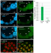
The repression of apoptosis during ovarian endocycles is conserved in the genus Drosophila. (A) Drawing of a longitudinal section through a stage-10 egg chamber. Somatic follicle cells (magenta) surround the germline nurse cells and oocyte. (B) An ovariole with egg chambers that mature through 14 stages (S) as they migrate posteriorly (left to right) down the ovariole (King, 1970). Germline and somatic follicle stem cells and their daughters (magenta) divide mitotically in the germarium. (C,D) TUNEL, PH3 and DAPI labeling of ovaries from D. melanogaster (Dmel) (C) and D. virilis (Dvir) (D) females 24 hours after irradiation. Arrowheads point to three of the many TUNEL-labeled follicle cells in stage 6 (S6) in both species. The germarium, follicle cells and nurse cells are indicated. See Table 1 for a complete list of species analyzed. Scale bars: 50 μm. FC, follicle cell; g, germarium; NC, nurse cell.
Fig. 2.
Fig. 2.
Notch signaling is required to repress apoptosis in follicle cells. (A-A′′) Presenilin (Psn) mutant follicle cell clone in a stage-8 egg chamber identified by the absence of green fluorescent protein (GFP; not green) (A). Psn mutant clones had small nuclei (A′) and retained the expression of the immature follicle cell marker Fasciclin III (A′-A′′), indicating that they had failed to switch into the endocycle and differentiate. (B-C′′) After irradiation, some Psn mutant follicle cells labeled for TUNEL (B-B′′) or anti-cleaved Caspase 3 (C-C′′), whereas wild-type endocycling follicle cells in the same chamber did not. (D) Quantification of cleaved Caspase labeling in Psn mutant (Psn-) and control (Psn+) follicle cells in the same egg chambers with (IR) and without (No IR) irradiation (n=3 biological replicates of ten PsnC1 clones each, representing ∼2000 total cells, **P=0.003 by Student’s t-test). Scale bars: 25 μm.
Fig. 3.
Fig. 3.
Induction of precocious endocycles in follicle cells. (A) Western blot for Cyclin A (CycA) and β-actin loading control from ovarian extracts of y w control (lane 1), hsp70:GAL4; UAS:CycARNAi without (lane 2) or with (lane 3) seven pulses of heat induction. (B,C) Anti-PH3 and DAPI labeling in control (B) and CycARNAi-expressing (C) ovaries. (D) Total DAPI fluorescence in nuclei of follicle cells of stage 4 (n=25) and stage 10A (n=20). ***P<0.0001, **P=0.0002. (E) The percentage of PH3-positive cells in control (5.58%) and CycARNAi (0.18%) ovaries between stages 1 and 6 (two biological replicates representing in total 80 ovarioles and ∼14,520 wild-type and ∼5600 CycARNAi cells, ***P<0.0001 by Student’s t-test). (F,G) DAPI and BrdU labeling in control (F) and CycARNAi-expressing (G) ovaries. (H) Cyclin E/Cdk2 activity oscillates in induced endocycling cells (iECs). Anti-MPM-2 labeling of a stage-4 egg chamber with follicle cells in a precocious endocycle induced by fzr overexpression. Histone locus bodies are indicated by arrowheads. (I) A wild-type control clone of follicle cells expressing RFP in a stage-6-7 egg chamber (arrow). (J,J′) A clone of follicle cells expressing CycARNAi and RFP in a stage-6 egg chamber (arrow). Scale bars: 15 μm. NC, nurse cell.
Fig. 4.
Fig. 4.
iECs repress apoptosis downstream of ATM. (A-B′) DAPI (A,B) and anti-cleaved Caspase 3 labeling (A′,B′) of ovaries 24 hours after irradiation with 4000 rads of gamma rays from hsp70:GAL4; UAS:CycARNAi females without (A,A′) or with (B,B′) seven heat inductions. Arrows in A′ indicate two cells labeled with anti-cleaved Caspase 3. (C) The average percentage of anti-cleaved Caspase 3-positive follicle cells in control (9.05%) and CycARNAi-expressing (0.17%) ovaries between stages 1 and 6, 24 hours after irradiation (two biological replicates representing in total 80 ovarioles and ∼14,520 wild-type and ∼5600 CycARNAi cells, ***P<0.0001 by Student’s t-test). (D,D′) DAPI (D) and anti-cleaved Caspase 3 labeling (D′) of ovaries from hsp70:GAL4; UAS:fzr females 24 hours after irradiation with 4000 rads of gamma rays. (E,F) Anti-γH2Av labeling of repair foci within UAS:fzr iECs without (E) or with (F) irradiation. Scale bars: 15 μm for A-B′,D,D′; 5 μm for E,F.
Fig. 5.
Fig. 5.
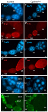
iECs repress apoptosis independently of Notch signaling and differentiation. (A-F′) Ovaries from hsp70:GAL4; UAS:CycARNAi females without (control A,A′,C,C′,E,E′) or with (B,B′,D,D′,F,F′) seven heat inductions were labeled with DAPI (A-F) and the indicated antibodies (A′-F′). The brightest FasIII labeling (E′,F′) corresponds to polar follicle cells. Scale bars: 15 μm.
Fig. 6.
Fig. 6.
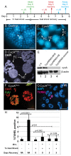
Recovery from CycARNAi expression results in polytene chromosomes and persistent repression of apoptosis. (A) Timeline of the CycA RNAi recovery experiment. Similar colors represent matched irradiation and assay 24 hours later. (B,C) Wide-field image of DAPI labeled egg chambers from wild-type control (B) or CycARNAi females on day 5 of recovery (C). Arrows in C point to three follicle cell (FC) nuclei with polytene chromosomes and one nurse cell (NC) nucleus. (D) Confocal image of DAPI labeled polytene chromosomes in follicle cells (FC) from a CycARNAi recovery female. (E) Cyclin A protein is undetectable upon recovery from CycARNAi expression. Western blot of ovarian protein extracts for Cyclin A and β-actin (loading control) proteins from y w control strain (lane 1), hsp70:GAL4: UAS:CycARNAi without heat induction (lane 2) or after seven heat inductions with 1 day (lane 3) or 5 days (lane 4) of recovery. (F) EdU labeling of polytene follicle cells on day 3 of recovery. (G) TUNEL labeling of one follicle cell undergoing apoptosis after IR on day 5 of recovery. (H) Most follicle cells do not apoptose upon recovery from CycA RNAi. Quantification of TUNEL-positive cells on days 0 and 3 of recovery with or without IR. Control females were not heat shocked and did not express CycARNAi (n=∼2800 heat shocked or 7260 control follicle cells per sample). Lines indicate P values for pairwise comparisons. The dotted line indicates the P value for the comparison of no IR treatment without heat shock to no IR treatment on day 3 of recovery from heat shock.
Fig. 7.
Fig. 7.
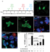
Recovery from fzr/Cdh1 overexpression results in an error-prone mitosis and apoptosis. (A) Experimental timeline for the UAS:fzr overexpression and recovery experiment. (B-D) Confocal image of anti-PH3 labeling of anaphase chromosomes in single follicle cells from control (B) or hsp70:GAL4; UAS:fzr females (C,D) on day 3 of recovery from heat shock. Arrowheads in D point to chromosome fragments. (E) Centrosome amplification and clustering in two follicle cells on day 3 of recovery labeled with DAPI and antibodies against the centrosome protein Centrosomin (Cnn). (F) TUNEL labeling (arrow) of one polyploid follicle cell undergoing spontaneous apoptosis on day 3 of recovery. (G) iECs recovering from fzr overexpression regain apoptotic competence. Quantification of TUNEL-positive cells on days 0 and 3 of recovery with or without IR. Control females were not heat shocked and did not express UAS:fzr (n=∼2800 heat shocked or 7260 control follicle cells per sample). Lines indicate P values for pairwise comparisons. The dotted line indicates the P value for the comparison of no IR treatment without heat shock to no IR treatment on day 3 of recovery from heat shock. Scale bars: 5 μm.
Fig. 8.
Fig. 8.
Endocycles repress apoptosis and contribute to aneuploidy. Notch signaling normally induces follicle cells to switch from mitotic cycles to endocycles and is associated with the repression of the apoptotic response to DNA damage. Genetic ablation of mitosis by knockdown of Cyclin A or overexpression of fzr results in induced endocycling cells (iECs), which repress apoptosis independently of Notch signaling or differentiation. Both developmental endocycling cells and iECs repress the apoptotic response to DNA damage downstream of ATM. Upon recovery from fzr overexpression, iECs regain apoptotic competence and undergo an error-prone polyploid mitosis. Although this error-prone mitosis probably results in severe aneuploidy and the death of many of these cells, some may survive.

Similar articles

Cited by

References

    1. Bachtrog D., Thornton K., Clark A., Andolfatto P. (2006). Extensive introgression of mitochondrial DNA relative to nuclear genes in the Drosophila yakuba species group. Evolution 60, 292–302 - PubMed
    1. Bartkova J., Rezaei N., Liontos M., Karakaidos P., Kletsas D., Issaeva N., Vassiliou L. V., Kolettas E., Niforou K., Zoumpourlis V. C., et al. (2006). Oncogene-induced senescence is part of the tumorigenesis barrier imposed by DNA damage checkpoints. Nature 444, 633–637 - PubMed
    1. Berry D. L., Baehrecke E. H. (2007). Growth arrest and autophagy are required for salivary gland cell degradation in Drosophila. Cell 131, 1137–1148 - PMC - PubMed
    1. Boveri T. (2008). Concerning the origin of malignant tumours by Theodor Boveri. Translated and annotated by Henry Harris. J. Cell Sci. 121 Suppl., S1–S84 - PubMed
    1. Brand A. H., Perrimon N. (1993). Targeted gene expression as a means of altering cell fates and generating dominant phenotypes. Development 118, 401–415 - PubMed

Publication types

MeSH terms

LinkOut - more resources