

Intratumoral hepatic stellate cells as a poor prognostic marker and a new treatment target for hepatocellular carcinoma
- PMID: 24278260
- PMCID: PMC3835887
- DOI: 10.1371/journal.pone.0080212
Intratumoral hepatic stellate cells as a poor prognostic marker and a new treatment target for hepatocellular carcinoma
Abstract
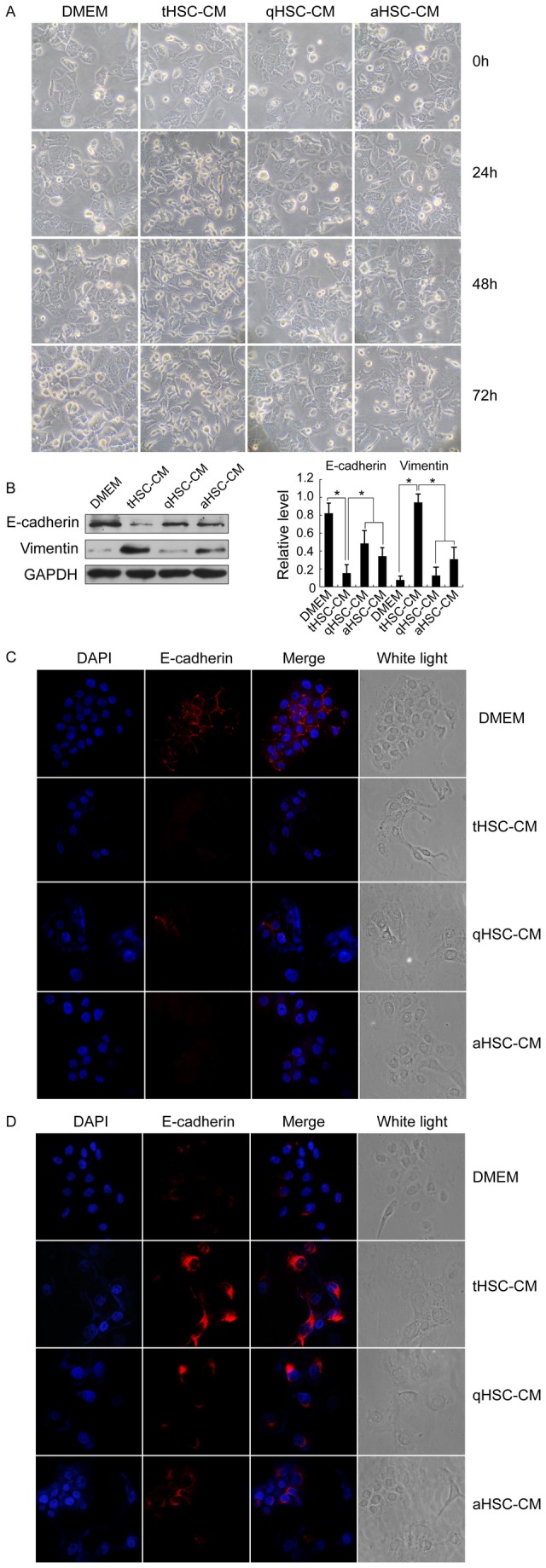
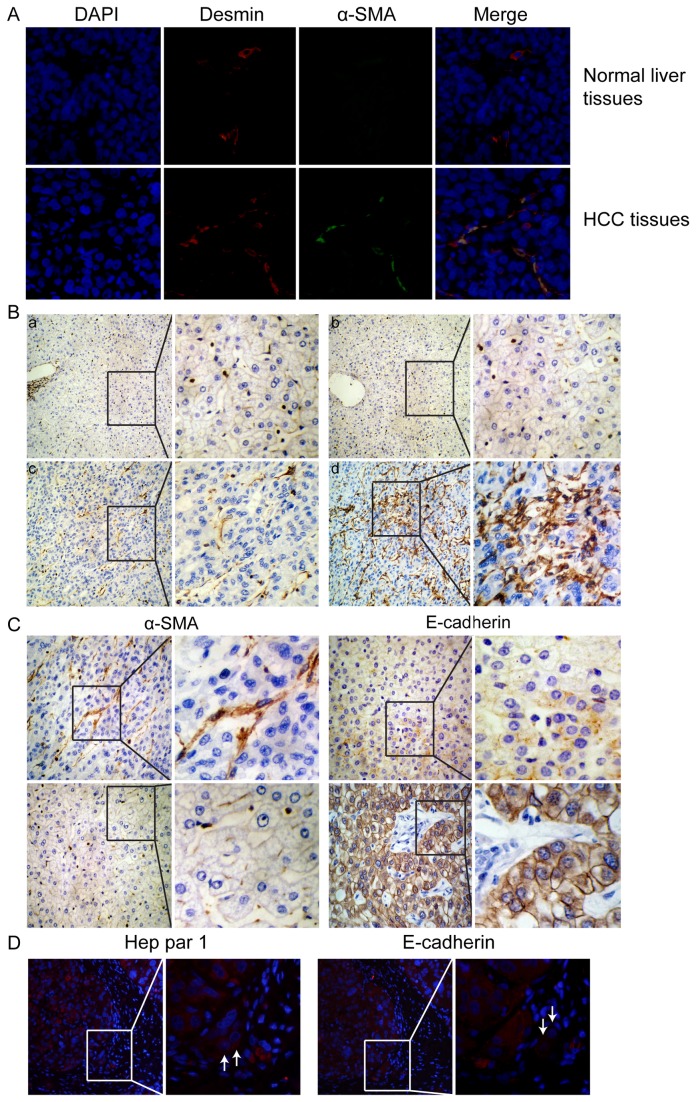
Hepatic stellate cells (HSCs), a specialized stromal cytotype in the liver, have been demonstrated to actively contribute to hepatocellular carcinoma (HCC) development. However, the previous studies were performed using HSC cell lines, and the prognostic value of intratumoral HSCs (tHSCs) was unclear. Here we isolated tHSCs from fresh human HCC tissues, and analyzed the abilities of tHSCs to promote HCC progression by using in vitro assays for cell viability, migration and invasion as well as epithelial-mesenchymal transition (EMT) phenotype. 252 HCC patients who underwent hepatectomy were enrolled for analysis of tHSCs and E-cadherin expression in tumor tissues, and 55 HCC patients for analysis of tHSCs in tumor tissues and circulating tumor cells (CTCs) in blood. Prognostic factors were then identified. The results showed that coculture of tHSCs with HCC cells had a stronger effect on HCC cell viability, migration and invasion, accompanied with the acquisition of epithelial-mesenchymal transition (EMT) phenotype. In vivo cotransplantation of HCC cells with tHSCs into nude mice more efficiently promoted tumor formation and growth. Icaritin, a known apoptosis inducer of HSCs, was demonstrated to effectively inhibit tHSC proliferation in vitro and tHSC-induced HCC-promoting effects in vivo. Clinical evidence indicated that tHSCs were rich in 45% of the HCC specimens, tHSC-rich subtypes were negatively correlated either with E-cadherin expression in tumor tissues (r = -0.256, p < 0.001) or with preoperative CTCs in blood (r = -0.287, p = 0.033), and were significantly correlated with tumor size (p = 0.027), TNM staging (p = 0.018), and vascular invasion (p = 0.008). Overall and recurrence-free survival rates of tHSC-rich patients were significantly worse than those for tHSC-poor patients. Multivariate analysis revealed tHSC-rich as an independent factor for overall and recurrence-free survival. In conclusion, tHSCs provide a promising prognostic biomarker and a new treatment target for HCC.
Conflict of interest statement
Figures


Similar articles
-
Inhibition of T-cell responses by intratumoral hepatic stellate cells contribute to migration and invasion of hepatocellular carcinoma.Clin Exp Metastasis. 2011 Oct;28(7):661-74. doi: 10.1007/s10585-011-9399-3. Epub 2011 Jun 30. Clin Exp Metastasis. 2011. PMID: 21717117
-
T-cell apoptosis induced by intratumoral activated hepatic stellate cells is associated with lung metastasis in hepatocellular carcinoma.Oncol Rep. 2013 Sep;30(3):1175-84. doi: 10.3892/or.2013.2571. Epub 2013 Jun 27. Oncol Rep. 2013. PMID: 23807027
-
Periostin involved in the activated hepatic stellate cells-induced progression of residual hepatocellular carcinoma after sublethal heat treatment: its role and potential for therapeutic inhibition.J Transl Med. 2018 Nov 6;16(1):302. doi: 10.1186/s12967-018-1676-3. J Transl Med. 2018. PMID: 30400797 Free PMC article.
-
Hepatic Stellate Cell: A Potential Target for Hepatocellular Carcinoma.Curr Mol Pharmacol. 2020;13(4):261-272. doi: 10.2174/1874467213666200224102820. Curr Mol Pharmacol. 2020. PMID: 32091349 Review.
-
Cancer Stem Cell and Hepatic Stellate Cells in Hepatocellular Carcinoma.Technol Cancer Res Treat. 2023 Jan-Dec;22:15330338231163677. doi: 10.1177/15330338231163677. Technol Cancer Res Treat. 2023. PMID: 36938618 Free PMC article. Review.
Cited by
-
The biological and clinical importance of epithelial-mesenchymal transition in circulating tumor cells.J Cancer Res Clin Oncol. 2015 Feb;141(2):189-201. doi: 10.1007/s00432-014-1752-x. Epub 2014 Jun 26. J Cancer Res Clin Oncol. 2015. PMID: 24965746 Review.
-
Role of TRP Channels in Liver-Related Diseases.Int J Mol Sci. 2023 Aug 7;24(15):12509. doi: 10.3390/ijms241512509. Int J Mol Sci. 2023. PMID: 37569884 Free PMC article. Review.
-
Midkine promotes hepatocellular carcinoma metastasis by elevating anoikis resistance of circulating tumor cells.Oncotarget. 2017 May 16;8(20):32523-32535. doi: 10.18632/oncotarget.15808. Oncotarget. 2017. PMID: 28430645 Free PMC article.
-
In vitro and in vivo study: Ethanolic extract leaves of Azadirachta indica Juss. variant of Indonesia and Philippines suppresses tumor growth of hepatocellular carcinoma by inhibiting IL-6/STAT3 signaling.F1000Res. 2022 Apr 29;11:477. doi: 10.12688/f1000research.109557.1. eCollection 2022. F1000Res. 2022. PMID: 37829248 Free PMC article.
-
Hepatic stellate cells - from past till present: morphology, human markers, human cell lines, behavior in normal and liver pathology.Rom J Morphol Embryol. 2020 Jul-Sep;61(3):615-642. doi: 10.47162/RJME.61.3.01. Rom J Morphol Embryol. 2020. PMID: 33817704 Free PMC article. Review.
References
Publication types
MeSH terms
Substances
Grants and funding
LinkOut - more resources
Full Text Sources
Other Literature Sources
Medical
