. 2013 Nov-Dec;15(6):426-41.
doi: 10.1111/ede.12052.

Deficiency of zebrafish fgf20a results in aberrant skull remodeling that mimics both human cranial disease and evolutionarily important fish skull morphologies

Affiliations

Deficiency of zebrafish fgf20a results in aberrant skull remodeling that mimics both human cranial disease and evolutionarily important fish skull morphologies

W James Cooper et al. Evol Dev. 2013 Nov-Dec.

Abstract

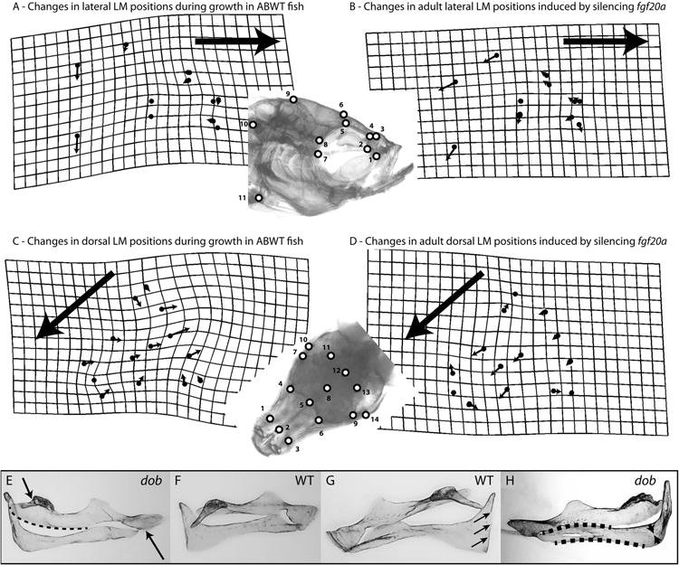
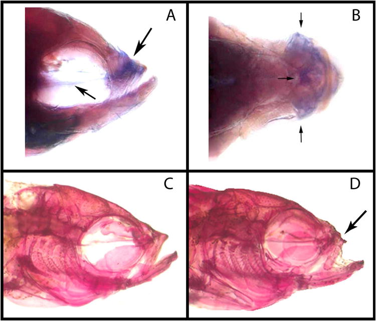
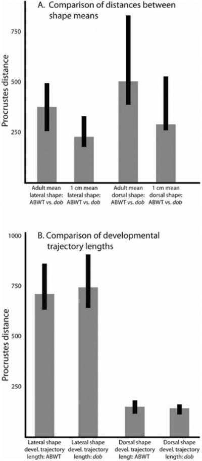
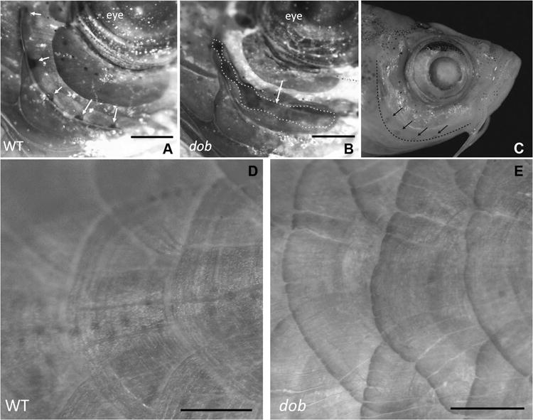
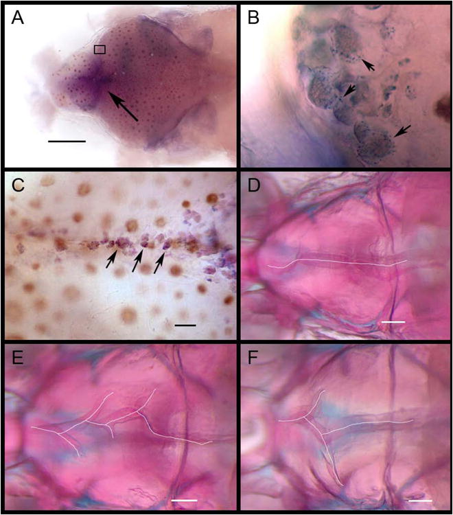
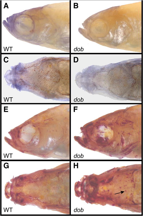
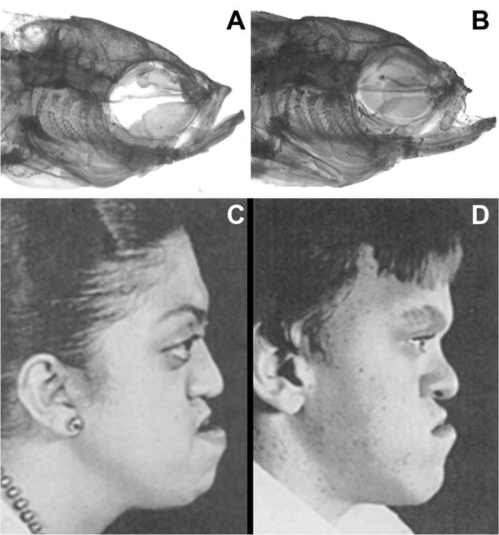
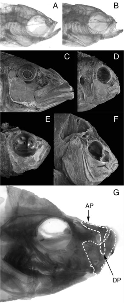
The processes that direct skull remodeling are of interest to both human-oriented studies of cranial dysplasia and evolutionary studies of skull divergence. There is increasing awareness that these two fields can be mutually informative when natural variation mimics pathology. Here we describe a zebrafish mutant line, devoid of blastema (dob), which does not have a functional fgf20a protein, and which also presents cranial defects similar to both adaptive and clinical variation. We used geometric morphometric methods to provide quantitative descriptions of the effects of the dob mutation on skull morphogenesis. In combination with "whole-mount in situ hybridization" labeling of normal fgf20a expression and assays for osteoblast and osteoclast activity, the results of these analyses indicate that cranial dysmorphologies in dob zebrafish are generated by aberrations in post-embryonic skull remodeling via decreased osteoblasotgenesis and increased osteoclastogenesis. Mutational effects include altered skull vault geometries and midfacial hypoplasia that are consistent with key diagnostic signs for multiple human craniofacial syndromes. These phenotypic shifts also mimic changes in the functional morphology of fish skulls that have arisen repeatedly in several highly successful radiations (e.g., damselfishes and East-African rift-lake cichlids). Our results offer the dob/fgf20a mutant as an experimentally tractable model with which to examine post-embryonic skull development as it relates to human disease and vertebrate evolution.


Figures

Figure 1
Figure 1
Depictions of skull and upper jaw bone shape transformations that occurred as a result of normal development or which were induced by silencing fgf20a signaling. The lateral and dorsal LM analyzed in this study are depicted in the inserts. The grids and vectors in panels A-D describe the differences between the mean LM configurations (i.e., consensus configurations calculated after Procrustes superimposition of the LM data) of 2 datasets. Deformation grids depict the minimal amount of “bending” needed to transform one mean LM configuration to another and were calculated using a thin-plate spline method. Vectors denote the direction and magnitude of positional differences between homologous LM in the 2 mean LM configurations being compared. Deformation grids and vectors were calculated using TwoGroupMac7. Large arrows indicate the anterior direction in panels A-D. A. Developmental changes in lateral ABWT skull shape that occur between 1 cm and adult (1 cm ABWT to adult ABWT transformation of the LM configuration). B. Changes in lateral adult skull shape induced by the loss of fgf20a signaling (adult ABWT to adult dob transformation of the LM configuration). C. Developmental changes in dorsal WT skull shape that occur between 1 cm and adult (1 cm ABWT to adult ABWT transformation of the LM configuration). D. Changes in dorsal adult skull shape induced by the loss of fgf20a signaling (adult ABWT to adult dob transformation of the LM configuration). E-H. The maxillae and premaxillae in these panels are shown in dorsal view. Maxillae and premaxillae were left articulated during their removal from the specimens. E and F. The maxillary shape differences depicted in in these panels are indicative of the shape changes induced by the loss of fgf20a expression. E. Maxilla from a dob specimen. Loss of fgf20a expression induces shortening and contortion of the premaxillad wing (upper arrow), a pronounced posterior-medial expansion of articular head (dotted line), and a reduction and contortion of the shank (lower arrow). F. ABWT maxilla. G and H. The premaxillary shape differences depicted in in these panels are indicative of the shape changes induced by the loss of fgf20a expression. G. ABWT premaxilla with a straight medial edge on the ascending arm (arrows). H. Premaxilla from a dob specimen. Loss of fgf20a expression induces a reduction in the anterior and posterior curvature of the dentigerous arm (dotted lines), a pronounced reduction of the area of bone in the angle between the dentigerous and ascending arms (arrows) and irregularity in the shape of the medial edge on the ascending arm.
Figure 2
Figure 2
Expression labeling of fgf20a in a ABWT zebrafish, and head morphology of ABWT and dob fish. Lateral (A) and dorsal (B) views of fgf20a expression labeling (indicated by arrows) in the midface (upper arrow in panel A, all arrows in panel B) and the parasphenoid bone (lower arrow in panel A) of a ABWT adult zebrafish. C. The head of an adult ABWT zebrafish. D. The head of an adult dob zebrafish.
Figure 3
Figure 3
Comparisons of Procrustes distances between shape means and along developmental trajectories. A. Comparisons of the Procrustes distances between the mean shapes of ABWT and dob at two developmental stages for both the lateral and dorsal LM data. B. Comparisons of developmental trajectory lengths (measured as the Procrustes distances between mean 1 cm skull shapes and adult skull shapes) between ABWT and dob fish for both the lateral and dorsal LM data.
Figure 4
Figure 4
The effect of the loss of fgf20a expression on zebrafish mineralized tissue anatomy. A. Lateral view of a ABWT specimen with normal lateral line formation in the preopercle bone. Lateral line pores are indicated by arrows. B. Lateral line canals are incompletely fused in dob fish, resulting in gaping clefts in the canals (dotted line/arrow). For A and B dorsal is up and anterior is to the right. C. ABWT zebrafish head showing the edge of the preopercle bone (dashed line) and lateral line pores in this bone (arrows). D. Normal body scales in a ABWT fish. E. Distorted body scales with irregular posterior margins in a dob fish. The scales depicted in D and E are located along the posterior flank of the fish. Dorsal is up and anterior is to the right. Scale bars equal 500μm.
Figure 5
Figure 5
Labeling of ABWT fgf20a expression and cranial suture morphology in ABWT and dob zebrafish. A. Dorsal view of labeled fgf20a expression in the calvaria of a ABWT fish. Arrow indicates the frontal fontanel. Scale bar = 500 μm. B. Magnified view of the boxed area in A showing labeled fgf20a expression in putative osteoblasts (some indicated by arrows) along the anterior growing edge of the right frontal bone. C. Labeled fgf20a expression in putative osteoblasts within the developing interfrontal suture of a ABWT fish. Some indicated by arrows. Scale bar = 100 μm. D. Normal interfrontal suture anatomy in a ABWT zebrafish. E and F. Aberrant branched suturing in dob zebrafish. Scale bar = 200 μm in D-F.
Figure 6
Figure 6
Osteoblast and osteoclast activity in representative ABWT fish (left column) and dob fish (right column). A. Lateral view of ABWT osteoblast activity labeled with alkaline phosphatase (AP). B. Lateral view of osteoblast activity labeled with AP in a dob fish. C. Dorsal view of ABWT osteoblast activity labeled with AP. D. Dorsal view of osteoblast activity labeled with AP in a dob fish. E. Lateral view of ABWT osteoclast activity labeled with tartrate resistant acid phosphatase (TRAP). F. Lateral view of osteoclast activity labeled with TRAP in a dob fish. G. Dorsal view of ABWT osteoclast activity labeled with TRAP. H. Dorsal view of osteoclast activity labeled with TRAP in a dob fish. Arrow indicates a possible bone absorption pit.
Figure 7
Figure 7
A. Normal head morphology in a ABWT zebrafish. B. Aberrant head morphology in a dob zebrafish with midfacial hypoplasia (arrow) similar to that seen in C and D. C. Adolescent female with Crouzon's syndrome. D. Adolescent male with Apert's syndrome. The images in C and D are from Plastic Surgery: Pediatric Plastic Surgery, 1st Edition, Volume 4, pp. 3022 and 3044, with permission (McCarthy 1990).
Figure 8
Figure 8
Comparative differences in the functional morphology of fish feed. A. Adult ABWT zebrafish. B. Adult dob zebrafish. C. Tyrannochromis macrostoma, a highly piscivorous cichlid from Lake Malawi in Eastern Africa. This fish uses large jaws and fast bite speeds to feed in the water column (pelagic feeding). D. Labeotropheus fuelleborni, a highly herbivorous cichlid from Lake Malawi in Eastern Africa. This fish uses small jaws and strong bites to scrape tough algae from rocks (benthic feeding). C and D depict fishes that represent the extremes of skull morphology that have evolved among the Lake Malawi cichlids. The anatomical differences that differentiate these skulls define the largest aspect of skull shape variation that has evolved among these fishes. E. Teixeirichthys jordani, a highly planktivorous (pelagic feeding) damselfish from the Indo-West Pacific. F. Hypsypops rubicundus, a hard-biting, benthic-feeding damselfish from the Eastern Pacific. E and F depict fishes that represent the extremes of skull morphology that have evolved among the damselfishes. The anatomical differences that differentiate these skulls define the largest aspect of skull shape variation that has evolved in this lineage. G. Caprichromis orthognathus, a specialized predator of the eggs and fry of mouthbrooding cichlids endemic to Lake Malawi. This fish has a highly reduced upper jaw relative to the lower jaw. Note the similarity of the reduced upper jaws in C. orthognathus and the dob zebrafish (panel B). AP – Ascending process of the premaxillary bone in the upper jaw. DP – dentigerous process of the premaxillary bone in the upper jaw. The dentigerous process articulates directly with the lower jaw via a ligamentous connection.

Similar articles

Cited by

References

    1. Adams CE, Fraser D, Huntingford FA, Greer RB, Askew CM, Walker AF. Trophic polymorphism amongst Arctic charr from Loch Rannoch, Scotland. Journal of Fish Biology. 1998;52:1259–1271.
    1. Adams DC, Otárola-Castillo E. geomorph: an r package for the collection and analysis of geometric morphometric shape data. Methods in Ecology and Evolution. 2013;4:393–399.
    1. Albertson RC, Cresko W, Detrich HW, Postlethwait JH. Evolutionary mutant models for human disease. Trends in Genetics. 2009;25:74–81. - PMC - PubMed
    1. Albertson RC, Yelick PC. Zebrafish: 2nd Edition Cellular and Developmental Biology. Elsevier Academic Press Inc; San Diego: 2004. Morphogenesis of the jaw: Development beyond the embryo; pp. 437–454. - PubMed
    1. Albertson RC, Yelick PC. Fgf8 haploinsufficiency results in distinct cranlofacial defects in adult zebrafish. Developmental Biology. 2007;306:505–515. - PMC - PubMed

Publication types

Substances

LinkOut - more resources