. 2013 Nov 18;8(11):e80976.
doi: 10.1371/journal.pone.0080976. eCollection 2013.

Ubiquitin-specific proteases 25 negatively regulates virus-induced type I interferon signaling

Affiliations

Ubiquitin-specific proteases 25 negatively regulates virus-induced type I interferon signaling

Huijuan Zhong et al. PLoS One. .

Abstract

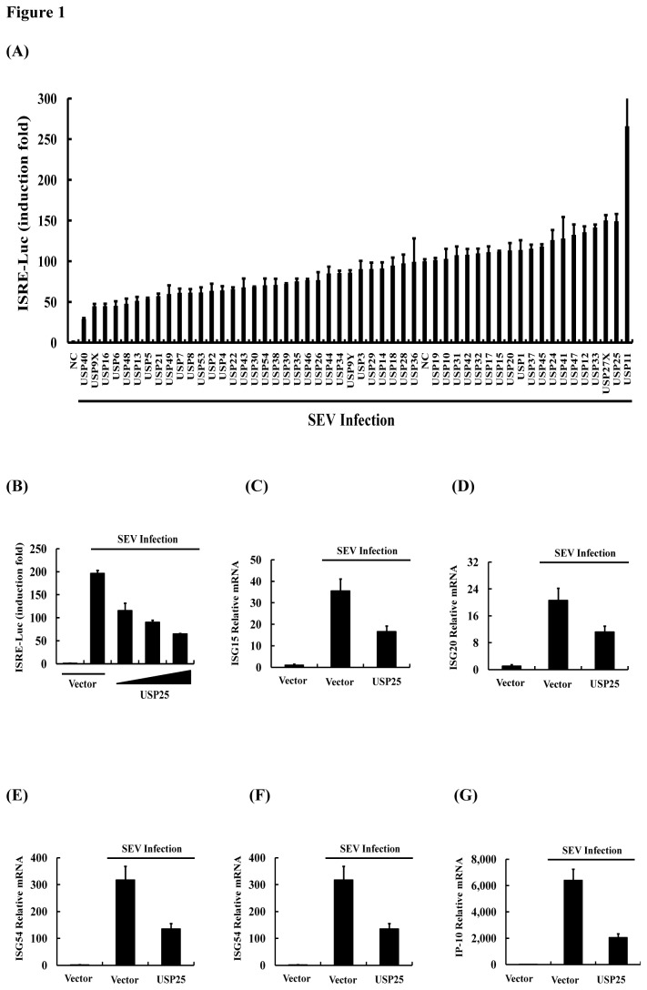
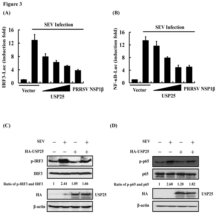
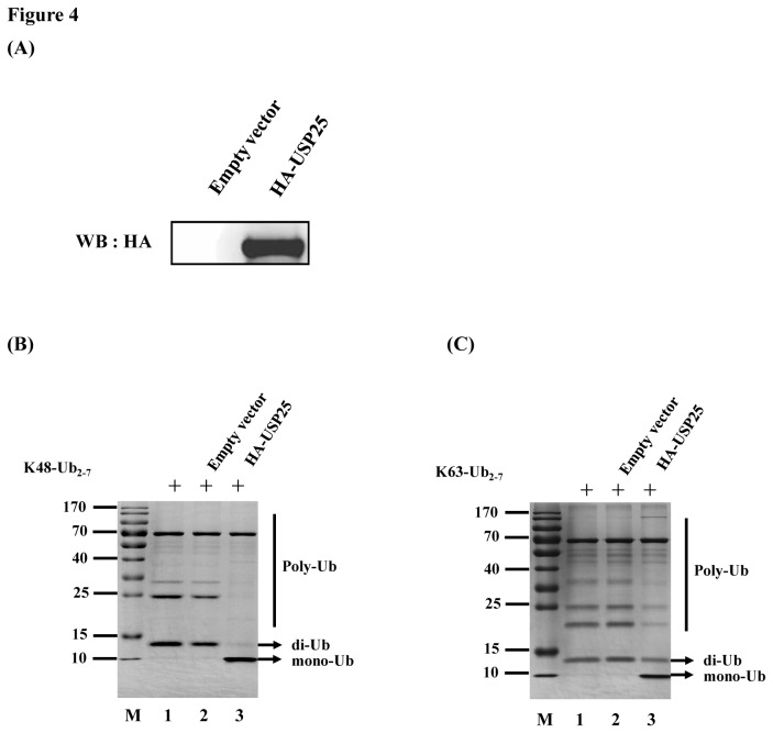
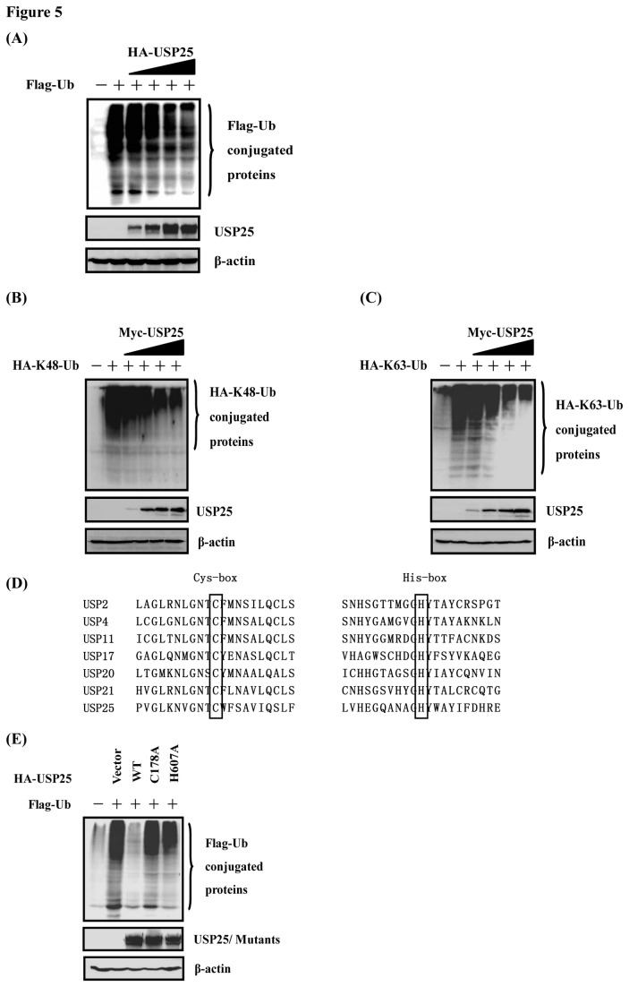
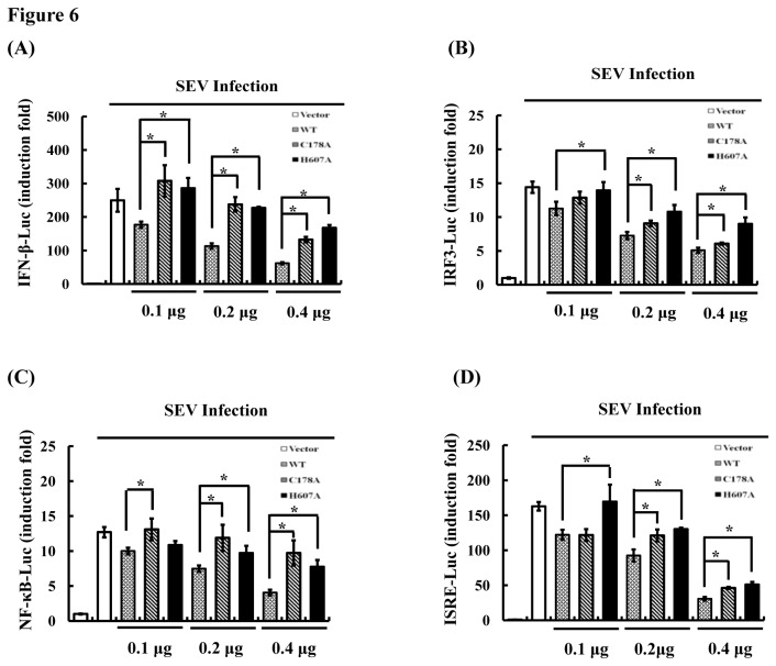
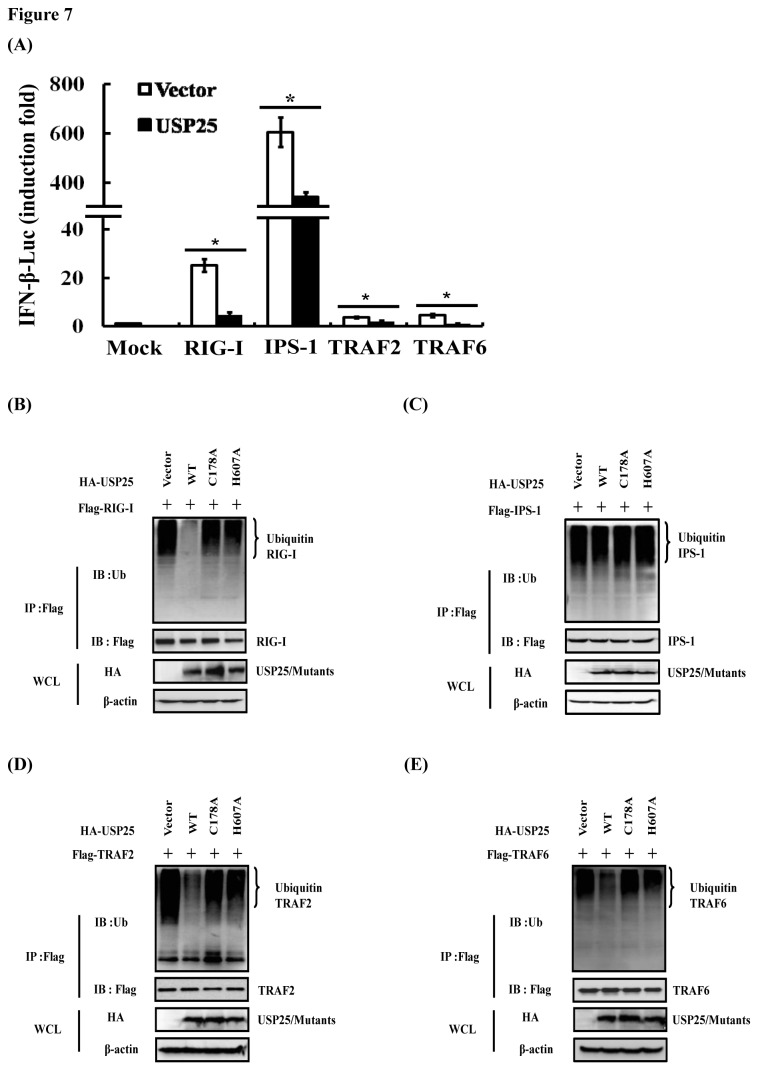
Ubiquitination and deubiquitination have emerged as critical regulatory processes in the virus-triggered type I interferon (IFN) induction pathway. In this study, we carried out a targeted siRNA screen of 54 ubiquitin-specific proteases (USPs) and identified USP25 as a negative regulator of the virus-triggered type I IFN signaling pathway. Overexpression of USP25 inhibited virus-induced activation of IFN-β, interferon regulation factor 3 (IRF3) and nuclear factor-kappa B (NF-κB), as well as the phosphorylation of IRF3 and NF-κB subunit p65. Furthermore, Knockdown of USP25 potentiated virus-induced induction of the IFN-β. In addition, detailed analysis demonstrated that USP25 cleaved lysine 48- and lysine 63-linked polyubiquitin chains in vitro and in vivo, and its deubiquitinating enzyme (DUB) activity, were dependent on a cysteine residue (Cys178) and a histidine residue (His607). USP25 mutants lacking DUB activity lost the ability to block virus-induced type I IFN to some degree. Mechanistically, USP25 deubiquitinated retinoic acid-inducible gene I (RIG-I), tumornecrosis factor (TNF) receptor-associated factor 2 (TRAF2), and TRAF6 to inhibit RIG-I-like receptor-mediated IFN signaling. Our findings suggest that USP25 is a novel DUB negatively regulating virus-induced type I IFN production.


Conflict of interest statement

Competing Interests: The authors have declared that no competing interests exist.

Figures

Figure 1
Figure 1. USP25 negatively regulates virus-induced activation of ISRE and the expression of ISGs.
(A) A siRNA screen for USPs functions on SEV-induced activation of the ISRE promoter. HEK-293T cells were transfected with an ISRE luciferase reporter (0.1 μg) and control Renilla luciferase reporter (0.02 μg) vectors and NC (control siRNA) or specific siRNA pools (three siRNAs per gene) of members of USPs subclass of deubiquitinase for 24 h, and then infected or mock-infected with SEV for 16 h before luciferase assays were performed. (B) USP25 inhibited SEV-induced activation of ISRE. ISRE luciferase reporter (0.1 μg) and control Renilla luciferase reporter (0.02 μg) vectors were co-transfected into HEK-293T cells with either an empty vector (1 μg) or increasing amounts of USP25 (0.25, 0.5, or 1 μg) for 24 h. Cells were then either untreated or treated with SEV for 16 h before the relative luciferase activity was measured and normalized with the Renilla activity. Error bars indicate ± SD in three independent experiments. (C–G) USP25 significantly reduced the transcription of multiple ISGs. Empty vector (1 μg) or expression plasmid of USP25 (1 μg) were transfected into HEK-293T cells for 24 h and either untreated or treated with SEV for 16 h before real-time RT-PCR was performed.
Figure 2
Figure 2. USP25 significantly inhibits virus-induced type I IFN signaling.
(A) USP25 inhibited SEV-induced activation of the IFN-β promoter in a dose-dependent manner. IFN-β luciferase reporter (0.1 μg) and control Renilla luciferase reporter (0.02 μg) vectors were co-transfected into HEK-293T cells with an empty vector (1 μg) or increasing amounts of USP25 (0.25, 0.5, or 1 μg) for 24 h. Cells were then either untreated or treated with SEV for 16 h before the relative luciferase activity was measured and normalized with the Renilla activity. Error bars indicate ± SD in three independent experiments. (B) USP25 significantly inhibited the transcription of IFN-β. Empty vector (1 μg) or expression plasmid of USP25 (1 μg) were transfected into HEK-293T cells for 24 h and either untreated or treated with SEV for 16 h before real-time RT-PCR was performed. (C) USP25 inhibited poly (I:C)-induced activation of the IFN-β promoter in a dose-dependent manner. IFN-β luciferase reporter (0.1 μg) and control Renilla luciferase reporter (0.02 μg) vectors were co-transfected into HEK-293T cells with either an empty vector (1 μg) or increasing amounts of USP25 (0.25, 0.5, or 1 μg) for 24 h. Cells were then either untreated or treated with 1 μg of poly (I:C) for 16 h before the relative luciferase activity was measured and normalized with the Renilla activity. Error bars indicate ± SD in three independent experiments. (D, E) Effects of USP25 siRNA on endogenous USP25. HEK-293T cells were transfected with the indicated siRNA (20 nm each) for 24 h, and cell lysates were analyzed by quantitative reverse transcription (RT)-PCR (D) or immunoblots with antibodies against USP25 and beta-actin (E). (F) Effects of USP25 siRNA on SEV-induced activation of the IFN-β promoter. HEK-293T cells were transfected with an IFN-β luciferase reporter (0.1 μg) and control Renilla luciferase reporter (0.02 μg) vectors and the indicated siRNA plasmids (20 nm each) for 24 h and then infected with SEV or uninfected for 16 h before luciferase assays were performed. Error bars indicate ± SD in three independent experiments. *P < 0.05 compared with cells transfected with Control followed by SEV infection. Significant differences between groups were determined by one-way ANOVA followed by Dunnett’s multiple comparisons test.
Figure 3
Figure 3. USP25 inhibits SEV-induced activation of IRF3 and NF-κB.
(A) IRF3 luciferase reporter (0.1 μg) and control Renilla luciferase reporter (0.02 μg) vectors were co-transfected into HEK-293T cells with either an empty vector (1 μg) or increasing amounts of USP25 (0.25, 0.5, or 1 μg) or porcine reproductive and respiratory syndrome virus (PRRSV) NSP1β which was a positive control for 24 h. Cells were then either untreated or treated with SEV for 16 h before the relative luciferase activity was measured and normalized with the Renilla activity. Error bars indicate ± SD in three independent experiments. (B) Reporter assays were performed similarly as in A, except that different reporter plasmids were used. (C, D) USP25 inhibited SEV-induced IRF3 and p65 phosphorylation. HEK-293T cells were transfected with either an empty vector (3 μg) or expression plasmid of USP25 (3 μg) for 24 h. Cells were then either untreated or treated with SEV for 16 h and subsequently immunoblotted with the antibodies indicated. Quantification of the intensity of each band is listed under each band. Total protein of IRF3 and p65 was used as an internal control.
Figure 4
Figure 4. Processing of K48- and K63-linked polyubiquitin chains by USP25 in vitro.
(A) Analysis for purified HA-tagged USP25 conjugated proteins. The protein was obtained from USP25-transfected or mock-transfected HEK-293T cells using a HA tagged Protein PURIFICATION KIT (MBL) and analyzed for HA-tagged USP25-conjugated proteins by western blotting (WB) with an anti-HA antibody. (B) In vitro K48-linked polyubiquitin deconjugation assay. K48-linked polyubiquitin was incubated with the protein obtained from mock-transfected (lane 2) or USP25-transfected (lane 3) HEK-293T cells at 37°C for 1 h before being analyzed by SDS-PAGE. Lane 1, uncleaved K48-linked polyubiquitin chain (K48-Ub2–7). M, molecular mass markers, including 170-, 130-, 100-, 70-, 55-, 40-, 35-, 25-, 15-, and 10-kDa bands. (C) In vitro K63-linked polyubiquitin deconjugation assay. The experiment was performed similarly as in B, except that the K63-linked polyubiquitin chain (K63-Ub2–7) was used.
Figure 5
Figure 5. USP25 has a dose-dependent deubiquitinating activity in vivo.
(A) HEK-293T cells grown in 60-mm dishes were transfected with Flag-tagged Ub expression plasmids (1 μg), along with increasing quantities (0.25, 0.5, 1, or 2 μg) of plasmid encoding USP25 using Lipofectamine 2000. Cell lysates were prepared at 30 h post-transfection and analyzed for Ub-conjugated proteins by Western blotting with an anti-Flag antibody. Western blotting with anti-HA antibodies shows expression of USP25, and Western blotting for beta-actin served as a protein loading control. (B, C) USP25 effectively cleaved both K48 and K63 Ub linkages in vivo. The experiment was performed similarly to that described for panel A except that HA-K48-Ub or HA-K63-Ub was used in lieu of Flag-Ub and different expression plasmids of USP25 were used. (D) Mapping of the putative sites are associated with the DUB activity of USP25. Black boxes indicate conserved residues tested in this study. The sequences were derived from GenBank entries with the following accession numbers: USP2, NM_004205; USP4, NM_003363; USP11, NM_004651; USP17, NM_201402 and XM_352721; USP20, NM_001110303; USP21, NM_001014443; and USP25, NM_013396. (E) Cysteine 178 (C178) and histidine 607 (H607) are deubiquitination active sites of USP25. Expression vectors encoding pcDNA3.1-Flag-Ub and control vectors or expression vectors encoding HA-USP25-WT, -C178A and -H607A were co-transfected into HEK-293T cells. USP25-WT and deletion mutant proteins in cell lysates were immunoprecipitated with anti-HA antibodies under denaturing conditions and immunoblotted with anti-Flag antibodies to detect the presence of Ub-conjugated proteins. Western blotting for beta-actin served as a protein loading control.
Figure 6
Figure 6. The deubiquitinating activity of USP25 is involved in virus-induced type I IFN signaling.
(A) The deubiquitinating activity of USP25 was involved in the transcription of IFN-β. IFN-β luciferase reporter (0.1 μg) and control Renilla luciferase reporter (0.02 μg) vectors were co-transfected into HEK-293T cells with either an empty vector or increasing amounts of USP25-WT or deletion mutants for 24 h. Cells were then either untreated or treated with SEV for 16 h before the relative luciferase activity was measured and normalized with the Renilla activity. Error bars indicate ± SD in three independent experiments. *P < 0.05 for all pairwise comparisons by one-way ANOVA followed by Dunnett’s multiple comparisons test. (B–D) The deubiquitinating activity of USP25 was involved in the activation of IRF3 (B), NF-κB (C) and ISRE (D). Reporter assays were performed similarly as in A, except that different reporter plasmids were used. *P < 0.05 for all pairwise comparisons by one-way ANOVA followed by Dunnett’s multiple comparisons test.
Figure 7
Figure 7. USP25 deubiquitinates RIG-I, TRAF2 and TRAF6.
(A) USP25 inhibited RIG-I-, IPS-1-, TRAF2-, and TRAF6-mediated activation of the IFN-β promoter. HEK-293T cells were transfected with an IFN-β promoter reporter (0.1 μg) and either an empty vector or the indicated plasmids encoding the RIG-I, IPS-1, TRAF2, and TRAF6 expression vector (1 μg each) for 30 h before luciferase assays were performed. *p < 0.05 for all pairwise comparisons by one-way ANOVA followed by Dunnett’s multiple comparisons test. (B–E) HEK-293T cells were co-transfected with the indicated plasmids encoding the RIG-I (B), IPS-1 (C), TRAF2 (D), or TRAF6 (E) expression vector (4 μg) and HA-USP25WT/ HA-USP25C178A/ HA-USP25H607A (4 μg) using Lipofectamine 2000. The immunoprecipitates (IP) were analyzed by immunoblots (IB) with anti-ubiquitin (top panels) and anti-Flag (middle panels). The levels of the transfected USP25/mutants were detected by immunoblots with anti-HA (bottom panels). The input tagged proteins were verified with the indicated antibodies.

Similar articles

Cited by

References

    1. Kawai T, Akira S (2006) Innate immune recognition of viral infection. Nat Immunol 7: 131-137. doi:10.1038/ni1303. PubMed: 16424890. - DOI - PubMed
    1. Darnell JE Jr., Kerr IM, Stark GR (1994) Jak-STAT pathways and transcriptional activation in response to IFNs and other extracellular signaling proteins. Science 264: 1415-1421. doi:10.1126/science.8197455. PubMed: 8197455. - DOI - PubMed
    1. Hubackova S, Novakova Z, Krejcikova K, Kosar M, Dobrovolna J et al. (2010) Regulation of the PML tumor suppressor in drug-induced senescence of human normal and cancer cells by JAK/STAT-mediated signaling. Cell Cycle 9: 3085-3099. doi:10.4161/cc.9.15.12521. PubMed: 20699642. - DOI - PubMed
    1. Kato H, Takeuchi O, Sato S, Yoneyama M, Yamamoto M et al. (2006) Differential roles of MDA5 and RIG-I helicases in the recognition of RNA viruses. Nature 441: 101-105. doi:10.1038/nature04734. PubMed: 16625202. - DOI - PubMed
    1. Pichlmair A, Schulz O, Tan CP, Näslund TI, Liljeström P et al. (2006) RIG-I-mediated antiviral responses to single-stranded RNA bearing 5'-phosphates. Science 314: 997-1001. doi:10.1126/science.1132998. PubMed: 17038589. - DOI - PubMed

Publication types

MeSH terms

Grants and funding

This work was supported by the National Natural Sciences Foundation of China (31225027, 31121004), and the Fundamental Research Funds for the Central Universities (2013PY043). The funders had no role in study design, data collection and analysis, decision to publish, or preparation of the manuscript.