

Effects of Calcium Gluconate, a Water Soluble Calcium Salt on the Collagen-Induced DBA/1J Mice Rheumatoid Arthritis
- PMID: 24244814
- PMCID: PMC3819902
- DOI: 10.4062/biomolther.2013.040
Effects of Calcium Gluconate, a Water Soluble Calcium Salt on the Collagen-Induced DBA/1J Mice Rheumatoid Arthritis
Abstract
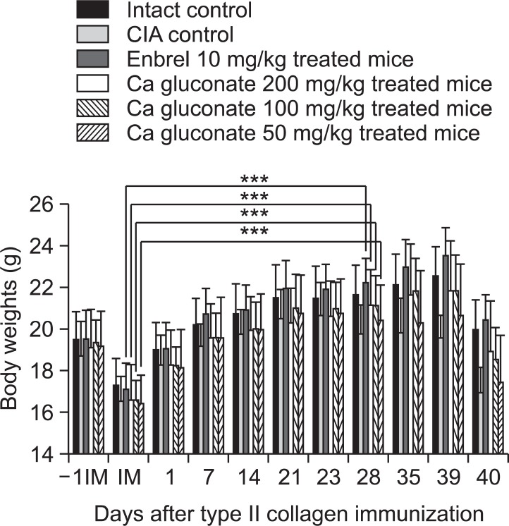
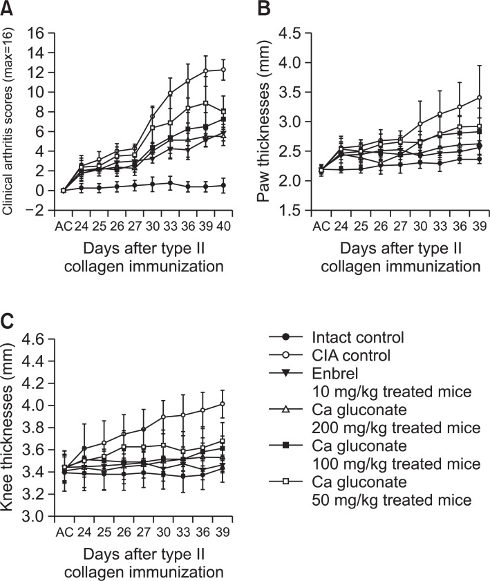
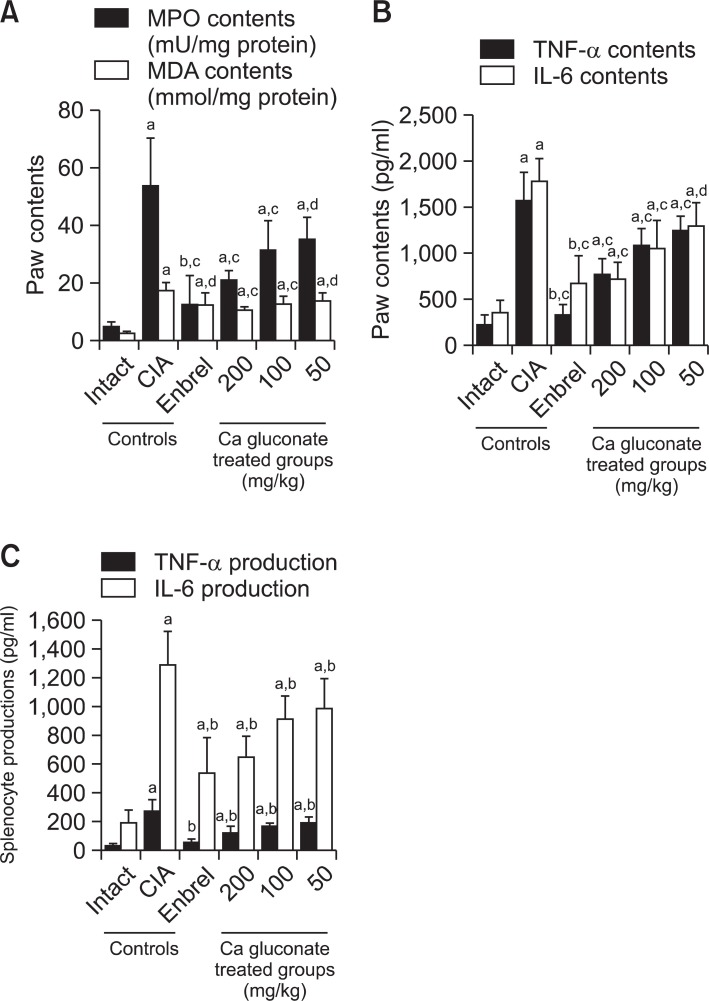
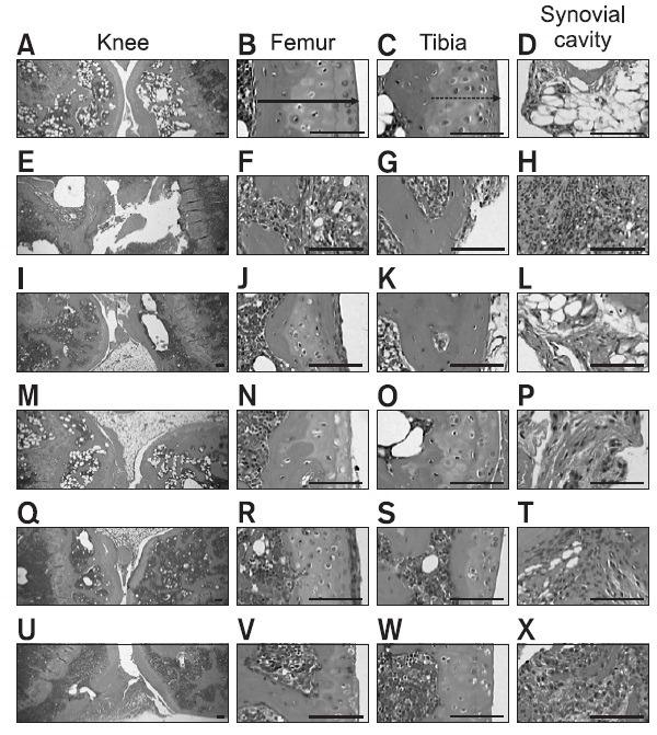
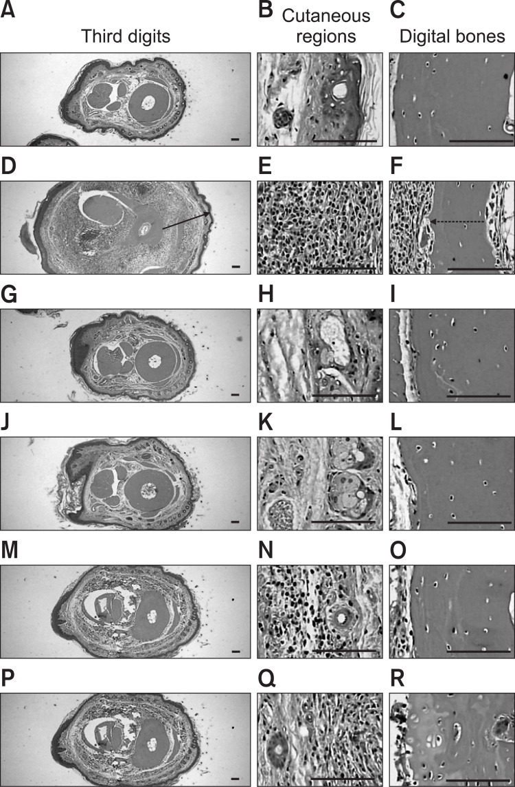
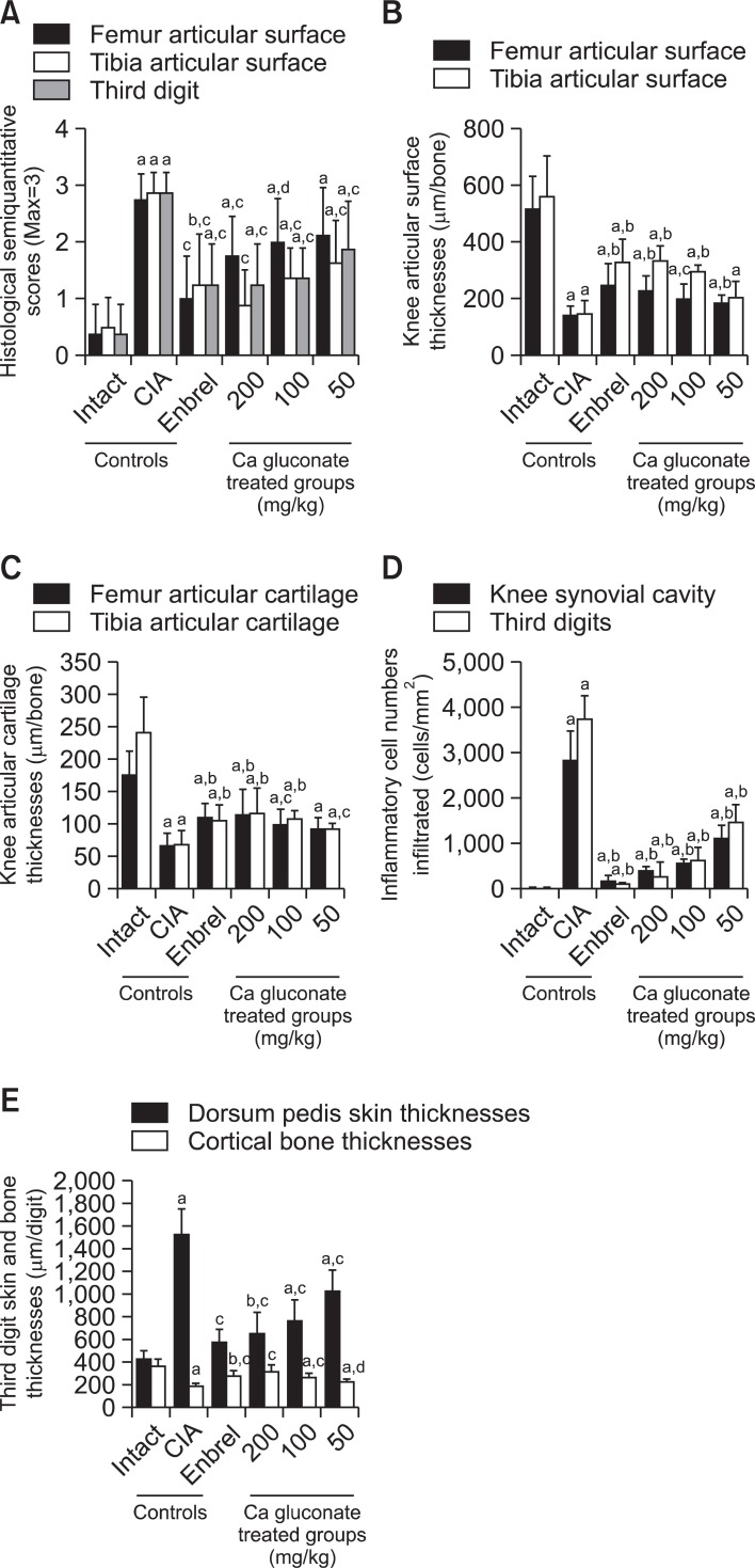
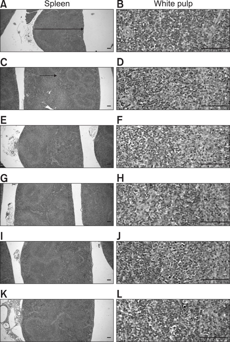
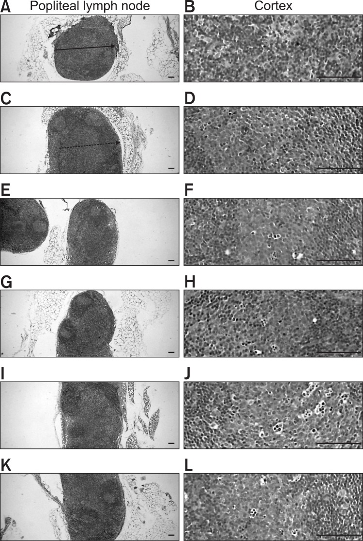
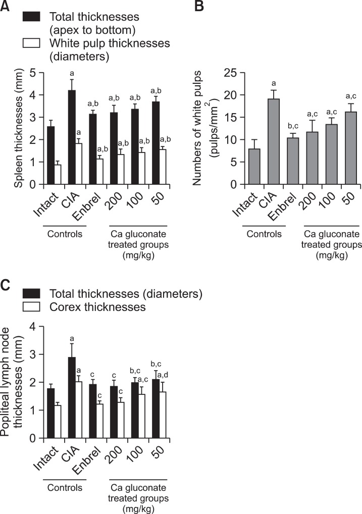
This study examined the effects of calcium (Ca) gluconate on collagen-induced DBA mouse rheumatoid arthritis (CIA). A single daily dose of 200, 100 or 50 mg/kg Ca gluconate was administered orally to male DBA/1J mice for 40 days after initial collagen immunization. To ascertain the effects administering the collagen booster, CIA-related features (including body weight, poly-arthritis, knee and paw thickness, and paw weight increase) were measured from histopathological changes in the spleen, left popliteal lymph node, third digit and the knee joint regions. CIA-related bone and cartilage damage improved significantly in the Ca gluconate- administered CIA mice. Additionally, myeloperoxidase (MPO) levels in the paw were reduced in Ca gluconate-treated CIA mice compared to CIA control groups. The level of malondialdehyde (MDA), an indicator of oxidative stress, decreased in a dosedependent manner in the Ca gluconate group. Finally, the production of IL-6 and TNF-α, involved in rheumatoid arthritis pathogenesis, were suppressed by treatment with Ca gluconate. Taken together, these results suggest that Ca gluconate is a promising candidate anti-rheumatoid arthritis agent, exerting anti-inflammatory, anti-oxidative and immunomodulatory effects in CIA mice.
Keywords: Anti-inflammation; Anti-oxidation; Calcium gluconate; Immunomodulation; Rheumatoid arthritis.
Figures
Similar articles
-
The Effects of Platycodin D, a Saponin Purified from Platycodi Radix, on Collagen-Induced DBA/1J Mouse Rheumatoid Arthritis.Evid Based Complement Alternat Med. 2014;2014:954508. doi: 10.1155/2014/954508. Epub 2014 Jan 6. Evid Based Complement Alternat Med. 2014. PMID: 24511322 Free PMC article.
-
Protection against cartilage and bone destruction by systemic interleukin-4 treatment in established murine type II collagen-induced arthritis.Arthritis Res. 1999;1(1):81-91. doi: 10.1186/ar14. Epub 1999 Oct 26. Arthritis Res. 1999. PMID: 11056663 Free PMC article.
-
Anti-arthritic and anti-inflammatory effects of (-)-Epicatechin-3-O-β-d-allopyranoside, a constituent of Davallia formosana.Phytomedicine. 2019 Jan;52:12-22. doi: 10.1016/j.phymed.2018.09.192. Epub 2018 Sep 18. Phytomedicine. 2019. PMID: 30599891
-
Potent Antiarthritic Properties of Phloretin in Murine Collagen-Induced Arthritis.Evid Based Complement Alternat Med. 2016;2016:9831263. doi: 10.1155/2016/9831263. Epub 2016 Dec 4. Evid Based Complement Alternat Med. 2016. PMID: 28044086 Free PMC article.
-
Soufeng sanjie formula alleviates collagen-induced arthritis in mice by inhibiting Th17 cell differentiation.Chin Med. 2021 May 13;16(1):39. doi: 10.1186/s13020-021-00448-9. Chin Med. 2021. PMID: 33985537 Free PMC article.
Cited by
-
The Effects of Topical Application of Polycal (a 2:98 (g/g) Mixture of Polycan and Calcium Gluconate) on Experimental Periodontitis and Alveolar Bone Loss in Rats.Molecules. 2016 Apr 22;21(4):527. doi: 10.3390/molecules21040527. Molecules. 2016. PMID: 27110759 Free PMC article.
-
Effects of various parameters on solution-mediated phase transformation of calcium d-gluconate: an approach to obtain pure metastable monohydrate.RSC Adv. 2023 Apr 19;13(18):12175-12183. doi: 10.1039/d3ra01424j. eCollection 2023 Apr 17. RSC Adv. 2023. PMID: 37091620 Free PMC article.
-
Design, synthesis and evaluation of (R)-3-(7-(methyl(7H-pyrrolo[2,3-d]pyrimidin-4-yl)amino)-5-azaspiro[2.4]heptan-5-yl)-3-oxopropanenitrile as a JAK1-selective inhibitor.Medchemcomm. 2018 Jan 15;9(3):477-489. doi: 10.1039/c7md00568g. eCollection 2018 Mar 1. Medchemcomm. 2018. PMID: 30108938 Free PMC article.
References
-
- Bracken W. M., Cuppage F., McLaury R. L., Kirwin C., Klaassen C. D. Comparative effectiveness of topical treatments for hydrofluoric acid burns. J. Occup. Med. (1985);27:733–739. - PubMed
-
- Brahn E., Banquerigo M. L., Firestein G. S., Boyle D. L., Salzman A. L., Szabo C. Collagen induced arthritis: reversal by mercaptoethylguanidine, a novel antiinflammatory agent with a combined mechanism of action. J. Rheumatol. (1998);25:1785–1793. - PubMed
-
- Cavallini M., de Boccard F., Corsi M. M., Fassati L. R., Baruffaldi Preis F. W. Serum pro-inflammatory cytokines and chemical acid burns in rats. Ann. Burns Fire Disasters. (2004);17:84–87.
LinkOut - more resources
Full Text Sources
Other Literature Sources
Research Materials
Miscellaneous
