

Effect of MWCNT size, carboxylation, and purification on in vitro and in vivo toxicity, inflammation and lung pathology
- PMID: 24225053
- PMCID: PMC3830505
- DOI: 10.1186/1743-8977-10-57
Effect of MWCNT size, carboxylation, and purification on in vitro and in vivo toxicity, inflammation and lung pathology
Abstract
Background: Several properties of multi-walled carbon nanotubes (MWCNT) have the potential to affect their bioactivity. This study examined the in vitro and in vivo outcomes of the influence of diameter, length, purification and carboxylation (in vitro testing only) of MWCNT.
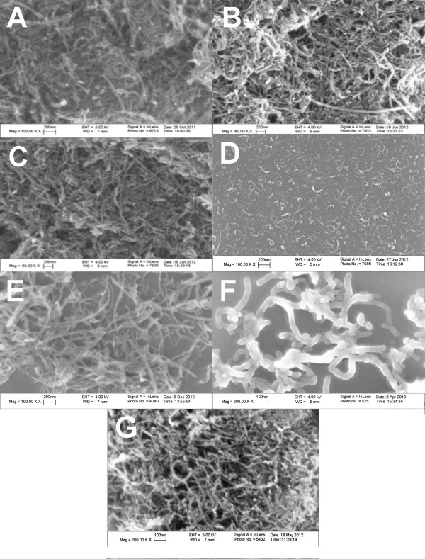
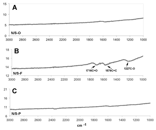
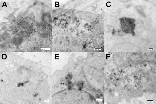
Methods: Three original 'as received' MWCNT that varied in size (diameter and length) were purified and functionalized by carboxylation. The resulting MWCNT were characterized and examined for cytotoxicity and inflammasome activation in vitro using THP-1 cells and primary alveolar macrophages from C57BL/6 mice. Oropharyngeal aspiration administration was used to deliver original MWCNT and in vivo bioactivity and lung retention was examined at 1 and 7 days.
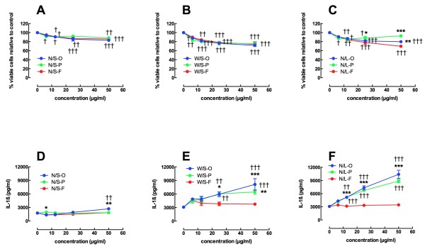
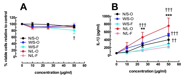
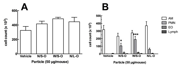
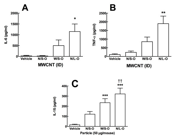
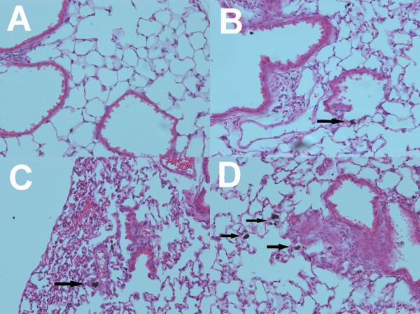
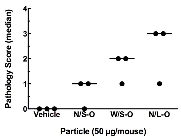
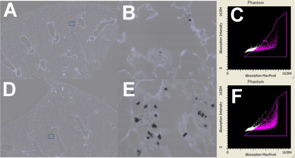
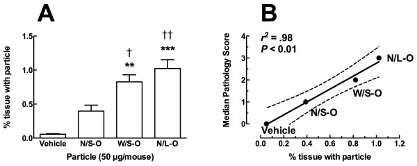
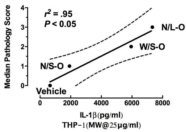
Results: Studies with THP-1 macrophages demonstrated that increased length or diameter corresponded with increased bioactivity as measured by inflammasome activation. Purification had little effect on the original MWCNT, and functionalization completely eliminated bioactivity. Similar results were obtained using alveolar macrophages isolated from C57BL/6 mice. The in vivo studies demonstrated that all three original MWCNT caused similar neutrophil influx at one day, but increasing length or diameter resulted in the lavaged cells to release more inflammatory cytokines (IL-6, TNF-α, and IL-1β) ex vivo. Seven-day histology revealed that, consistent with the in vitro results, increasing width or length of MWCNT caused more severe pathology with the longest MWCNT causing the most severe inflammation. In addition, the same two larger MWCNT were retained more in the lung at 7 days.
Conclusions: Taken together, the results indicated that in vitro and in vivo bioactivity of MWCNT increased with diameter and length. Purification had no significant modifying effect from the original MWCNT. Functionalization by carboxylation completely eliminated the bioactive potential of the MWCNT regardless of size in in vitro testing.
Figures
Similar articles
-
Mouse pulmonary dose- and time course-responses induced by exposure to nitrogen-doped multi-walled carbon nanotubes.Inhal Toxicol. 2020 Jan;32(1):24-38. doi: 10.1080/08958378.2020.1723746. Epub 2020 Feb 7. Inhal Toxicol. 2020. PMID: 32028803 Free PMC article.
-
The Effects of Varying Degree of MWCNT Carboxylation on Bioactivity in Various In Vivo and In Vitro Exposure Models.Int J Mol Sci. 2018 Jan 25;19(2):354. doi: 10.3390/ijms19020354. Int J Mol Sci. 2018. PMID: 29370073 Free PMC article.
-
Purification and sidewall functionalization of multiwalled carbon nanotubes and resulting bioactivity in two macrophage models.Inhal Toxicol. 2013 Mar;25(4):199-210. doi: 10.3109/08958378.2013.775197. Inhal Toxicol. 2013. PMID: 23480196 Free PMC article.
-
Synthesis, characterization, and bioactivity of carboxylic acid-functionalized titanium dioxide nanobelts.Part Fibre Toxicol. 2014 Sep 2;11:43. doi: 10.1186/s12989-014-0043-7. Part Fibre Toxicol. 2014. PMID: 25179214 Free PMC article.
-
The impact of multi-walled carbon nanotubes (MWCNTs) on macrophages: contribution of MWCNT characteristics.Sci China Life Sci. 2018 Nov;61(11):1333-1351. doi: 10.1007/s11427-017-9242-3. Epub 2018 May 22. Sci China Life Sci. 2018. PMID: 29797182 Review.
Cited by
-
Comparative in Vitro Cytotoxicity of Realistic Doses of Benchmark Multi-Walled Carbon Nanotubes towards Macrophages and Airway Epithelial Cells.Nanomaterials (Basel). 2019 Jul 6;9(7):982. doi: 10.3390/nano9070982. Nanomaterials (Basel). 2019. PMID: 31284615 Free PMC article.
-
Mouse pulmonary dose- and time course-responses induced by exposure to nitrogen-doped multi-walled carbon nanotubes.Inhal Toxicol. 2020 Jan;32(1):24-38. doi: 10.1080/08958378.2020.1723746. Epub 2020 Feb 7. Inhal Toxicol. 2020. PMID: 32028803 Free PMC article.
-
Peroxisome Proliferator-activated Receptor-γ Deficiency Exacerbates Fibrotic Response to Mycobacteria Peptide in Murine Sarcoidosis Model.Am J Respir Cell Mol Biol. 2019 Aug;61(2):198-208. doi: 10.1165/rcmb.2018-0346OC. Am J Respir Cell Mol Biol. 2019. PMID: 30741559 Free PMC article.
-
Length, but Not Reactive Edges, of Cup-stack MWCNT Is Responsible for Toxicity and Acute Lung Inflammation.Toxicol Pathol. 2018 Jan;46(1):62-74. doi: 10.1177/0192623317732303. Epub 2017 Sep 25. Toxicol Pathol. 2018. PMID: 28946794 Free PMC article.
-
Respiratory and systemic impacts following MWCNT inhalation in B6C3F1/N mice.Part Fibre Toxicol. 2021 Mar 26;18(1):16. doi: 10.1186/s12989-021-00408-z. Part Fibre Toxicol. 2021. PMID: 33771183 Free PMC article.
References
-
- Donaldson K, Aitken R, Tran L, Stone V, Duffin R, Forrest G, Alexander A. Carbon nanotubes: a review of their properties in relation to pulmonary toxicology and workplace safety. Toxicol Sci. 2006;92(1):5–22. - PubMed
-
- Murphy FA, Poland CA, Duffin R, Al-Jamal KT, Ali-Boucetta H, Nunes A, Byrne F, Prina-Mello A, Volkov Y, Li S. et al.Length-dependent retention of carbon nanotubes in the pleural space of mice initiates sustained inflammation and progressive fibrosis on the parietal pleura. Am J Pathol. 2011;178(6):2587–2600. - PMC - PubMed
-
- Palomaki J, Valimaki E, Sund J, Vippola M, Clausen PA, Jensen KA, Savolainen K, Matikainen S, Alenius H. Long, needle-like carbon nanotubes and asbestos activate the NLRP3 inflammasome through a similar mechanism. ACS Nano. 2011;5(9):6861–6870. - PubMed
-
- Fenoglio I, Aldieri E, Gazzano E, Cesano F, Colonna M, Scarano D, Mazzucco G, Attanasio A, Yakoub Y, Lison D. et al.Thickness of multiwalled carbon nanotubes affects their lung toxicity. Chem Res Toxicol. 2012;25(1):74–82. - PubMed
-
- Qu GB, Bai YH, Zhang Y, Jia Q, Zhang WD, Yan B. The effect of multiwalled carbon nanotube agglomeration on their accumulation in and damage to organs in mice. Carbon N Y. 2009;47(8):2060–2069.
Publication types
MeSH terms
Substances
Grants and funding
LinkOut - more resources
Full Text Sources
Other Literature Sources
