

Comment
doi: 10.1038/jid.2013.483.
Epub 2013 Nov 12.
Merkel cell polyomavirus-positive Merkel cell carcinoma requires viral small T-antigen for cell proliferation
Affiliations
- PMID: 24217011
- PMCID: PMC3989379
- DOI: 10.1038/jid.2013.483
Item in Clipboard
Comment
Merkel cell polyomavirus-positive Merkel cell carcinoma requires viral small T-antigen for cell proliferation
J Invest Dermatol.
2014 May.
No abstract available
Figures
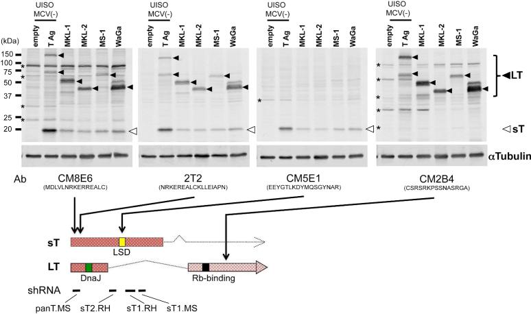
Detection of Merkel cell polyomavirus (MCV) small T (sT) antigen expression by multiple MCV T antigen antibodies. MCV-positive MCC cells (MKL-1, MKL-2, MS-1 and WaGa) and MCV-negative UISO cells transfected with MCV genomic T antigen gene or empty vector as positive and negative controls, were immunoblotted with multiple MCV T antigen antibodies. αTubulin was used as a loading control. Both large T (LT, closed arrows) and small T (sT, open arrows) were detected by CM8E6 and 2T2, sT by CM5E1, and LT by CM2B4. Asterisks indicate non-specific bands. Peptide sequences used for monoclonal antibody production and shRNA targeting sites are shown in the bottom diagram of T antigen transcripts with a DnaJ (green box), an Rb-binding (black box) as well as large T stabilization (LSD, yellow box (Kwun et al., 2013)) domains.
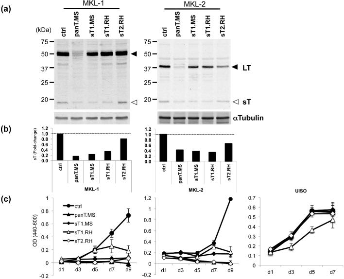
Merkel cell polyomavirus (MCV) small T (sT) antigen knockdown inhibits MCV-positive Merkel cell carcinoma (MCC) cell proliferation. (a) MCV-positive MCC cell lines, MKL-1 and MKL-2, were transduced with pLKO.1-based lentiviral shRNAs targeting both LT and sT (panT.MS) or sT alone (sT1.MS, sT1.RH, and sT2.RH) as described (Houben et al., 2010). Both LT (closed arrows) and sT (open arrows) proteins are detected by 2T2. (b) Expression of LT and sT was quantitated by LI-COR IR immunoblotting system using αTubulin for normalization. Relative sT expression to sh ctrl is shown. (c) shRNA-transduced MCV-positive (MKL-1 and MKL-2) cells and MCV-negative (UISO) cells were subjected to Wst-1 cell proliferation assay. Error bars indicate standard deviation.
Comment in
-
Response to Shuda et al.J Invest Dermatol. 2014 May;134(5):1481-1482. doi: 10.1038/jid.2013.486. Epub 2013 Nov 12. J Invest Dermatol. 2014. PMID: 24217012 No abstract available.
Comment on
-
Merkel cell polyomavirus-positive Merkel cell carcinoma cells do not require expression of the viral small T antigen.J Invest Dermatol. 2013 Aug;133(8):2059-64. doi: 10.1038/jid.2013.82. Epub 2013 Feb 25. J Invest Dermatol. 2013. PMID: 23439392
Similar articles
-
Response to Shuda et al.J Invest Dermatol. 2014 May;134(5):1481-1482. doi: 10.1038/jid.2013.486. Epub 2013 Nov 12. J Invest Dermatol. 2014. PMID: 24217012 No abstract available.
-
Merkel cell polyomavirus-positive Merkel cell carcinoma cells do not require expression of the viral small T antigen.J Invest Dermatol. 2013 Aug;133(8):2059-64. doi: 10.1038/jid.2013.82. Epub 2013 Feb 25. J Invest Dermatol. 2013. PMID: 23439392
-
Merkel cell polyomavirus in Merkel cell carcinogenesis: small T antigen-mediates c-Jun phosphorylation.Virus Genes. 2016 Jun;52(3):397-9. doi: 10.1007/s11262-016-1304-3. Epub 2016 Mar 19. Virus Genes. 2016. PMID: 26995220
-
Merkel cell polyomavirus: a newly discovered human virus with oncogenic potential.Virology. 2013 Jan 5;435(1):118-30. doi: 10.1016/j.virol.2012.09.029. Virology. 2013. PMID: 23217622 Free PMC article. Review.
-
A cornucopia of human polyomaviruses.Nat Rev Microbiol. 2013 Apr;11(4):264-76. doi: 10.1038/nrmicro2992. Epub 2013 Mar 11. Nat Rev Microbiol. 2013. PMID: 23474680 Free PMC article. Review.
Cited by
-
Adapting the Stress Response: Viral Subversion of the mTOR Signaling Pathway.Viruses. 2016 May 24;8(6):152. doi: 10.3390/v8060152. Viruses. 2016. PMID: 27231932 Free PMC article. Review.
-
The Role of the Large T Antigen in the Molecular Pathogenesis of Merkel Cell Carcinoma.Genes (Basel). 2024 Aug 27;15(9):1127. doi: 10.3390/genes15091127. Genes (Basel). 2024. PMID: 39336718 Free PMC article. Review.
-
Characterization of molecular mechanisms driving Merkel cell polyomavirus oncogene transcription and tumorigenic potential.PLoS Pathog. 2023 Aug 30;19(8):e1011598. doi: 10.1371/journal.ppat.1011598. eCollection 2023 Aug. PLoS Pathog. 2023. PMID: 37647312 Free PMC article.
-
Merkel cell polyomavirus and associated Merkel cell carcinoma.Tumour Virus Res. 2022 Jun;13:200232. doi: 10.1016/j.tvr.2021.200232. Epub 2021 Dec 15. Tumour Virus Res. 2022. PMID: 34920178 Free PMC article. Review.
-
The Merkel Cell Polyomavirus T Antigens Function as Tumor Promoters in Murine Skin.Cancers (Basel). 2021 Jan 9;13(2):222. doi: 10.3390/cancers13020222. Cancers (Basel). 2021. PMID: 33435392 Free PMC article.
References
-
- Angermeyer S, Hesbacher S, Becker JC, et al. Merkel cell polyomavirus-positive merkel cell carcinoma cells do not require expression of the viral small T antigen. The Journal of investigative dermatology. 2013;133:2059–64. - PubMed
-
- Hahn WC, Counter CM, Lundberg AS, et al. Creation of human tumour cells with defined genetic elements. Nature. 1999;400:464–8. - PubMed
-
- Houben R, Ortmann S, Schrama D, et al. Activation of the MAP kinase pathway induces apoptosis in the Merkel cell carcinoma cell line UISO. The Journal of investigative dermatology. 2007;127:2116–22. - PubMed
Publication types
MeSH terms
Substances
Grants and funding
LinkOut - more resources
Full Text Sources
Other Literature Sources
Medical
