Review
. 2014 Jul 10;21(2):293-312.
doi: 10.1089/ars.2013.5718. Epub 2014 Feb 4.

microRNAs in cancer cell response to ionizing radiation

Affiliations
Review

microRNAs in cancer cell response to ionizing radiation

Jennifer R Czochor et al. Antioxid Redox Signal. .

Abstract

Significance: microRNAs (miRNA) have been characterized as master regulators of the genome. As such, miRNAs are responsible for regulating almost every cellular pathway, including the DNA damage response (DDR) after ionizing radiation (IR). IR is a therapeutic tool that is used for the treatment of several types of cancer, yet the mechanism behind radiation response is not fully understood.

Recent advances: It has been demonstrated that IR can alter miRNA expression profiles, varying greatly from one cell type to the next. It is possible that this variation contributes to the range of tumor cell responsiveness that is observed after radiotherapy, especially considering the extensive role for miRNAs in regulating the DDR. In addition, individual miRNAs or miRNA families have been shown to play a multifaceted role in the DDR, regulating multiple members in a single pathway.

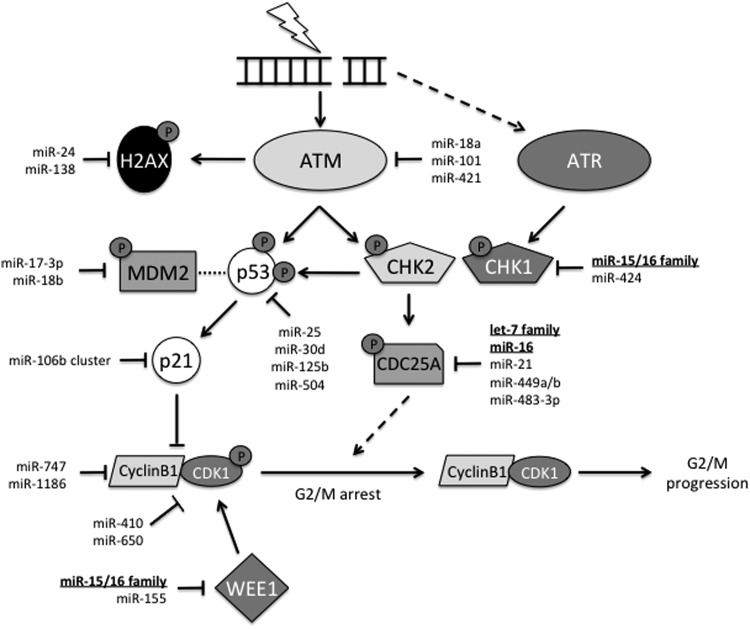
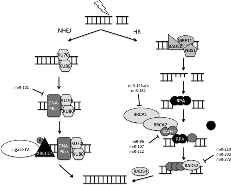
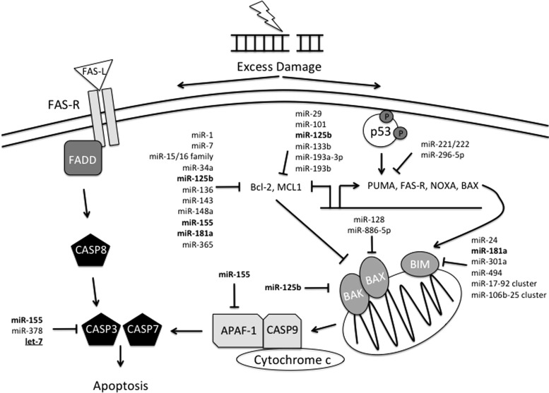
Critical issues: In this review, we will discuss the effects of radiation on miRNA expression as well as explore the function of miRNAs in regulating the cellular response to radiation-induced damage. We will discuss the importance of miRNA regulation at each stage of the DDR, including signal transduction, DNA damage sensing, cell cycle checkpoint activation, DNA double-strand break repair, and apoptosis. We will focus on emphasizing the importance of a single miRNA targeting several mediators within a pathway.

Future directions: miRNAs will continue to emerge as critical regulators of the DDR. Understanding the role of miRNAs in the response to IR will provide insights for improving the current standard therapy.


Figures

<b>FIG. 1.</b>
FIG. 1.
miRNA biogenesis and radiation response. miRNAs are transcribed in the nucleus by RNA polymerase II (RNA pol II) generating a primary miRNA transcript (pri-miR). DROSHA and Pasha cleave the pri-miR into the precursor miRNA (pre-miRNA) hairpin. As a hairpin, Exportin 5 exports the pre-miRNA into the cytoplasm, where it is further cleaved by an enzyme called Dicer and its binding partner TRBP/PACT. This final cleavage step results in a duplex RNA structure. The mature miRNA is separated from the passenger strand and loaded into the RISC. Along with RISC, the miRNA binds to the 3′ UTR of a target gene and mediates translational repression, or in some cases, mRNA degradation. In response to IR, miRNA expression is dysregulated. Although it remains to be proved, it seems likely that IR will result in modification of the biogenesis pathway. Importantly, when AGO2 and Dicer are repressed, the DDR is inhibited (67). DDR, DNA damage response; IR, ionizing radiation; miRNA, microRNA; RISC, RNA-induced silencing complex; AGO2, Argonaute-2.
<b>FIG. 2.</b>
FIG. 2.
miRNAs are differentially regulated in response to IR. On exposure to IR, miRNA expression is often dysregulated. IR induces some miRNAs, while others are repressed, a decision likely dependent on the target genes involved. A summary of the miRNAs discussed in this review whose expression changes in response to IR can be found in this diagram. On the left and right are lists of miRNAs whose induction or repression, respectively, have been observed. In the center are miRNAs where both induction and repression have been observed in different cell types. Interestingly, the largest group of miRNAs exists in the center, illustrating how different the miRNA profile can be from one cell type to the next. Bold miRNAs function in multiple aspects of the DDR. Additional underlining identifies miRNA families that are involved in almost every stage of the DDR.
<b>FIG. 3.</b>
FIG. 3.
PI3K/AKT and MAPK signal transduction pathway activation in response to IR. IR activates the EGFR family of RTKs, which triggers both PI3K/AKT and MAPK signaling. On the left, activation of RAS by the RTK leads to activation of RAF-1. Active RAF-1 initiates a cascade of phosphorylation-dependent activation, first of MEK1/2 followed by ERK1/2. ERK1/2 activation results in the activation of p90rsk, which promotes the transcription of anti-apoptotic factors, including miRNAs such as miR-21. Importantly, miR-21 targets PTEN, which inhibits the PI3K/AKT pathway, promoting PI3K/AKT signaling. On the right, the RTK activates PI3K, which, in turn, phosphorylates AKT. Activated AKT promotes the transcription of anti-apoptotic factors and inhibits the transcription of pro-apoptotic factors. miRNA regulation of the FOXO proteins is shown. Other members of the apoptotic pathway will be discussed later in Figure 7. miRNA regulation is extensive in both these pathways. miRNAs that have been bolded indicate miRNAs which have multiple targets in the same pathway. Additional underlining identifies miRNAs that are involved in almost every pathway of the DDR. Dotted arrow indicates potential for a positive feedback loop. ERK1/2, extracellular signal-regulated kinase 1/2; MAPK, mitogen-activated protein kinase; MEK1/2, MAPK kinase 1/2; PI3K, phosphoinositide 3-kinase; PTEN, phosphatase and tensin homolog; RAS; RTK, receptor tyrosine kinase.
<b>FIG. 4.</b>
FIG. 4.
G1/S cell cycle checkpoint activation in response to radiation-induced DNA damage. IR induces DNA DSBs. ATM and ATR are recruited to sites of DNA damage, but ATM is specific for DSBs. ATM activates several down-stream mediators by phosphorylation, including H2AX, p53, and the CHK proteins, typically CHK2. Activated CHK2 phosphorylates CDC25A, targeting it for degradation. In the absence of CDC25A, Cyclin E and CDK2 are impaired, resulting in cell cycle arrest. CHK2 is also capable of activating p53 by phosphorylation. Concurrently, activated p53 is unable to bind to MDM2 (indicated by dotted line), leading to its accumulation. Accumulation of p53 stimulates p21 expression. p21 binds to both cyclin/CDK complexes and inhibits them from promoting progression through the cell cycle. miRNAs regulate almost every member of this checkpoint. Bold miRNAs indicate multiple targets within the pathway. Underlined miRNA families are seen throughout the DDR. Dotted arrow indicates limited secondary activation of ATR in response to DSBs. ATM, ataxia telangiectasia mutated; ATR, ataxia telangiectasia and Rad3 related; CDK, cyclin-dependent kinase.
<b>FIG. 5.</b>
FIG. 5.
G2/M cell cycle checkpoint activation in response to radiation-induced DNA damage. Similar to the G1/S checkpoint, IR induces DNA DSBs that are recognized by the MRN complex (not pictured), which activates ATM and ATR. ATM phosphorylates CHK2, which targets CDC25A for degradation. CDC25A is necessary for the removal of an inhibitory phosphate on CDK1. WEE1 kinase is responsible for this phosphorylation of CDK1. Degradation of CDC25A inhibits progression through the cell cycle. In addition, ATM activates p53, preventing its association with MDM2. p53 accumulation promotes p21 expression. p21 inhibits cyclin B1/CDK1, inducing cell cycle arrest. As with G1/S, miRNA regulation is observed in almost every member of the G2/M checkpoint. Bold miRNAs are multifaceted within the pathway. Underlined miRNAs play a role in almost every aspect of the DDR from signal transduction to apoptosis.
<b>FIG. 6.</b>
FIG. 6.
miRNA regulation of radiation-induced DNA DSB repair via NHEJ or HR. After cell cycle checkpoint activation, radiation-induced DSBs are repaired by one of two pathways; NHEJ or HR. NHEJ occurs throughout the cell cycle, while HR occurs only during S and early G2 phase when a homologous template is available. NHEJ begins with recognition of the DSB by KU70 and KU80 (XRCC6 and XRCC5 in humans). DNA-PK, including its catalytic subunit DNA-PKcs, is recruited to the site of damage, and phosphorylates the DNA Ligase IV cofactor XRCC4. After end-processing by a yet-unidentified nuclease, DNA Ligase IV ligates the two ends together, often resulting in deletions of DNA. On the other hand, HR is less error prone, as it utilizes a homologous template to repair the break. The MRN complex binds the DSB and facilitates resection of the DNA, resulting in single-strand overhangs. RPA binds to the single-stranded DNA and protects it from nuclease activity. RAD51, with the help of RAD52 and BRCA1/2, displaces RPA to coat the single-stranded DNA, forming a nucleoprotein filament to initiate the homology search. RAD54 facilitates the removal of RAD51 once a homologous template has been located to enable DNA synthesis and ligation to repair the gap. miRNAs target key members of these pathways, namely DNA-PKcs, BRCA1/2, RAD51, and RAD52. HR, homologous recombination; NHEJ, non-homologous end-joining.
<b>FIG. 7.</b>
FIG. 7.
miRNA regulation of radiation-induced apoptosis. Excessive radiation-induced DNA damage leads to activation of apoptosis. Here, we depict two mechanisms by which apoptosis is initiated in response to IR. Activated p53 transcriptionally activates pro-apoptotic factors, including PUMA, FAS-R, and BAX, while inhibiting transcription of anti-apoptotic factors Bcl-2 and MCL1. Activation of pro-apoptotic factors BAX, BAK, and BIM in the mitochondria, ultimately, results in cytochrome c release into the cytoplasm. Cytochrome c forms a complex with APAF-1 and caspase 9, which activates downstream caspases 3 and 7. Activation of caspase 3 and 7 can also be accomplished via death-receptor signaling. FAS-R, a death-receptor, is activated by its ligand, FAS-L, resulting in caspase 8 activation and subsequent activation of caspases 3 and 7. miRNA regulation is significantly involved in the regulation of apoptosis signaling, especially with regard to the Bcl-2-like family of pro- and anti-apoptotic factors. miRNAs in bold regulate multiple targets within the abridged pathway depicted here. Underlined miRNAs are involved throughout DDR. FAS-R, Fas receptor.

Similar articles

Cited by

References

    1. Adlakha YK. and Saini N. MicroRNA-128 downregulates Bax and induces apoptosis in human embryonic kidney cells. Cell Mol Life Sci 68: 1415–1428, 2011 - PMC - PubMed
    1. Afanasyeva EA, Mestdagh P, Kumps C, Vandesompele J, Ehemann V, Theissen J, Fischer M, Zapatka M, Brors B, Savelyeva L, Sagulenko V, Speleman F, Schwab M, and Westermann F. MicroRNA miR-885-5p targets CDK2 and MCM5, activates p53 and inhibits proliferation and survival. Cell Death Differ 18: 974–984, 2011 - PMC - PubMed
    1. Ambs S, Prueitt RL, Yi M, Hudson RS, Howe TM, Petrocca F, Wallace TA, Liu CG, Volinia S, Calin GA, Yfantis HG, Stephens RM, and Croce CM. Genomic profiling of microRNA and messenger RNA reveals deregulated microRNA expression in prostate cancer. Cancer Res 68: 6162–6170, 2008 - PMC - PubMed
    1. Anastasov N, Hofig I, Vasconcellos IG, Rappl K, Braselmann H, Ludyga N, Auer G, Aubele M, and Atkinson MJ. Radiation resistance due to high expression of miR-21 and G2/M checkpoint arrest in breast cancer cells. Radiat Oncol 7: 206, 2012 - PMC - PubMed
    1. Babar IA, Czochor J, Steinmetz A, Weidhaas JB, Glazer PM, and Slack FJ. Inhibition of hypoxia-induced miR-155 radiosensitizes hypoxic lung cancer cells. Cancer Biol Ther 12: 908–914, 2011 - PMC - PubMed

LinkOut - more resources