. 2013 Oct 18;8(10):e77445.
doi: 10.1371/journal.pone.0077445. eCollection 2013.

Cooperative functions of ZnT1, metallothionein and ZnT4 in the cytoplasm are required for full activation of TNAP in the early secretory pathway

Affiliations

Cooperative functions of ZnT1, metallothionein and ZnT4 in the cytoplasm are required for full activation of TNAP in the early secretory pathway

Shigeyuki Fujimoto et al. PLoS One. .

Abstract

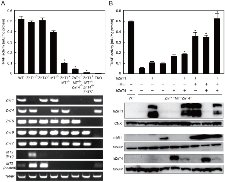
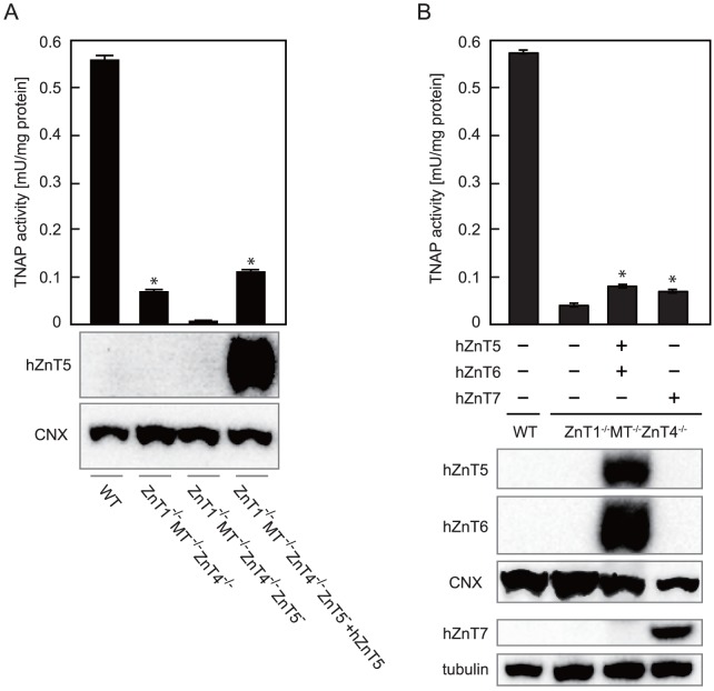
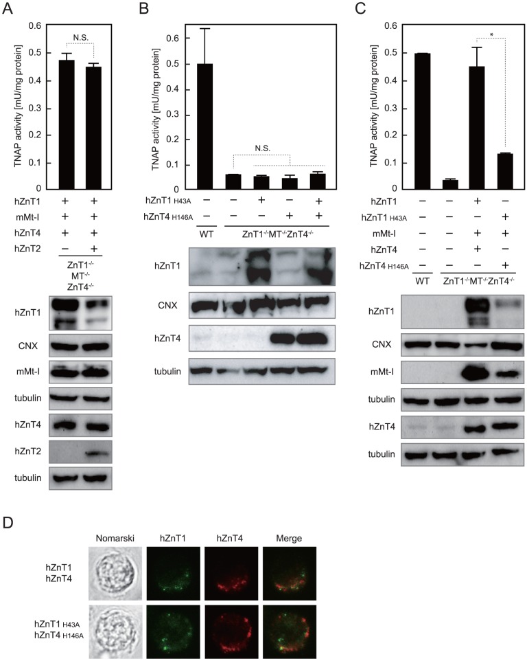
The activation process of secretory or membrane-bound zinc enzymes is thought to be a highly coordinated process involving zinc transport, trafficking, transfer and coordination. We have previously shown that secretory and membrane-bound zinc enzymes are activated in the early secretory pathway (ESP) via zinc-loading by the zinc transporter 5 (ZnT5)-ZnT6 hetero-complex and ZnT7 homo-complex (zinc transport complexes). However, how other proteins conducting zinc metabolism affect the activation of these enzymes remains unknown. Here, we investigated this issue by disruption and re-expression of genes known to be involved in cytoplasmic zinc metabolism, using a zinc enzyme, tissue non-specific alkaline phosphatase (TNAP), as a reporter. We found that TNAP activity was significantly reduced in cells deficient in ZnT1, Metallothionein (MT) and ZnT4 genes (ZnT1(-/-) MT(-/-) ZnT4(-/-) cells), in spite of increased cytosolic zinc levels. The reduced TNAP activity in ZnT1(-/-) MT(-/-) ZnT4(-/-) cells was not restored when cytosolic zinc levels were normalized to levels comparable with those of wild-type cells, but was reversely restored by extreme zinc supplementation via zinc-loading by the zinc transport complexes. Moreover, the reduced TNAP activity was adequately restored by re-expression of mammalian counterparts of ZnT1, MT and ZnT4, but not by zinc transport-incompetent mutants of ZnT1 and ZnT4. In ZnT1(-/-) MT(-/-) ZnT4(-/-) cells, the secretory pathway normally operates. These findings suggest that cooperative zinc handling of ZnT1, MT and ZnT4 in the cytoplasm is required for full activation of TNAP in the ESP, and present clear evidence that the activation process of zinc enzymes is elaborately controlled.


Conflict of interest statement

Competing Interests: The authors have declared that no competing interests exist.

Figures

Figure 1
Figure 1. Cytosolic zinc levels are increased in ZnT1 −/− MT −/− ZnT4 −/− cells.
(A) Zinc sensitivity of DT40 cells deficient in cZnT1, cMT and/or cZnT4 genes. Cells were grown in the presence of the indicated concentrations of ZnSO4 for 72 h and the number of living cells was counted (plotted as a percentage of living cells at 40 µM ZnSO4 for each group of cells). •, wild-type (WT); ○, MT −/−; □, ZnT1 −/−; △, ZnT1 −/− MT −/−; ▪, ZnT1 −/− MT −/− ZnT4 −/− cells. Each value is the mean of two independent experiments. (B) Effects of zinc on MT-I luciferase reporter expression in WT cells and ZnT1 −/− MT −/− ZnT4 −/− cells. Both cell types were transiently transfected and cultured in the presence of 0, 25 or 50 µM ZnSO4 for 12 h. Relative activity of luciferase is shown (the luciferase activity of WT cells cultured without ZnSO4 is defined as 1). Each value is the mean ± SD of three independent experiments (*P<0.01).
Figure 2
Figure 2. ZnT1, MT and ZnT4 are all involved in the activation process of TNAP.
(A) TNAP activity decreased in DT40 cells deficient in genes coding for cZnT1, cMT and/or cZnT4 (upper panel). TNAP activity of total cellular protein prepared from the indicated cells is expressed as the mean ± SD of three independent experiments (*P<0.01 vs. WT cells). Disruption of each gene was confirmed by RT-PCR using the appropriate primers (lower panel). cMT2 expression is shown in duplicate (first and nested) to show significant or moderate induction of cMT2 mRNA expression in ZnT1 −/− or ZnT4 −/− cells. Confirmation of cTNAP expression is also shown. TKO; ZnT5 ZnT6 −/− ZnT7 −/− cells. (B) Re-expression of hZnT1, mMt-I and/or hZnT4 restored TNAP activity in ZnT1 −/− MT −/− ZnT4 −/− cells (upper panel). Expression of FLAG-hZnT1, mMt-I and hZnT4-HA was confirmed by immunoblot analysis using total cellular or membrane proteins prepared from the indicated cells. Tubulin and calnexin (CNX) are shown as loading controls (lower panels). TNAP activity is expressed as the mean ± SD of three independent experiments (*P<0.01 vs. ZnT1 −/− MT −/− ZnT4 −/− cells).
Figure 3
Figure 3. The zinc transport complexes cannot fully operate for TNAP activation in ZnT1 −/− MT −/− ZnT4 −/− cells.
(A) The residual TNAP activity in ZnT1 −/− MT −/− ZnT4 −/− cells was dependent on zinc-loading by the ZnT5-ZnT6 hetero-complex. TNAP activity of the total cellular protein prepared from the indicated cells is expressed as the mean ± SD of three independent experiments (*P<0.01 vs. ZnT1 −/− MT −/− ZnT4 −/− ZnT5 −/− cells, upper panel). Expression of FLAG-hZnT5 in ZnT1 −/− MT −/− ZnT4 −/− ZnT5 cells was confirmed by immunoblot analysis using membrane proteins. Calnexin (CNX) is shown as a loading control (lower panels). (B) Over-expression of hZnT5 and hZnT6 or hZnT7 failed to restore the reduced TNAP activity in ZnT1 −/− MT −/− ZnT4 −/− cells. TNAP activity of the total cellular protein prepared from the indicated cells is expressed as the mean ± SD of three independent experiments (*P<0.01 vs. WT cells, upper panel). Expression of FLAG-hZnT5 and HA-hZnT6 or HA-hZnT7 in ZnT1 −/− MT −/− ZnT4 −/− cells was confirmed by immunoblot analysis as in A (lower panels).
Figure 4
Figure 4. Reduction of the cytosolic zinc levels in ZnT1−/−MT−/−ZnT4−/− cells did not restore reduced TNAP activity.
(A) Exogenous expression of hZnT2 reduced the cytosolic zinc levels in ZnT1 −/− MT −/− ZnT4 −/− cells. ZnT1 −/− MT −/− ZnT4 −/− cells or ZnT1 −/− MT −/− ZnT4 −/− cells stably expressing hZnT2-HA were transiently transfected with MT-I luciferase as in Fig. 1B. Relative activity of luciferase is shown (the luciferase activity of ZnT1 −/− MT −/− ZnT4 −/− cells stably expressing hZnT2 cultured without ZnSO4 is defined as 1). Each value is the mean ± SD of three independent experiments (*P<0.01). (B) TNAP activity of the total cellular protein prepared from the indicated cells is expressed as the mean ± SD of three independent experiments (N.S., not significant, upper panel). Expression of hZnT2 was confirmed by immunoblot analysis using total cellular proteins. Tubulin is shown as a loading control (lower panels). (C) The change in cytosolic zinc levels in WT and ZnT1 −/− MT −/− ZnT4 −/− cells during zinc-deficient culture. Both cell lines were transiently transfected with MT-I luciferase, cultured in normal medium for 9 h, and then cultured in zinc-deficient medium for the indicated period of time prior to measuring luciferase activity. (D) The change in TNAP activity in WT and ZnT1 −/− MT −/− ZnT4 −/− cells during zinc-deficient culture. Both cells cultured in normal medium were washed once and then cultured in zinc-deficient medium for the indicated period. TNAP activity is expressed as the mean ± SD of three independent experiments. In the lower panels of C and D, relative rates of change are plotted with values of WT or ZnT1 −/− MT −/− ZnT4 −/− cells at time 0 h defined as 1. 1M4; ZnT1 −/− MT −/− ZnT4 −/− cells.
Figure 5
Figure 5. TNAP activity is restored by high zinc supplementation in ZnT1 −/− MT −/− ZnT4 −/− cells.
The indicated cells were cultured in medium supplemented with 0, 25 or 50 µM ZnSO4 for 40 h. TNAP activity is the mean ± SD of three independent experiments (N.S., not significant). Note that TNAP activity in ZnT5 ZnT6 −/− ZnT7 −/− (TKO) cells was never restored.
Figure 6
Figure 6. Zinc transport activities of ZnT1 and ZnT4 are required for TNAP activation.
(A) Expression of hZnT2 did not significantly affect TNAP activity restored by re-expression of all of hZnT1, mMt-I and hZnT4 in ZnT1 −/− MT −/− ZnT4 −/− cells (upper panel). Expression of FLAG-hZnT1, mMt-I, hZnT4-HA and hZnT2-FLAG was confirmed by immunoblot analysis using total cellular or membrane proteins prepared from the indicated cells (lower panels). (B) Re-expression of zinc transport-incompetent mutants hZnT1 (FLAG-hZnT1 H43A) or/and hZnT4 (hZnT4-HA H146A) failed to significantly restore the reduced TNAP activity in ZnT1 −/− MT −/− ZnT4 −/− cells (upper panel). Expression of FLAG-hZnT1 and hZnT4-HA mutants was confirmed by immunoblot analysis (lower panels). (C) Expression of hZnT1 and hZnT4 mutants failed to adequately restore reduced TNAP activity, even if mMt-I was expressed (upper panel). Expression of FLAG-hZnT1 H43A mutant, mMt-I and hZnT4-HA H146A mutant was confirmed by immunoblot analysis (lower panels). In A–C, TNAP activity is expressed as the mean ± SD of three independent experiments (*P<0.01, N.S., not significant, upper panels), and tubulin and calnexin (CNX) are shown as loading controls (lower panels). (D) The subcellular localization of hZnT1, hZnT4 (upper panels) and their mutants (lower panels) expressed in ZnT1 −/− MT −/− ZnT4 −/− cells. Nomarski, FLAG-hZnT1 (green), hZnT4-HA (red) and the merged images are shown.
Figure 7
Figure 7. Homeostasis of the secretory pathway is not significantly impaired in ZnT1−/−MT−/−ZnT4−/− cells.
(A) Surface IgM expression was not impaired in ZnT1 −/− MT −/− ZnT4 −/− cells. WT and ZnT1 −/− MT −/− ZnT4 −/− cells were fixed and immunostained without permeabilization. (B) The cell surface proteins biotinylated with membrane-impermeable reagent were almost the same between WT and ZnT1 −/− MT −/− ZnT4 −/− cells. Surface refers to the solubilized proteins captured using streptavidin beads, while input refers to the aliquot of the biotinylated proteins before avidin capture (that is total cell lysate). In the surface panel, lectin blotting using WGA detects cell surface glycosylated proteins, and CBB staining detects total cell surface proteins. 1M4; ZnT1 −/− MT −/− ZnT4 −/− cells. (C) Secretory Cypridina luciferase expression was not significantly different between WT and ZnT1 −/− MT −/− ZnT4 −/− cells. Both cells transiently transfected with secretory Cypridina luciferase reporter were cultured for 4 h after the medium change. Relative activity of Cypridina luciferase in the spent medium is shown (the luciferase activity/total cellular proteins in WT cells is defined as 1). Each value is the mean ± SD of three independent experiments (N.S., not significant).

Similar articles

Cited by

References

    1. Vallee BL, Falchuk KH (1993) The biochemical basis of zinc physiology. Physiol Rev 73: 79–118. - PubMed
    1. Maret W (2012) New perspectives of zinc coordination environments in proteins. J Inorg Biochem 111: 110–116. - PubMed
    1. Andreini C, Banci L, Bertini I, Rosato A (2006) Counting the zinc-proteins encoded in the human genome. J Proteome Res 5: 196–201. - PubMed
    1. Andreini C, Bertini I, Cavallaro G (2011) Minimal functional sites allow a classification of zinc sites in proteins. PLoS One 6: e26325. - PMC - PubMed
    1. Maret W, Li Y (2009) Coordination dynamics of zinc in proteins. Chem Rev 109: 4682–4707. - PubMed

Publication types

MeSH terms

Grants and funding

This work was supported by grants-in-aid from the Ministry of Education, Culture, Sports, Science and Technology of Japan and by the Mochida Memorial Foundation for Medical and Pharmaceutical Research, the Suzuken Memorial Foundation, the Takeda Science Foundation and the Japan Foundation for Applied Enzymology (to T. Kambe). The funders had no role in study design, data collection and analysis, decision to publish, or preparation of the manuscript.