Clinical Trial
. 2013 Oct;9(10):e1003751.
doi: 10.1371/journal.ppat.1003751. Epub 2013 Oct 31.

IRF-4-mediated CIITA transcription is blocked by KSHV encoded LANA to inhibit MHC II presentation

Affiliations
Clinical Trial

IRF-4-mediated CIITA transcription is blocked by KSHV encoded LANA to inhibit MHC II presentation

Qiliang Cai et al. PLoS Pathog. 2013 Oct.

Abstract

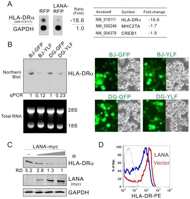
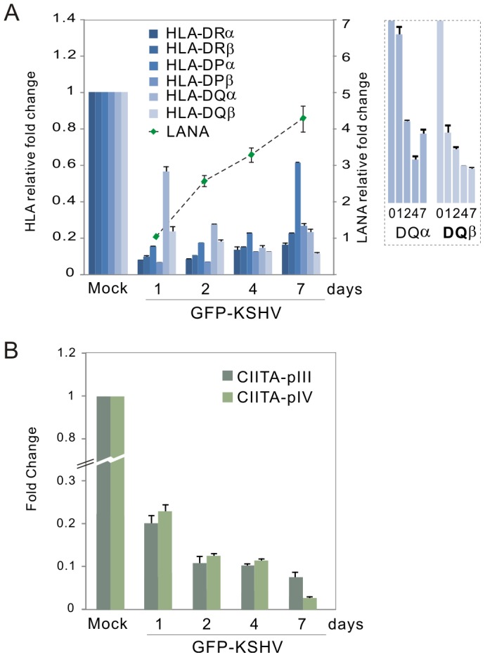
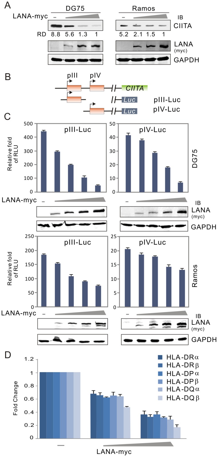
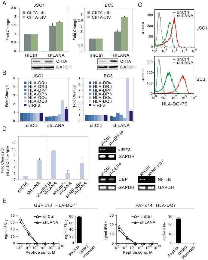
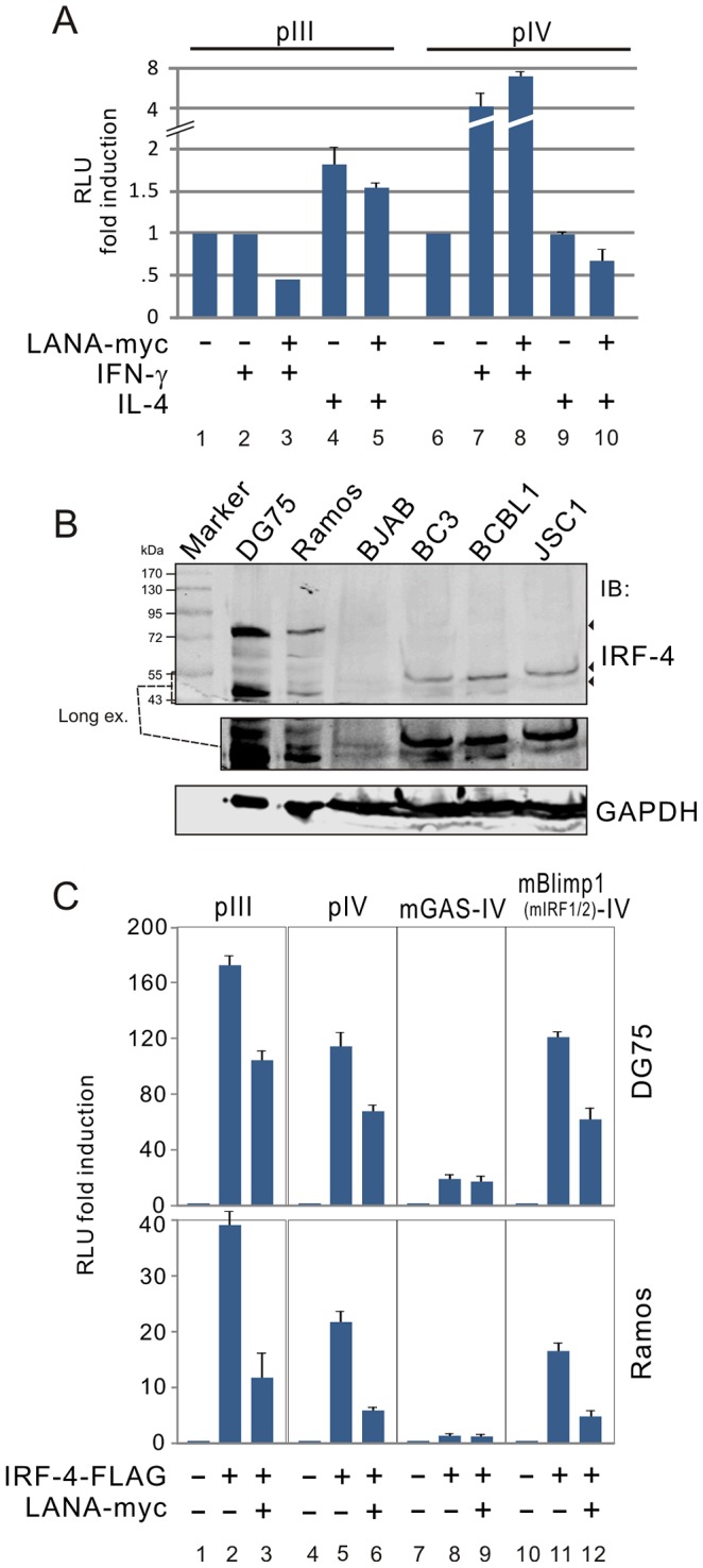
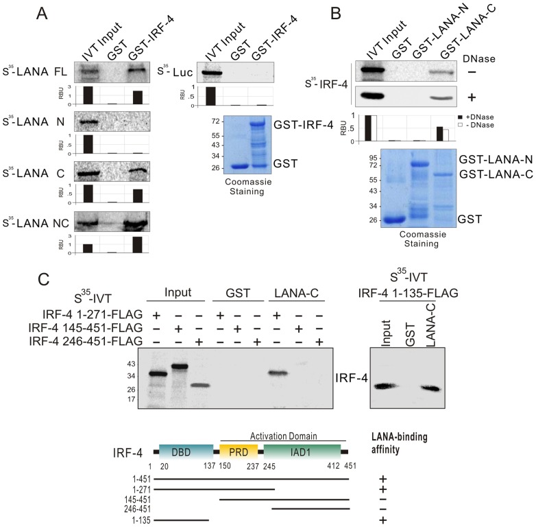
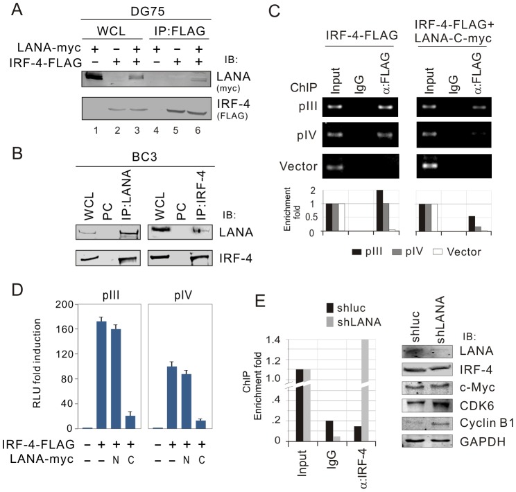
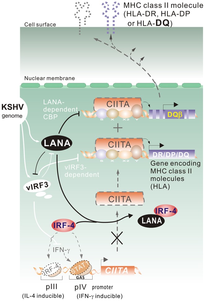
Peptides presentation to T cells by MHC class II molecules is of importance in initiation of immune response to a pathogen. The level of MHC II expression directly influences T lymphocyte activation and is often targeted by various viruses. Kaposi's sarcoma-associated herpesvirus (KSHV) encoded LANA is known to evade MHC class I peptide processing, however, the effect of LANA on MHC class II remains unclear. Here, we report that LANA down-regulates MHC II expression and presentation by inhibiting the transcription of MHC II transactivator (CIITA) promoter pIII and pIV in a dose-dependent manner. Strikingly, although LANA knockdown efficiently disrupts the inhibition of CIITA transcripts from its pIII and pIV promoter region, the expression of HLA-DQβ but no other MHC II molecules was significantly restored. Moreover, we revealed that the presentation of HLA-DQβ enhanced by LANA knockdown did not help LANA-specific CD4+ T cell recognition of PEL cells, and the inhibition of CIITA by LANA is independent of IL-4 or IFN-γ signaling but dependent on the direct interaction of LANA with IRF-4 (an activator of both the pIII and pIV CIITA promoters). This interaction dramatically blocked the DNA-binding ability of IRF-4 on both pIII and pIV promoters. Thus, our data implies that LANA can evade MHC II presentation and suppress CIITA transcription to provide a unique strategy of KSHV escape from immune surveillance by cytotoxic T cells.


Conflict of interest statement

The authors have declared that no competing interests exist.

Figures

Figure 1
Figure 1. HLA-DRα is down-regulated in LANA-expressing B lymphoma cells.
(A) The mRNA level of HLA-DRα was reduced in LANA-expressing BJAB cells. The total RNA of BJAB cells with LANA-RFP or vector RFP stable expression were subjected to microarray assay. Representative data and average ratio of HLA-DRα, CIITA and CREB from LANA-RFP and RFP microarray assay are shown. (B) Northern analysis of HLA-DRα transcript. The total RNA of BJAB and DG75 cells individually transduced with lentiviruses carrying LANA (YLF) or vector (GFP) alone was subjected to northern blot as described in material and method. The relative density of HLA-DRα transcript is verified by quantitative PCR. The efficiency of lentivirus transduction is shown at the right panels. (C) LANA inhibits the expression of HLA-DRα. Ten million of DG75 cells were electroporated with pA3M-LANA (5, 10, 15 µg) or Vector pA3M. At 48-hr post-transfection, the cell lysate were separated by SDS-PAGE and analyzed by Western blot using antibodies HLA-DRα or myc(9E10). Detection of GAPDH was used as an internal control. (D) The surface expression of HLA-DR proteins on DG75 cells transfected with LANA-myc or vector was determined by FACS using PE-labeled mAb against HLA-DR.
Figure 2
Figure 2. Kinetics of MHC class II expression in PBMCs with KSHV primary infection.
Quantitative real-time PCR analysis of (A) MHC II and LANA transcripts, and (B) CIITA pIII and pIV promoter transcripts, in PBMC with GFP-KSHV infection. The levels of HLA-DQαβ were enlarged on the right panel A. Total RNA was isolated from cells with GFP-KSHV infection for 0, 1, 2, 4 and 7 days. Real-time PCR was performed as described in Materials and Methods. The relative levels of MHCII, LANA and CIITA transcripts were calculated by the cycle threshold (ΔΔCt) values and shown by the fold change compared to mock (day 0) after normalized with GAPDH internal control. All samples were tested in triplicate and the calculation of the mean and standard deviation from two separate experiments. The mock cells without or with GFP-KSHV infection indicated by the relative intensity of LANA were quantified by qPCR shown in the figure.
Figure 3
Figure 3. LANA expression represses CIITA expression in B lymphoma cells.
(A) The level of CIITA expression was reduced by LANA in a dose-dependent manner. Ten million of DG75 or Ramos cells were transfected with a different dose of DNA construct expressing LANA. At 24 hr post-transfection, cells were harvested and lysed for immunoblotting assays as indicated in the figure. GAPDH blot was used as loading control. The relative density (RD) of CIITA proteins was quantified and shown in the figure. (B) Schematic representation of CIITA promoter reporters (pIII-Luc and pIV-Luc) with luciferase. (C) DG75 and Ramos cells were individually co-transfected CIITA promoter (pIII and pIV) driving luciferase reporter plasmid with either pA3M-LANA or pA3M vector. At 24-hrs post-transfection, cells were harvested and subjected to reporter assay. The results were presented by the RLU (relative luciferase unit) fold compared to pGL3-basic with vector alone. Data is presented as means±SD of three independent experiments. The expression of LANA were detected by immunoblotting and shown at the bottom panels. (D) Quantitative real-time PCR analysis of MHCII in DG75 cells transfected with different dose LANA as in panel A.
Figure 4
Figure 4. MHCII molecules and CIITA transcript are dramatically enhanced in a LANA-knocked down manner.
(A) The relative levels of CIITA pIII and pIV transcripts in PEL cells with LANA or control knockdown. Total RNA from PEL (JSC1 and BC3) cells with LANA (shLANA) or control firefly luciferase knockdown (shCtrl) were extracted for RT-PCR analysis. Data from two repeat experiments. The protein levels of CIITA detected by western blotting were shown at the bottom panel. (B) The levels of partial MHCII transcripts and vIRF3 were increased in PEL cells with LANA knockdown. (C) Cytometric profile of HLA-DQ expression in PEL cells with or without LANA knockdown. PEL cells with LANA or control knockdown were individually divided and subjected to PE-conjugated HLA-DQ antibodies staining. The FACS result is one representative experiment. (D) The level of HLA-DQβ transcripts in JSC1 PEL cells with LANA or LANA combined with vIRF3, CBP or NF-κB knockdown. The knockdown efficiency of vIRF3, CBP or NF-κB was presented at right panel by agarose gel after RT-PCR. (E) CD4+ T cell recognition of JSC1 primary effusion lymphoma cells transduced with LANA-specific or control shRNA lentivirus. Transduced JSC1 cells were sensitized with ten-fold dilutions of the HLA-DQ7 presented peptides GSPTVFTSGLPAFVS or PAFVSSPTLPVAPIP; peptides are identified in the figure by their first three amino acids. Target cells were incubated in triplicate with cognate epitope-specific CD4+ T cell clones for 18 hours and IFN-γ secreted from these cells into the culture supernatant estimated by ELISA. In parallel T cell clones were assayed against HLA-matched Epstein-Barr virus transformed B cell line LCL sensitized with the peptide or the peptide solvent, or an HLA mismatched B cell line (right panel). Data shown is one representative experiment of two.
Figure 5
Figure 5. LANA blocks the transcriptional activity of IRF-4 on both pIII and pIV CIITA promoters.
(A) The inhibition of LANA on CIITA promoter is independent on IL-4 or IFNγ-induced signaling.HEK293 cells were co-transfected pA3M-LANA with pIII or pIV luciferase reporter as indicated in the figure. At 24 hr post-transfection, cells were treated with IL-4 (20 ng/ml), IFNγ (400 ng/ml) or untreated for 36 hours, followed by reporter assays. (B) Expression of IRF-4 in KSHV negative B lymphoma and positive PEL cells. Immunoblotting (IB) analysis was performed using 100 µg of whole-cell lysates from KSHV negative B lymphoma (DG75, Ramos and BJAB) and positive PEL (BC3, BCBL1, JSC1) cell lines. Immunoblots were probed with anti-IRF-4 antibody. The membrane was reblotted with anti-GAPDH antibody to verify protein-loading homogeneity. The position of IRF-4 molecular weight around 43–55 kDa with long exposure was presented at the middle panel. (C) LANA blocks IRF-4-mediated transcription levels of both pIII and pIV. DG75 and Ramos cells were individually co-transfected CIITA promoter (pIII, pIV, and pIV with GAS or Blimp1(IRF1/2) binding-site mutation) driving luciferase reporter plasmid with different combination of pA3F-IRF-4, pA3M-LANA and vector. At 24 hr post-transfection, cells lysates were subjected to luciferase reporter assays. The results were presented by the RLU (relative luciferase unit) fold compared to each reporter with vector alone. Data is presented as means±SD of three independent experiments.
Figure 6
Figure 6. IRF-4 binds with LANA through its DNA-binding domain in vitro.
(A) IRF-4 binds to C-terminal domain of LANA in vitro. The 35S-radiolabeled in vitro-translated proteins of LANA truncated mutants were pre-cleared with GST bead, followed by incubation with GST or GST-IRF-4 beads. The bound protein mixtures were resolved by appropriate SDS-PAGE, and protein species detected by autoradiography. 5% of in vitro translated protein is used as input. The quantification of relative amount of bound proteins (RBU) is shown at the bottom. (B) The 35S-radiolabeled in vitro-translated full length IRF-4 was pulled down by truncated mutants of LANA fusion with GST (GST-LANA N1–340 and C945–1162) in the presence or absence of DNase I (20 U/ml) treatment. Coomassie blue staining of purified GST-LANA is shown at the bottom panels. (C) LANA binds to N-terminal domain of IRF-4 in vitro. The 35S-radiolabeled in vitro-translated proteins IRF-4 with FLAG tag (1–135, 1–271, 145–451 and 246–451) was pulled down by GST or GST-LANA C. Schematics illustrate different structural domains of IRF-4 with LANA-binding ability.
Figure 7
Figure 7. Endogenous IRF-4 interacts with LANA and results in failure of the DNA binding capacity on CIITA promoter.
(A) Exogenous IRF-4 interacts with LANA. DG75 cells were co-transfected IRF-4-FLAG with pA3M-LANA or vector control. At 48-hr post-transfection, the whole cell lysates (WCL) were subjected to directly immunoblotting (IB) or immunoprecipitation (IP) with anti-FLAG (M2) antibody followed by immunoblotting with indicated antibodies. (B) Endogenous IRF-4 associates with LANA. Cell lysates of five million of PEL cells (BC3) were subjected to directly immunoblotting or immunoprecipitation with anti-IRF-4 antibody followed by immunoblotting with indicated antibodies. Normal serum IgG was used for preclear (PC). (C) LANA reduces DNA-binding ability of IRF-4 on pIII and pIV promoter. DG75 cells were co-transfected IRF-4-FLAG with or without LANA-C930–1162-myc in the presence of pIII, pIV or pGL3-basic vector alone. Chromatin immunoprecipitation assays were performed with anti-FLAG(M2) or normal IgG antibodies control. The DNA-binding ability of IRF-4 was detected by standard (upper) and quantitative real-time (lower) PCR with primers targeting ampicillin gene of pGL3 plasmid. (D) The carboxyl terminus of LANA dramatically blocks the transcriptional activity of IRF-4 on both pIII and pIV promoter. Ten million DG75 cells were individually co-transfected with different combination of IRF-4-FLAG, LANA1-340-myc (N), LANA 930–1162(C) or vector alone as indicated in the figure. At 24 hr post-transfection, cells lysates were subjected to luciferase reporter assays. The results were presented by the RLU (relative luciferase unit) fold compared to each reporter with vector alone. Data is presented as means±SD of three independent experiments. (E) Knockdown of LANA allows IRF-4 to bind to the CIITA promoter. ChIP analysis of endogenous CIITA pIII promoter in BC3 cells with or without LANA knockdown was conducted using normal mouse IgG, α-IRF-4 agarose, and subjected to qPCR analysis using primers indicated in Figure 2. The protein levels of IRF-4 and its downstream genes in BC3 cells with or without LANA knockdown are shown at the right panel.
Figure 8
Figure 8. A model depicting the role of LANA on MHC II expression and presentation.
In the KSHV-infected cells, the major latent antigen LANA interacts with IRF-4 and reduces the DNA-binding ability of IRF-4 on the IL-4-inducible pIII and IFN-γ-inducible pIV promoter of CIITA, which inhibits the transcriptional activity of CIITA on MHC class II promoter. Meanwhile, associated with this is LANA-mediated inhibition of MHC class II expression and presentation (particularly HLA-DQβ) and other viral genes (like vIRF3) expression in terms of cooperative regulation of host MHC II molecules during viral pathogenesis.

Similar articles

Cited by

References

    1. Waldburger JM, Suter T, Fontana A, Acha-Orbea H, Reith W (2001) Selective abrogation of major histocompatibility complex class II expression on extrahematopoietic cells in mice lacking promoter IV of the class II transactivator gene. J Exp Med 194: 393–406. - PMC - PubMed
    1. Matechak EO, Killeen N, Hedrick SM, Fowlkes BJ (1996) MHC class II-specific T cells can develop in the CD8 lineage when CD4 is absent. Immunity 4: 337–347. - PubMed
    1. Steidl C, Shah SP, Woolcock BW, Rui L, Kawahara M, et al. (2011) MHC class II transactivator CIITA is a recurrent gene fusion partner in lymphoid cancers. Nature 471: 377–381. - PMC - PubMed
    1. Honey K, Rudensky A (2001) The pIV-otal class II transactivator promoter regulates major histocompatibility complex class II expression in the thymus. J Exp Med 194: F15–18. - PMC - PubMed
    1. Chang CH, Fontes JD, Peterlin M, Flavell RA (1994) Class II transactivator (CIITA) is sufficient for the inducible expression of major histocompatibility complex class II genes. J Exp Med 180: 1367–1374. - PMC - PubMed

Publication types

MeSH terms