Clinical Trial
. 2013 Oct;9(10):e1003738.
doi: 10.1371/journal.ppat.1003738. Epub 2013 Oct 31.

Viral escape from HIV-1 neutralizing antibodies drives increased plasma neutralization breadth through sequential recognition of multiple epitopes and immunotypes

Affiliations
Clinical Trial

Viral escape from HIV-1 neutralizing antibodies drives increased plasma neutralization breadth through sequential recognition of multiple epitopes and immunotypes

Constantinos Kurt Wibmer et al. PLoS Pathog. 2013 Oct.

Abstract

Identifying the targets of broadly neutralizing antibodies to HIV-1 and understanding how these antibodies develop remain important goals in the quest to rationally develop an HIV-1 vaccine. We previously identified a participant in the CAPRISA Acute Infection Cohort (CAP257) whose plasma neutralized 84% of heterologous viruses. In this study we showed that breadth in CAP257 was largely due to the sequential, transient appearance of three distinct broadly neutralizing antibody specificities spanning the first 4.5 years of infection. The first specificity targeted an epitope in the V2 region of gp120 that was also recognized by strain-specific antibodies 7 weeks earlier. Specificity for the autologous virus was determined largely by a rare N167 antigenic variant of V2, with viral escape to the more common D167 immunotype coinciding with the development of the first wave of broadly neutralizing antibodies. Escape from these broadly neutralizing V2 antibodies through deletion of the glycan at N160 was associated with exposure of an epitope in the CD4 binding site that became the target for a second wave of broadly neutralizing antibodies. Neutralization by these CD4 binding site antibodies was almost entirely dependent on the glycan at position N276. Early viral escape mutations in the CD4 binding site drove an increase in wave two neutralization breadth, as this second wave of heterologous neutralization matured to recognize multiple immunotypes within this site. The third wave targeted a quaternary epitope that did not overlap any of the four known sites of vulnerability on the HIV-1 envelope and remains undefined. Altogether this study showed that the human immune system is capable of generating multiple broadly neutralizing antibodies in response to a constantly evolving viral population that exposes new targets as a consequence of escape from earlier neutralizing antibodies.


Conflict of interest statement

The authors have declared that no competing interests exist.

Figures

Figure 1
Figure 1. CAP257 broadly neutralizing antibodies develop sequentially in three distinct waves.
A) Longitudinal neutralization of the autologous CAP257 virus (black) and 37 heterologous viruses neutralized by CAP257 plasma at titers >1∶100. The ID50 titers (y-axis) are shown versus weeks p.i. (x-axis). Three peaks in heterologous neutralization titers at 67, 122, and 213 weeks p.i. are indicated with dotted lines. Heterologous viruses are colored according to subtype (A = green, B = blue, C = red). B) A summary of the three waves of heterologous neutralization defined by a representative virus, superimposed over the neutralization kinetics shown in Figure 1A. Wave 1 was subtype C specific and is colored red. Wave 2 neutralized viruses from all three clades and is colored green. Wave 3 is colored brown. C) Adsorption of heterologous neutralization at the peak of each of the three waves. Percentage inhibition (y-axis) is shown versus plasma dilution (x-axis). Untreated plasma is shown in black, blank beads in grey and beads coated with recombinant proteins are shown in red (wave 1), blue/green (wave 2) or brown (wave 3).
Figure 2
Figure 2. The first wave of broadly neutralizing antibodies targets residues in the V2 region.
A) Longitudinal neutralization of ConC V2 mutants. ConC wild-type (wt) is shown in red. V2 mutants F159A, N160A, R166A, K168A, K169E, K171A, and I181A that abrogated wave 1 neutralization are shown in purple. The D167N mutation that enhanced wave 1 neutralization is shown in orange, while the L165A mutation that resulted in universal neutralization sensitivity is shown in grey. The timing of wave 1 (red), wave 2 (green), and wave 3 (brown) neutralization is summarized above as horizontal lines, while the peak titers at each wave are indicated with dotted lines. ID50 titers (y-axis) are shown versus weeks p.i. (x-axis). B) The dependence of CAP257 wave 1 neutralizing antibodies (at 67 weeks p.i.) on V2 residues in ConC, compared to monoclonal antibodies PGT145, CH01-04, and PG9/16. Complete abrogation of neutralization is colored red, 2–10 fold reductions in IC50 are colored yellow, and >10 fold reductions in IC50 are colored orange.
Figure 3
Figure 3. Escape from V2 neutralizing antibodies drives the formation/exposure of broadly neutralizing antibody epitopes in the CD4bs.
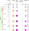
A) Amino acid sequence alignment of the CAP257 B- and C- strands in the V1/V2 sub-domain of gp120, from twelve time points. The number of envelopes per unique V2 sequence is shown on the right. The timing of wave 1 (red), wave 2 (green), and wave 3 (brown) neutralization is summarized to the left with vertical lines. Potential N-linked glycans are shaded grey, and the presence (grey slices) or absence (red slices) of the N160 glycan within the population at each time point is shown with pie charts to the right. B) CAP257 develops a strain-specific V2 response prior to wave 1 broadly neutralizing antibodies. Neutralization of an autologous virus amplified from 174 weeks p.i. (CAP257 3 yr), is shown in grey. The V1/V2 region of this virus was back-mutated to the earliest known sequence (CAP257 3 yr(V1/V2s)) shown in black. Longitudinal neutralization of the N167D, N160D/S, and K169E mutants is shown in orange, purple, and pink respectively. The timing of wave 1 (red), wave 2 (green), and preceding strain-specific V2 (black) neutralization is summarized above with horizontal lines. ID50 titers (y-axis) are shown versus weeks p.i. (x-axis). C) Wave 2 neutralization of Q842 (green) or RHPA (blue) wild-type (wt) viruses, and their N160K mutants (purple). The timing of wave 1 (red), wave 2 (green), and wave 3 (brown) neutralization is summarized above as in Figure 1B. ID50 titers (y-axis) are shown versus weeks p.i. (x-axis).
Figure 4
Figure 4. Accumulating escape mutations from wave 2 broadly neutralizing antibodies occur in the CD4 binding site.
Amino acid sequence alignment of the CAP257 D loop and β23 regions of gp120 from seven time points. The number of envelopes per unique sequence is shown on the right. The timing of wave 1 (red), wave 2 (green), and wave 3 (brown) neutralization is summarized to the left with vertical lines. Amino acids contacting CD4 (as described in [57]) are indicated above the sequence alignment, with ○ denoting backbone only contacts, formula image denoting side chain contacts, and • denoting main chain and side chain contacts. Potential N-linked glycans are shaded grey. The frequency of escape mutations at position 279 (orange slices), in the N276/T278 glycosylation sequon (purple slices), or at position 456 (pink slices) for each time point is shown with pie charts.
Figure 5
Figure 5. CAP257 broadly neutralizing antibodies bind a glycan dependent epitope in the CD4bs, also targeted by mAb HJ16.
A) Effect of escape mutations T278A, N279D, and R456W in the CD4bs on wave 2 neutralization. ID50 titers at 122 weeks p.i. (y-axis) are shown for Q842 and RHPA (black bars), or the corresponding triple mutants (red bars). B) Adsorption of peak wave 2 titers (122 weeks p.i.) with wild-type or mutant gp120 proteins. The residual neutralizing activity in the adsorbed plasma samples is shown as ID50 titers (y-axis) for the heterologous viruses Q842 and RHPA. Untreated plasma is shown in black, plasma adsorbed with blank beads in grey, plasma adsorbed with ConC gp120 in white, plasma adsorbed with D368R mutant gp120 in yellow, plasma adsorbed with D474A/M475A/R476A triple mutant gp120 in brown, and plasma adsorbed with RSC3 protein in orange. C) Effect of glycosylation on the CAP257 wave 2 epitope in RHPA. The fold reduction in titer (y-axis) of adsorbed samples from 122 weeks p.i. are shown for CAP257 wave 2, HJ16, and VRC01. Plasma adsorbed with blank beads is shown in grey, gp120 core in white, and deglycosylated gp120 core in purple. D) The dependence of CAP257 wave 2 neutralizing antibodies (at 122 weeks p.i.) on D-loop and β23 residues/glycans in RHPA, compared to the monoclonal antibodies HJ16, VRC01, and 10E8. The effect of N276 glycan mutations on CAP257 plasma is boxed in purple, and the presence or absence of a glycan at N276 for each mutation is indicated. Fold effects between 2–10 are yellow, 10–100 colored orange, and >100 colored red. Triple* is the triple mutant described in A. E) Effect of glycosylation on the HJ16 epitope. The ELISA OD at 450 nm is shown (y-axis) versus antibody concentration (x-axis). Binding to glycosylated or deglycosylated gp120 core is shown with solid or open circles respectively. The monoclonal antibodies tested were VRC01 (green), b12 (yellow), HJ16 (purple), 2G12 (blue), and CAP88-3468L (grey).
Figure 6
Figure 6. Changes in the fine specificity of CAP257 CD4bs antibodies in response to autologous escape mutations.
The longitudinal neutralization of Q842 (green) or RHPA (blue) wild-type (wt) viruses was compared to the neutralization of N279D (orange), T278A (purple), and R456W (pink) mutant viruses. The triple mutant (T278A/N279D/R456W) from Figure 5A is also shown in red. ID50 titers (y-axis) are shown versus weeks p.i. (x-axis). Dotted lines indicate the time points at which each mutation (D = N279D, A = T278A, W = R456W) first appears in the autologous sequences (colored as above).
Figure 7
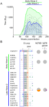
Figure 7. Maturation of the wave 2 CD4bs response results in the increased neutralization breadth of CAP257 plasma.
A) Summary of early (green) and late (blue) heterologous neutralization by wave 2 antibodies superimposed over the individual virus neutralization kinetics (grey). Viruses neutralized by V2 antibodies in wave 1 have been excluded. ID50 titers (y-axis) are shown versus weeks p.i. (x-axis). B) Amino acid sequence alignment of heterologous viruses depicted in (A). The timing of early or late heterologous neutralization is shown on the left with horizontal lines. The N276 glycan is shaded grey, and position 279 is boxed in orange. The frequency of the N279D mutation or disrupted N276 glycosylation within the two groups is shown with orange or purple pie slices respectively. The N-terminal region of the D-loop is boxed in blue.
Figure 8
Figure 8. Wave 3 neutralizing antibodies target a novel epitope.
A) Neutralization of Du156 wild-type (wt) virus by longitudinal CAP257 serum is shown in red. The timing of wave 1 (red), wave 2 (green), and wave 3 (brown) neutralization is summarized above as horizontal lines, with peak titers of each wave indicated with dotted lines. The Du156 N160K/T278A double mutant is shown in brown. Du156 triple mutants N160K/T278A/(R166A, K168A, K169E, K171A, or N332A) are shown in yellow. ID50 titers (y-axis) are shown versus weeks p.i. (x-axis). B) Adsorption of MPER binding antibodies. OD (450 nm) against the MPER peptide is shown (y-axis) versus plasma dilution (x-axis). Untreated plasma is shown in black, and MPER adsorbed plasma in brown. C) Adsorption of wave 3 neutralization by MPER (colored as above). ID50 neutralization titers at 213 weeks p.i. (y-axis) are shown for Du156 and Du156 N160K/T278A.
Figure 9
Figure 9. Accumulating resistance of CAP257 clones to broadly neutralizing monoclonal antibodies targeting V2 and the CD4bs.
The sensitivity of nine CAP257 clones from 7, 30, 54, 93, and 174 weeks p.i. to broadly neutralizing antibodies targeting the four known sites of vulnerability was measured in a TZM-bl assay. The timing of wave 1 (red), wave 2 (green), and wave 3 (brown) neutralization is summarized above with arrows. Increases in IC50 relative to the 7 week clone are colored as follows: 5–10 fold (yellow), >10 fold (orange), complete neutralization resistance (red).
Figure 10
Figure 10. Summary of the role of CAP257 viral evolution in shaping broadly neutralizing antibody responses.
The schema depicts the evolution of plasma neutralizing antibodies and viral escape mutations over 240 weeks. Each of the three waves of CAP257 broadly neutralizing antibodies is shown. Text boxes highlight the key events described herein. Env trimers (EMD-5447) were drawn in UCSF-Chimera, and spheres were used to approximate the location of escape mutations. 1) Strain-specific antibodies (dotted orange line) developed at 23 weeks, targeting a V2 epitope overlapping with known V2 antibodies (eg: PG9). 2) Strain-specific V2 antibodies were escaped by an N167D mutation (D being the global consensus at this site). 3) Broadly neutralizing antibodies targeting V2 (wave 1 – red curve) developed at 30 weeks. 4) Escape from wave 1 antibodies through deletion of the N160 glycan was associated with exposure of an epitope in the CD4bs. 5) At 67 weeks broadly neutralizing antibodies targeting the CD4bs (wave 2 – green curve) develop. Neutralization was N276 glycan dependent and sensitive to an N279D mutation. 6) The N279D change emerges at 93 weeks, significantly affecting wave 2 neutralization at this time point. 7) Wave 2 neutralization becomes independent of position 279, which was associated with increased neutralization breadth. 8) Mutations that delete the N276 glycan (as well as an R456W change) escape wave 2 antibodies. 9) CAP257 develops a third broadly neutralizing antibody specificity that could not be mapped to any of the four known antibody targets.
Figure 11
Figure 11. The R456 side chain stabilizes the CD4bs epitope through hydrogen bonding.
A diagram of the 93TH057 gp120 crystal structure (pdb file 4JKP) shown in an orientation similar to the angle of approach for CD4. The crystalized part of the N276 attached glycan (GlcNAc2Man4) is shown with purple spheres. The D-loop is shown in green, the V5 loop is shown in cyan, and the R456 residue is shown in pink. Oxygen atoms are colored red, and nitrogen atoms blue. The inset shows a magnified view of the interaction between R456 and residues in the D-loop or the β24 strand. Putative hydrogen bonds are shown with dotted orange lines. The image was created using The PyMOL Molecular Graphics System, Version 1.3r1edu, Schrödinger LLC.

Similar articles

Cited by

References

    1. Gray ES, Moore PL, Choge IA, Decker JM, Bibollet-Ruche F, et al. (2007) Neutralizing antibody responses in acute human immunodeficiency virus type 1 subtype C infection. J Virol 81: 6187–6196. - PMC - PubMed
    1. Richman DD, Wrin T, Little SJ, Petropoulos CJ (2003) Rapid evolution of the neutralizing antibody response to HIV type 1 infection. Proc Natl Acad Sci U S A 100: 4144–4149. - PMC - PubMed
    1. Li B, Decker JM, Johnson RW, Bibollet-Ruche F, Wei X, et al. (2006) Evidence for potent autologous neutralizing antibody titers and compact envelopes in early infection with subtype C human immunodeficiency virus type 1. J Virol 80: 5211–5218. - PMC - PubMed
    1. Wei X, Decker JM, Wang S, Hui H, Kappes JC, et al. (2003) Antibody neutralization and escape by HIV-1. Nature 422: 307–312. - PubMed
    1. Moog C, Fleury HJ, Pellegrin I, Kirn A, Aubertin AM (1997) Autologous and heterologous neutralizing antibody responses following initial seroconversion in human immunodeficiency virus type 1-infected individuals. J Virol 71: 3734–3741. - PMC - PubMed

Publication types