Structural biology of the Bcl-2 family and its mimicry by viral proteins
- PMID: 24201808
- PMCID: PMC3847314
- DOI: 10.1038/cddis.2013.436
Structural biology of the Bcl-2 family and its mimicry by viral proteins
Abstract
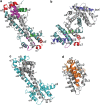
Intrinsic apoptosis in mammals is regulated by protein-protein interactions among the B-cell lymphoma-2 (Bcl-2) family. The sequences, structures and binding specificity between pro-survival Bcl-2 proteins and their pro-apoptotic Bcl-2 homology 3 motif only (BH3-only) protein antagonists are now well understood. In contrast, our understanding of the mode of action of Bax and Bak, the two necessary proteins for apoptosis is incomplete. Bax and Bak are isostructural with pro-survival Bcl-2 proteins and also interact with BH3-only proteins, albeit weakly. Two sites have been identified; the in-groove interaction analogous to the pro-survival BH3-only interaction and a site on the opposite molecular face. Interaction of Bax or Bak with activator BH3-only proteins and mitochondrial membranes triggers a series of ill-defined conformational changes initiating their oligomerization and mitochondrial outer membrane permeabilization. Many actions of the mammalian pro-survival Bcl-2 family are mimicked by viruses. By expressing proteins mimicking mammalian pro-survival Bcl-2 family proteins, viruses neutralize death-inducing members of the Bcl-2 family and evade host cell apoptosis during replication. Remarkably, structural elements are preserved in viral Bcl-2 proteins even though there is in many cases little discernible sequence conservation with their mammalian counterparts. Some viral Bcl-2 proteins are dimeric, but they have distinct structures to those observed for mammalian Bcl-2 proteins. Furthermore, viral Bcl-2 proteins modulate innate immune responses regulated by NF-κB through an interface separate from the canonical BH3-binding groove. Our increasing structural understanding of the viral Bcl-2 proteins is leading to new insights in the cellular Bcl-2 network by exploring potential alternate functional modes in the cellular context. We compare the cellular and viral Bcl-2 proteins and discuss how alterations in their structure, sequence and binding specificity lead to differences in behavior, and together with the intrinsic structural plasticity in the Bcl-2 fold enable exquisite control over critical cellular signaling pathways.
Figures


Similar articles
-
Structural insight into BH3 domain binding of vaccinia virus antiapoptotic F1L.J Virol. 2014 Aug;88(15):8667-77. doi: 10.1128/JVI.01092-14. Epub 2014 May 21. J Virol. 2014. PMID: 24850748 Free PMC article.
-
Structural biology of the Bcl-2 family of proteins.Biochim Biophys Acta. 2004 Mar 1;1644(2-3):83-94. doi: 10.1016/j.bbamcr.2003.08.012. Biochim Biophys Acta. 2004. PMID: 14996493 Review.
-
Vaccinia virus anti-apoptotic F1L is a novel Bcl-2-like domain-swapped dimer that binds a highly selective subset of BH3-containing death ligands.Cell Death Differ. 2008 Oct;15(10):1564-71. doi: 10.1038/cdd.2008.83. Epub 2008 Jun 13. Cell Death Differ. 2008. PMID: 18551131
-
A comparison of two strategies for affinity maturation of a BH3 peptide toward pro-survival Bcl-2 proteins.ACS Synth Biol. 2012 Mar 16;1(3):89-98. doi: 10.1021/sb200002m. Epub 2011 Oct 7. ACS Synth Biol. 2012. PMID: 23651073
-
Discoveries and controversies in BCL-2 protein-mediated apoptosis.FEBS J. 2016 Jul;283(14):2690-700. doi: 10.1111/febs.13527. Epub 2015 Oct 27. FEBS J. 2016. PMID: 26411300 Review.
Cited by
-
BHRF1, a BCL2 viral homolog, disturbs mitochondrial dynamics and stimulates mitophagy to dampen type I IFN induction.Autophagy. 2021 Jun;17(6):1296-1315. doi: 10.1080/15548627.2020.1758416. Epub 2020 May 13. Autophagy. 2021. PMID: 32401605 Free PMC article.
-
Sequence analysis of Epstein-Barr virus (EBV) early genes BARF1 and BHRF1 in NK/T cell lymphoma from Northern China.Virol J. 2015 Sep 4;12:135. doi: 10.1186/s12985-015-0368-3. Virol J. 2015. PMID: 26337172 Free PMC article.
-
Conformation of BCL-XL upon Membrane Integration.J Mol Biol. 2015 Jul 3;427(13):2262-70. doi: 10.1016/j.jmb.2015.02.019. Epub 2015 Feb 27. J Mol Biol. 2015. PMID: 25731750 Free PMC article.
-
Mastering Death: The Roles of Viral Bcl-2 in dsDNA Viruses.Viruses. 2024 May 30;16(6):879. doi: 10.3390/v16060879. Viruses. 2024. PMID: 38932171 Free PMC article. Review.
-
Contrasting roles for G-quadruplexes in regulating human Bcl-2 and virus homologues KSHV KS-Bcl-2 and EBV BHRF1.Sci Rep. 2022 Mar 23;12(1):5019. doi: 10.1038/s41598-022-08161-9. Sci Rep. 2022. PMID: 35322051 Free PMC article.
References
Publication types
MeSH terms
Substances
LinkOut - more resources
Full Text Sources
Other Literature Sources
Research Materials

