

Upregulated WAVE3 expression is essential for TGF-β-mediated EMT and metastasis of triple-negative breast cancer cells
- PMID: 24197660
- PMCID: PMC3888319
- DOI: 10.1007/s10549-013-2753-1
Upregulated WAVE3 expression is essential for TGF-β-mediated EMT and metastasis of triple-negative breast cancer cells
Abstract
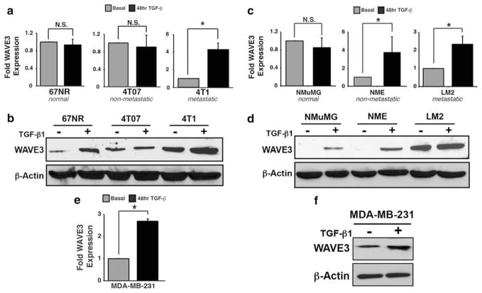
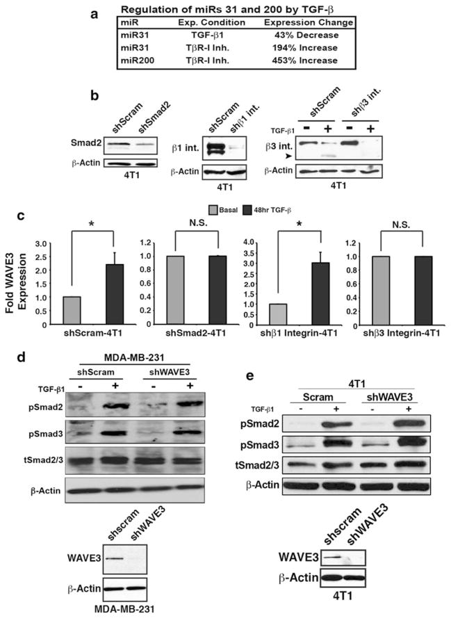
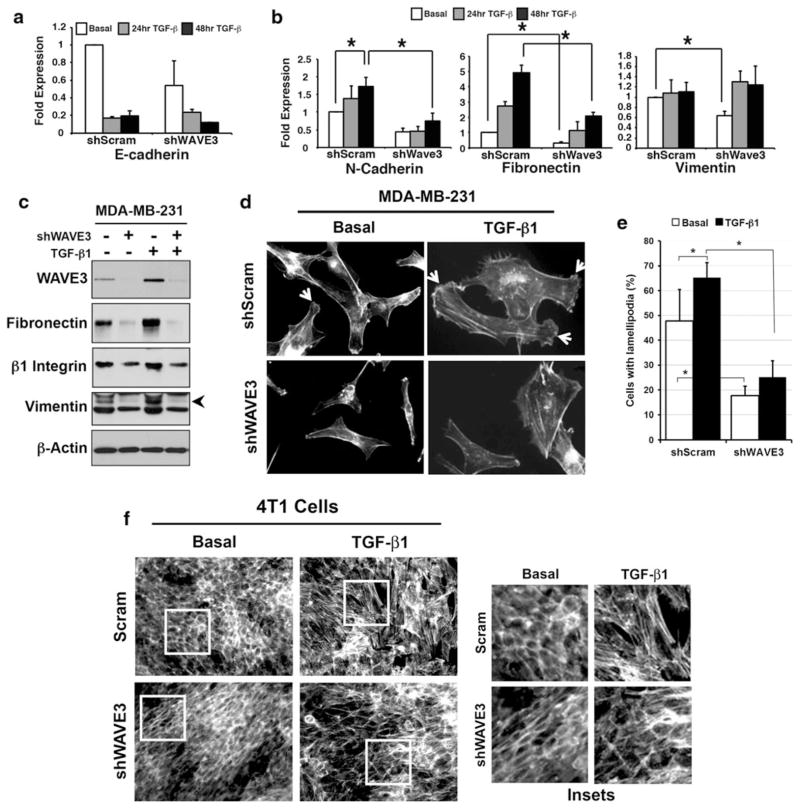
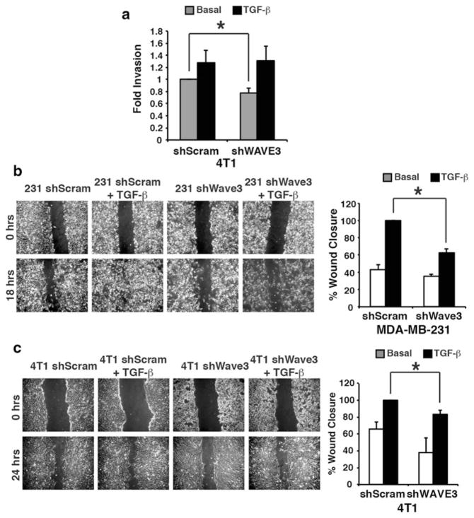
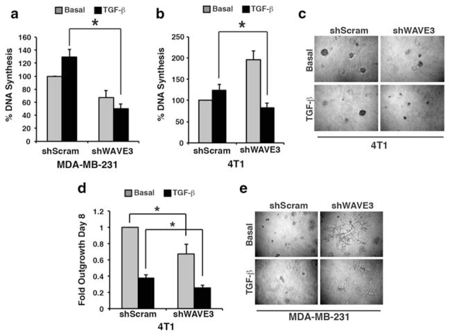
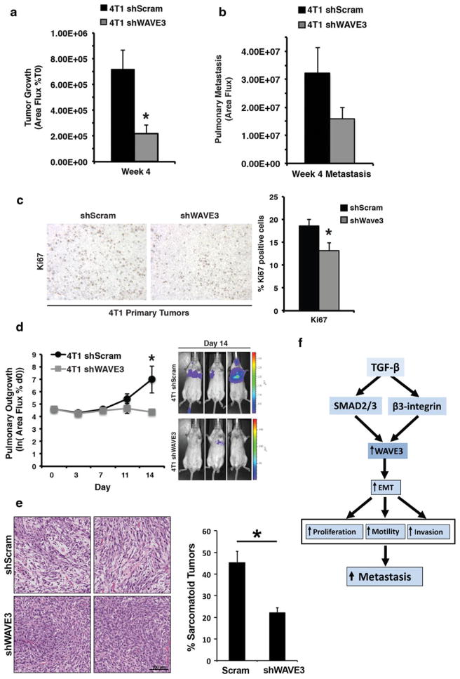
Breast cancer is the second leading cause of cancer death in women in the United States. Metastasis accounts for the death of ~90 % of these patients, yet the mechanisms underlying this event remain poorly defined. WAVE3 belongs to the WASP/WAVE family of actin-binding proteins that play essential roles in regulating cell morphology, actin polymerization, cytoskeleton remodeling, cell motility, and invasion. Accordingly, we demonstrated previously that WAVE3 promotes the acquisition of invasive and metastatic phenotypes by human breast cancers. Herein, we show that transforming growth factor-β (TGF-β) selectively and robustly induced the expression of WAVE3 in metastatic breast cancer cells, but not in their nonmetastatic counterparts. Moreover, the induction of WAVE3 expression in human and mouse triple-negative breast cancer cells (TNBCs) by TGF-β likely reflects its coupling to microRNA expression via a Smad2- and β3 integrin-dependent mechanism. We further demonstrate the requirement for WAVE3 expression in mediating the initiation of epithelial-mesenchymal transition (EMT) programs stimulated by TGF-β. Indeed, stable depletion of WAVE3 expression in human TNBC cells prevented TGF-β from inducing EMT programs and from stimulating the proliferation, migration, and the formation of lamellipodia in metastatic TNBC cells. Lastly, we observed WAVE3 deficiency to abrogate the outgrowth of TNBC cell organoids in 3-dimensional organotypic cultures as well as to decrease the growth and metastasis of 4T1 tumors produced in syngeneic Balb/C mice. Indeed, WAVE3 deficiency significantly reduced the presence of sarcomatoid morphologies indicative of EMT phenotypes in pulmonary TNBC tumors as compared to those detected in their parental counterparts. Collectively, these findings indicate the necessity for WAVE3 expression and activity during EMT programs stimulated by TGF-β; they also suggest that measures capable of inactivating WAVE3 may play a role in alleviating metastasis stimulated by TGF-β.
Conflict of interest statement
Figures
Similar articles
-
Phosphorylation of the proline-rich domain of WAVE3 drives its oncogenic activity in breast cancer.Sci Rep. 2021 Feb 16;11(1):3868. doi: 10.1038/s41598-021-83479-4. Sci Rep. 2021. PMID: 33594155 Free PMC article.
-
Targeted inactivation of β1 integrin induces β3 integrin switching, which drives breast cancer metastasis by TGF-β.Mol Biol Cell. 2013 Nov;24(21):3449-59. doi: 10.1091/mbc.E12-10-0776. Epub 2013 Sep 4. Mol Biol Cell. 2013. PMID: 24006485 Free PMC article.
-
WAVE3 promotes epithelial-mesenchymal transition of gastric cancer through upregulation of Snail.Cancer Gene Ther. 2014 Dec;21(12):499-506. doi: 10.1038/cgt.2014.52. Epub 2014 Nov 7. Cancer Gene Ther. 2014. PMID: 25378074
-
Role of WAVE3 as an of actin binding protein in the pathology of triple negative breast cancer.Cytoskeleton (Hoboken). 2024 Jul 18. doi: 10.1002/cm.21898. Online ahead of print. Cytoskeleton (Hoboken). 2024. PMID: 39021344 Review.
-
Elucidating the molecular signaling pathways of WAVE3.Ann Transl Med. 2020 Jul;8(14):900. doi: 10.21037/atm.2020.02.16. Ann Transl Med. 2020. PMID: 32793744 Free PMC article. Review.
Cited by
-
Common variants at 10p12.31, 10q21.1 and 13q12.13 are associated with sporadic pituitary adenoma.Nat Genet. 2015 Jul;47(7):793-7. doi: 10.1038/ng.3322. Epub 2015 Jun 1. Nat Genet. 2015. PMID: 26029870
-
WAVE3 upregulation in esophageal squamous cell carcinoma and its effect on the migration of human esophageal cancer cell lines in vitro.Mol Med Rep. 2020 Jul;22(1):465-473. doi: 10.3892/mmr.2020.11126. Epub 2020 May 5. Mol Med Rep. 2020. PMID: 32377706 Free PMC article.
-
WAVE3 Induces EMT and Promotes Migration and Invasion in Intrahepatic Cholangiocarcinoma.Dig Dis Sci. 2016 Jul;61(7):1950-60. doi: 10.1007/s10620-016-4102-9. Epub 2016 Mar 12. Dig Dis Sci. 2016. PMID: 26971088
-
Actin Cytoskeleton and Regulation of TGFβ Signaling: Exploring Their Links.Biomolecules. 2021 Feb 23;11(2):336. doi: 10.3390/biom11020336. Biomolecules. 2021. PMID: 33672325 Free PMC article. Review.
-
Ascending the PEAK1 toward targeting TGFβ during cancer progression: Recent advances and future perspectives.Cancer Cell Microenviron. 2016;3(1):e1162. doi: 10.14800/ccm.1162. Epub 2016 Jan 28. Cancer Cell Microenviron. 2016. PMID: 29392163 Free PMC article.
References
-
- Perou CM, Sorlie T, Eisen MB, van de Rijn M, Jeffrey SS, Rees CA, Pollack JR, Ross DT, Johnsen H, Akslen LA, Fluge O, Pergamenschikov A, Williams C, Zhu SX, Lonning PE, Borresen-Dale AL, Brown PO, Botstein D. Molecular portraits of human breast tumours. Nature. 2000;406(6797):747–752. - PubMed
-
- Carey L, Winer E, Viale G, Cameron D, Gianni L. Triple-negative breast cancer: disease entity or title of convenience? Nat Rev Clin Oncol. 2010;7(12):683–692. - PubMed
-
- Carey LA. Directed therapy of subtypes of triple-negative breast cancer. Oncologist. 2010;15(Suppl 5):49–56. - PubMed
-
- Siegel R, Naishadham D, Jemal CA. Cancer statistics. CA Cancer J Clin. 2012;62(1):10–29. - PubMed
Publication types
MeSH terms
Substances
Grants and funding
LinkOut - more resources
Full Text Sources
Other Literature Sources
