Review
. 2014 Jul;38(4):598-632.
doi: 10.1111/1574-6976.12052. Epub 2013 Dec 6.

Bitter-sweet symphony: glycan-lectin interactions in virus biology

Affiliations
Review

Bitter-sweet symphony: glycan-lectin interactions in virus biology

Wander Van Breedam et al. FEMS Microbiol Rev. 2014 Jul.

Abstract

Glycans are carbohydrate modifications typically found on proteins or lipids, and can act as ligands for glycan-binding proteins called lectins. Glycans and lectins play crucial roles in the function of cells and organs, and in the immune system of animals and humans. Viral pathogens use glycans and lectins that are encoded by their own or the host genome for their replication and spread. Recent advances in glycobiological research indicate that glycans and lectins mediate key interactions at the virus-host interface, controlling viral spread and/or activation of the immune system. This review reflects on glycan-lectin interactions in the context of viral infection and antiviral immunity. A short introduction illustrates the nature of glycans and lectins, and conveys the basic principles of their interactions. Subsequently, examples are discussed highlighting specific glycan-lectin interactions and how they affect the progress of viral infections, either benefiting the host or the virus. Moreover, glycan and lectin variability and their potential biological consequences are discussed. Finally, the review outlines how recent advances in the glycan-lectin field might be transformed into promising new approaches to antiviral therapy.

Keywords: DC-SIGN; antiviral; collectin; galectin; hemagglutinin; receptor-destroying enzyme.


Figures

Figure 1
Figure 1
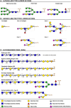
Classification and basic structure of common types of protein-linked glycosylation. (a) GlcNAcβ-Asn type N-linked glycans are covalently attached to the amide nitrogen atoms of Asn side chains and are almost exclusively found on Asn residues within the sequence Asn-X-Ser/Thr, in which X can be any amino acid except Pro. The nature of the glycan structures that decorate the common glycan core – the glycan part shown in a dashed box – dictates classification of N-linked glycans as high-mannose type, hybrid type or complex type glycans, examples of which are shown in the panel. (b) GalNAcα-Ser/Thr type O-linked glycans have a GalNAc residue α-linked to the oxygen atom of the hydroxyl group of Ser or Thr residues. Unlike for GlcNAcβ-Asn type N-linked protein glycosylation, there are no clear amino acid motifs that mark these O-linked glycosylation sites. A single GalNac residue linked to the Ser/Thr is termed the ‘Tn antigen’. Depending on the basic structure of the glycan core, more complex (extended) O-linked glycans are categorized into different ‘core types’. Cores 1–4 are the most common core structures, but also other core types exist. The Tn antigen and examples of extended core 1, 2, 3, and 4 O-glycans are shown in the panel. The distinct glycan cores are shown in dashed boxes. (c) Glycosaminoglycans (GAGs) are linear polysaccharide chains composed of repeated disaccharide subunits of a uronic acid/galactose residue and an amino sugar. Glycosaminoglycans are classified as hyaluronan (HA), heparan sulfate/heparin (HS), chondroitin sulfate (CS), dermatan sulfate (DS), or keratan sulfate (KS), depending on the structure of their basic disaccharide subunits (shown in square brackets) and further modification (e.g. sulfation at different positions) of the glycan chain. With exception of hyaluronan, all major glycosaminoglycan types are sulfated and occur covalently linked to proteins. HS, CS, and DS are found on Ser-linked xylose residues. Although no unambiguous consensus sequence for xylosylation exists, the Ser attachment site is consistently flanked by a Gly residue at its carboxy-terminal side. As depicted in the figure, heparan sulfate and heparin have the same basic structure. Although they share a common biosynthesis, heparin generally undergoes more extensive sulfation and epimerization of uronic acid to iduronic acid. Moreover, heparin is synthesized only in connective tissue mast cells as part of serglycin proteoglycans, whereas heparan sulfate is synthesized in virtually all mammalian cells. KS is found on Asn-linked N-glycan core structures (KS I) or Ser/Thr-linked O-glycan core 2 structures (KS II). Capping or further modification of the glycosaminoglycan chains – sulfation excepted – is not depicted (adapted from Varki et al., 2009).
Figure 2
Figure 2
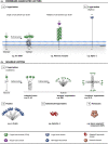
Classification and basic structure of major types of lipid-linked glycosylation. (a) Glycosphingolipids consist of a hydrophilic glycan moiety linked to a hydrophobic sphingolipid. In higher animals, a ceramide lipid molecule is initially modified with a β-linked glucose or galactose residue, after which further extension and modification of the glycan moiety can occur. Extension to larger glycan chains is common on ceramide-linked glucose residues, whereas further glycan extension on ceramide-linked galactose residues is more rare. Depending on their glycan core structure, glycosphingolipids are classified in ‘series’. The figure depicts a number of glycosphingolipid core structures. The key features that characterize each series are shown in dashed boxes. Core structures can be further modified with sialic acids or sulfate groups, which allows subclassification of glycosphingolipids as neutral (lacking charged carbohydrates or ionic groups), sialylated or sulfated. (b) Glycosylphosphatidylinositol (GPI) anchors are found in association with certain membrane proteins and serve as linkers between the protein and the lipid membrane. Glycosylphosphatidylinositol anchors have a common core structure comprising ethanolamine-PO4-6Manα1-2Manα1-6Manα1-4GlcNα1-6myo-inositol-1-PO4-lipid. Differential derivatization of this common core structure through lipid remodeling and modification of the glycan moiety can cause significant glycosylphosphatidylinositol anchor heterogeneity. The protein is linked to the glycosylphosphatidylinositol anchor via an amide linkage between the C-terminal carboxyl group of the protein and the amino group of phosphatidylethanolamine (adapted from Varki et al., 2009).
Figure 3
Figure 3
Schematic overview of different types of membrane-associated (a) and soluble (b) animal lectins that are considered in this review. The lectin domains are highlighted and listed in the key. C-type lectin/C-type lectin domain: Lectins are classified as C-type lectins based on their Ca2+-dependency and shared primary structure. In the C-type CRD, a Ca2+ ion is directly involved in carbohydrate binding by making coordination bonds to both the CRD surface and key hydroxyl groups of the carbohydrate. The C-type lectin family contains both membrane-associated (a.1) and soluble (b.1) lectins. The collectins are solubleC-type lectins characterized by the presence of collagen-like domains. R-type lectin domain: This term refers to a CRD that is structurally similar to the CRD in ricin, a toxin found in the plant Ricinus communis. I-type lectin/I-type lectin domain: I-type lectins are glycan-binding proteins that belong to the Ig superfamily, but are not antibodies or T-cell receptors. The ‘sialic acid-binding Ig-like lectin (siglec)’ family of membrane-associated lectins is currently the only well-characterized group of I-type lectins (a.2). Ficolin: Ficolins (b.2) are soluble lectins characterized by the presence of collagen-like domains and fibrinogen-like globular domains with a lectin activity. Galectin/S-type lectin (domain): Galectins (b.3) are soluble lectins that typically bind β-galactose-containing glycoconjugates and show primary structural homology in their CRDs. Galectins were initially referred to as S-type lectins to reflect their sulfhydryl dependency, the presence of cysteine residues and their solubility; however, at present, not all identified galectins fit this initial description anymore. Pentraxin/pentraxin domain: Pentraxins (b.4) are characterized by the presence of pentraxin domains, which contain an eight amino acid long conserved ‘pentraxin signature’ (HxCxS/TWxS, where x is any amino acid) and display an L-type (Legume-type) lectin fold. SAP is a soluble lectin that requires Ca2+ ions for carbohydrate ligand binding (adapted from Fujita, ; Varki et al., ; Bottazzi et al., 2010).
Figure 4
Figure 4
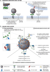
Schematic overview of how membrane-associated (a) and soluble (b) host lectins are implicated in antiviral defense. (a.1) Binding of virion-associated glycans with membrane-associated host lectins can lead to virus uptake, degradation, and presentation of viral antigens to cells of the adaptive immune system. Binding may trigger specific signaling that promotes an effective antiviral immunity. (a.2) Binding of virion-associated glycans with membrane-associated host lectins may promote direct presentation of the virus to immune cells in trans. Binding may trigger specific signaling that promotes an effective antiviral immunity. (b.1) Binding of soluble host lectins to virion-associated glycans may interfere directly with viral infection by destabilizing virions, blocking interaction of the virus with its receptors or interfering with other crucial steps in the infection process (e.g. membrane fusion). Soluble host lectins may also aggregate virions, which often negatively impacts viral infectivity (not depicted). (b.2) Soluble host lectins can act as opsonins: lectin binding to virion-associated glycans may facilitate viral uptake in immune cells via lectin receptors, leading to viral degradation and potential presentation of viral antigens to cells of the adaptive immune system. Lectin binding may also trigger complement deposition on the virus (through the lectin pathway) and facilitate viral uptake via complement receptors. (b.3) Detection of virion-associated glycans by soluble host lectins may trigger complement deposition on the virus (through the lectin pathway), which may directly inhibit viral infection and/or elicit lysis of the (enveloped) virus.
Figure 5
Figure 5
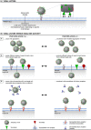
(a) illustrates how viral lectins promote target cell infection. (b) shows how many viruses that employ viral lectins also benefit from a matching receptor-destroying enzyme (RDE) activity, which provides a counterweight against (high avidity) lectin activity. (a) Interaction of viral lectins with glycosylated receptors on a target cell promotes viral entry and infection (attachment/internalization/fusion, depending on specific virus biology). (b) Although they clearly benefit the virus, the use of (high avidity) viral lectins comes with a price. For instance, viral lectin activity can cause virions to aggregate (b.1) and can impair efficient release of newly formed virions from (glycosylated) infected cells (b.2). Moreover, binding of viral lectins to nontarget cell-associated glycoconjugates (decoy receptors) can prevent the virus from efficiently targeting susceptible host cells (b.3). Intriguingly, several lectin-carrying viruses are also equipped with an RDE that matches the specificity of the viral lectin and provides a counterweight against lectin-mediated glycan binding. In fact, for viruses equipped with both viral lectins and RDEs, a functional balance between these molecules appears to be an important determinant of the viral (replicative) fitness.
Figure 6
Figure 6
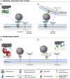
Schematic overview of how membrane-associated (a) and soluble (b) host lectins can be implicated in interactions that benefit the virus and facilitate viral infection and spread. (a.1) Binding of virion-associated glycans to membrane-associated host lectins can promote (cis-) infection of the lectin-expressing cell: host lectins may facilitate viral attachment, internalization, and fusion (depending on specific virus biology). Viral attachment to membrane-associated host lectins may trigger signaling mechanisms that facilitate viral infection, spread, and/or immune evasion. (a.2) Binding of virion-associated glycans to membrane-associated host lectins can promote presentation of the virus to susceptible target cells in trans, thereby facilitating target cell infection. Viral attachment to membrane-associated host lectins may trigger signaling mechanisms that facilitate viral infection, spread, and/or immune evasion. (b.1) Multivalent soluble host lectins may facilitate virus attachment and promote viral infection by crosslinking virus- and host cell-displayed glycans. (b.2) Virus recognition by soluble host lectins and subsequent association with target cell-expressed lectin receptors may promote cis-infection of target cells. In a similar manner, soluble host lectins may capture and concentrate virions on a cell surface for subsequent presentation to target cells in trans (not depicted). Moreover, lectin binding can trigger complement deposition on the virus (through the lectin pathway), which may potentially promote cis- or trans-infection via cell surface-expressed complement receptors (not depicted).

Similar articles

Cited by

References

    1. Aguilar HC, Matreyek KA, Filone CM. et al (2006) N-glycans on Nipah virus fusion protein protect against neutralization but reduce membrane fusion and viral entry. J Virol 80: 4878–4889. - PMC - PubMed
    1. Alexandre KB, Gray ES, Mufhandu H, McMahon JB, Chakauya E, O'Keefe BR, Chikwamba R, Morris L. (2012) The lectins griffithsin, cyanovirin-N and scytovirin inhibit HIV-1 binding to the DC-SIGN receptor and transfer to CD4(+) cells. Virology 423: 175–186. - PMC - PubMed
    1. Alymova IV, Taylor G, Portner A. (2005) Neuraminidase inhibitors as antiviral agents. Curr Drug Targets 5: 401–409. - PubMed
    1. Anders EM, Hartley CA, Reading PC, Ezekowitz RA. (1994) Complement-dependent neutralization of influenza virus by a serum mannose-binding lectin. J Gen Virol 75: 615–622. - PubMed
    1. Arrighi JF, Pion M, Wiznerowicz M. et al (2004) Lentivirus-mediated RNA interference of DC-SIGN expression inhibits human immunodeficiency virus transmission from dendritic cells to T cells. J Virol 78: 10848–10855. - PMC - PubMed

Publication types