. 2013 Nov 14;5(3):573-81.
doi: 10.1016/j.celrep.2013.09.041. Epub 2013 Oct 31.

A molecular mechanism regulating the timing of corticogeniculate innervation

Affiliations

A molecular mechanism regulating the timing of corticogeniculate innervation

Justin M Brooks et al. Cell Rep. .

Abstract

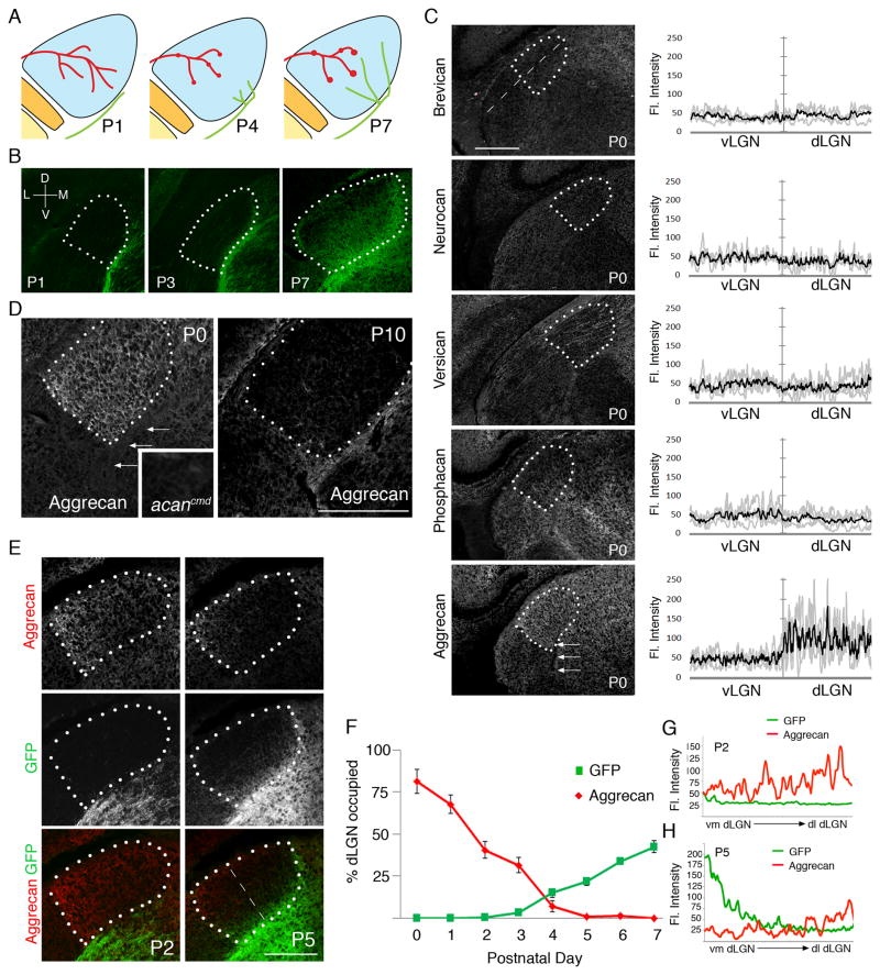
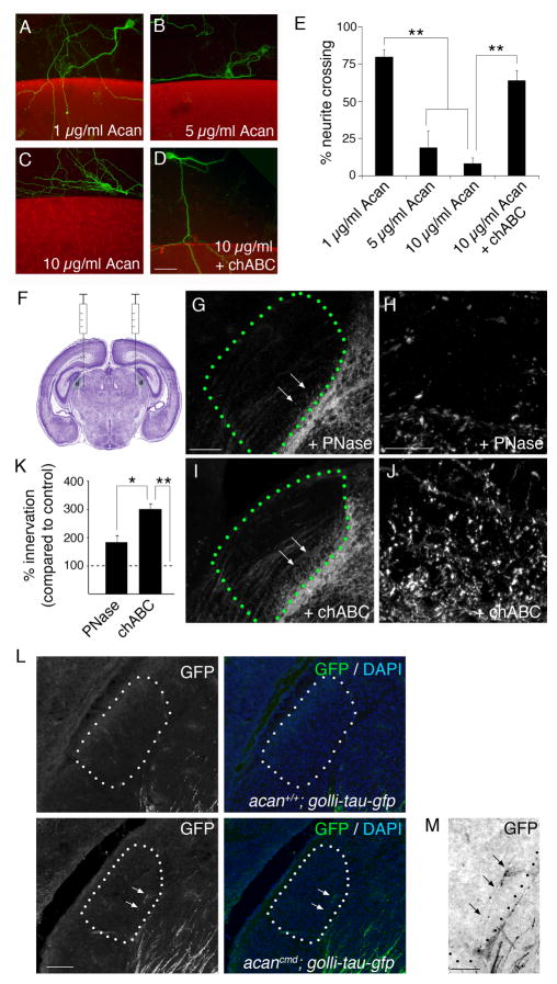
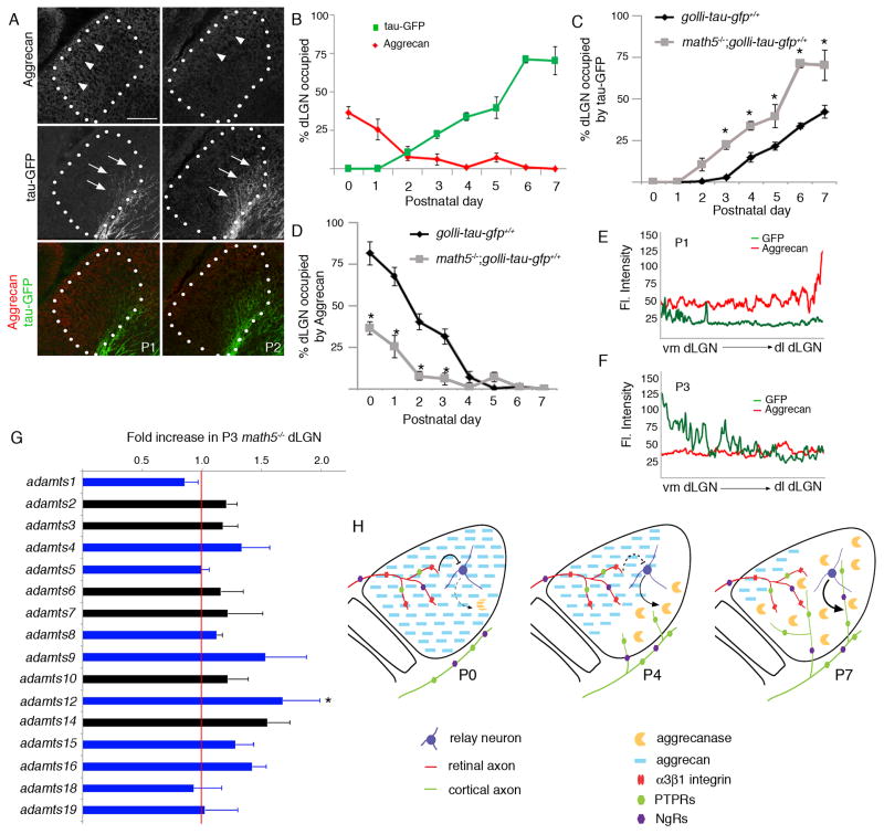
Neural circuit formation demands precise timing of innervation by different classes of axons. However, the mechanisms underlying such activity remain largely unknown. In the dorsal lateral geniculate nucleus (dLGN), axons from the retina and visual cortex innervate thalamic relay neurons in a highly coordinated manner, with those from the cortex arriving well after those from retina. The differential timing of retino- and corticogeniculate innervation is not a coincidence but is orchestrated by retinal inputs. Here, we identified a chondroitin sulfate proteoglycan (CSPG) that regulates the timing of corticogeniculate innervation. Aggrecan, a repulsive CSPG, is enriched in neonatal dLGN and inhibits cortical axons from prematurely entering the dLGN. Postnatal loss of aggrecan from dLGN coincides with upregulation of aggrecanase expression in the dLGN and corticogeniculate innervation and, it is important to note, is regulated by retinal inputs. Taken together, these studies reveal a molecular mechanism through which one class of axons coordinates the temporal targeting of another class of axons.


Figures

Figure 1
Figure 1
Developmental regulation of aggrecan in postnatal dLGN. A. Schematic depiction of the timing of retino- and corticogeniculate innervation. Retinal axons are shown in red; cortical axons are shown in green. Synapses are illustrated by red or green dots. B. Development of corticogeniculate projections in golli-tau-gfp transgenic mice. Projections from layer VI cortical neurons are labeled with tau-GFP in these mice. For all panels dLGN are encircled by white dots. D – dorsal; V – ventral; M- medial; L – lateral. C. Immunostaining revealed aggrecan but not other CSPGs was enriched in P0 mouse dLGN. Arrows depict eml. Fluorescent intensities were measured with a line scan along the ventrolateral to dorsomedial axis of LGN (see dashed line). Fluorescent intensities in vLGN and dLGN are plotted. Grey lines represent IR in individual animals (n=4) and black line represents mean of all experiments. D. Developmental regulation of aggrecan distribution during the first 2 weeks of postnatal dLGN development. Inset shows a lack of aggrecan-IR in dLGN of an aggrecan-deficient mutant (acancmd). E. Aggrecan-IR and GFP-IR in dLGN of P2 and P5 golli-tau-gfp transgenic mice. Note the first regions occupied by cortical axons lack aggrecan-IR. F. The percentage of dLGN occupied by aggrecan-IR was measured in golli-tau-gfp transgenic mice for the first 8 days of postnatal development. The percentage of cortical innervation in these mice was also quantified. Data are shown +/− SEM. G,H. Fluorescent intensities in E were measured with a line scan along the ventromedial (vm) to dorsolateral (dl) axis of LGN (see dashed line in E). Mean fluorescent intensities from 4 P2 (G) and P5 (H) golli-tau-gfp mice are plotted for GFP- and aggrecan-IR. Scale bars = 250 μm.
Figure 2
Figure 2
Aggrecan inhibits cortical axon outgrowth. A–D. Modified stripe assays demonstrate that high concentrations of aggrecan (Acan)(B,C) inhibit neurite outgrowth from layer VI neurons isolated from golli-tau-gfp transgenic mice. Pretreatment of 10 μg/ml aggrecan with chABC alleviated its growth-inhibitory properties (D). Aggrecan-containing substratum depicted in red; tau-GFP-expressing neurons shown in green. E. Quantification of the percentage of neurites capable of crossing into aggrecan-containing substrata shown in A–D. Data are shown +/−SEM: n>4 experiments in triplicate. ** Differ by p<0.01 by Tukey-Kramer Test. F. Schematic depiction of the site of our bilateral intrathalamic injections. Image of the cresyl violet stained brain modified from Allen Institute of Brain Science. G–J. GFP-labeled axon invasion of P3 dLGN following intrathalamic injection of PNase (G,H) or chABC (I,J) in P0 golli-tau-gfp transgenic mice. Delivery of chABC accelerated the rate of CG innervation. H,J. High magnification images of GFP-IR in areas highlighted by arrows in G and I respectively. K. Quantification of the percent dLGN innervated by GFP-containing cortical axons following injection of PNase or chABC. Data are normalized to data obtained from uninjected golli-tau-gfp littermates and are shown +/− SEM: n>4. ** chABC treatment differs from uninjected controls by p<0.0005 by Tukey-Kramer Test. *chABC treatment differs from PNase treatment by p<0.02. PNase treatment and uninjected controls were not statistically different. L,M. Cortical axons invade dLGN at P0 in acancmd; golli-tau-gfp mutants (see arrows). dLGN encircled by dots. Nuclei were labeled with DAPI to determine dLGN boundaries. H. High-magnification of tau-GFP labeled cortical axons in P0 dLGN shown in L. Signal has been inverted to enhance detection of thin caliber cortical axons. dLGN encircled by dots. Scale bar in D = 50 μm for A–D, in G = 150 μm for G,I, in H = 20 μm for H,J, in L = 100 μm, and in M = 50μm.
Figure 3
Figure 3
Aggrecanases are upregulated in postnatal dLGN. A. Microarray revealed that several members of the ADAMTS family of metalloproteinases are enriched in P3 dLGN compared to adjacent thalamic regions (vLGN). Expression of ADAMTS genes with known aggrecan-degrading activity are colored in blue. Red line represents no change in gene expression. Data are shown +/− SEM: n=3. * differs by p<0.05 by T=test; ** differs by p<0.01. B. Microarray demonstrates upregulation of several aggrecan-degrading ADAMTS members in dLGN from P3 to P8. Expression of ADAMTS genes with known aggrecan-degrading activity are colored in blue. Data are shown +/− SEM: n=3. * differs by p<0.05; ** differs by p<0.01. C. qPCR confirmed the upregulation of aggrecanases in dLGN from P2 to P14. Data are shown +/− SEM: n=3. * differs by p<0.001. D,E. In situ hybridization (ISH) of adamtsl5 mRNA in P3 and P14 LGN. dLGN encircled by green dots. F,G. Double-ISH revealed that adamts4 (F) and adamtsl5 (G) mRNAs are expressed by syt1-expressing neurons in dLGN. H. GFP-labeled axon invasion of P3 dLGN following intrathalamic injection of ADAMTS4. I. High magnification image of GFP-IR in area highlighted by arrow in H. J. Quantification of the percent dLGN innervated by GFP-containing cortical axons following injection of ADAMTS4. Data are normalized to data obtained from uninjected golli-tau-gfp littermates and are shown +/− SEM: n>4. * ADAMTS treatment differs from uninjected controls or PNase treatment by p<0.0001 by Tukey-Kramer Test. ADAMTS treatment and chABC treatment were not statistically different. Scale bar in D = 200 μm for D,E, in F = 75 μm for F,G, in H = 150 μm, and in I = 20 μm.
Figure 4
Figure 4
Retinal inputs influences the degradation of aggrecan in dLGN. A. Aggrecan-IR in math5−/ −; golli-tau-gfp mutant dLGN which lacks retinal inputs. Arrowheads depict remaining aggrecan-IR in the lateral aspect of dLGN. Arrows highlight GFP-labeled axons prematurely invading dLGN. Note that aggrecan is absent from sites of cortical axon invasion. dLGN are encircled by white dots. B. The percentage of dLGN occupied by aggrecan-IR was measured in math5−/ −; golli-tau-gfp mutants for the first 8 days of postnatal development. The percentage of dLGN occupied by tau-GFP-expressing cortical axons was also quantified. Dashed line represented the age at which the percent occupied by GFP and aggrecan were equal in controls (see Figure 1G). C,D. The percent dLGN occupied by tau-GFP-expressing axons or by aggrecan-IR was compared in math5−/ −; golli-tau-gfp mutants and golli-tau-gfp controls. Data are shown +/−SEM. * Differ with age-matched controls by P<0.01 by Tukey-Kramer test. E,F. Fluorescent intensities in A were measured with a line scan along the ventromedial (vm) to dorsolateral (dl) axis of LGN (see example in Figure 1G,H). Mean fluorescent intensities from 4 P1 and P3 math5−/ −;golli-tau-gfp mice are plotted for GFP- and aggrecan-IR. G. Microarray analysis revealed some adamts mRNAs are modestly upregulated in P3 dLGN in math5−/ − mutants (compared with wild-type controls). Expression of ADAMTS genes with known aggrecan-degrading activity are colored in blue. Red line represents no change in gene expression. Data are shown +/− SEM: n=3. * differs by p<0.05 by t-test. Scale bar = 100 μm. H. Schematic depicting the mechanism by which aggrecan controls the timing of corticogeniculate innervation. See discussion for details.

Similar articles

Cited by

References

    1. Alilain WJ, Horn KP, Hu H, Dick TE, Silver J. Functional regeneration of respiratory pathways after spinal cord injury. Nature. 2011;475:196–200. - PMC - PubMed
    1. Bickford ME, Slusarczyk A, Dilger EK, Krahe TE, Kucuk C, Guido W. Synaptic development of the mouse dorsal lateral geniculate nucleus. J Comp Neurol. 2010;518:622–635. - PMC - PubMed
    1. Condic ML, Snow DM, Letourneau PC. Embryonic neurons adapt to the inhibitory proteoglycan aggrecan by increasing integrin expression. J Neurosci. 1999;19:10036–10043. - PMC - PubMed
    1. Dickendesher TL, Baldwin KT, Mironova YA, Koriyama Y, Raiker SJ, Askew KL, Wood A, Geoffrey CG, Zheng B, Liepmann CD, et al. NgR1 and NgR3 are receptors for chondroitin sulfate proteoglycans. Nat Neurosci. 2012;15:703–712. - PMC - PubMed
    1. Fisher D, Xing B, Dill J, Li H, Hoang HH, Zhao Z, Yang XL, Bachoo R, Cannon S, Longo FM, et al. Leukocyte common antigen-related phosphatase is a functional receptor for chondroitin sulfate proteoglycan axon growth inhibitors. J Neurosci. 2011;31:14051–14066. - PMC - PubMed

Publication types

LinkOut - more resources