

Primary motor cortex activation and lateralization in patients with tumors of the central region
- PMID: 24179775
- PMCID: PMC3777836
- DOI: 10.1016/j.nicl.2013.01.002
Primary motor cortex activation and lateralization in patients with tumors of the central region
Abstract
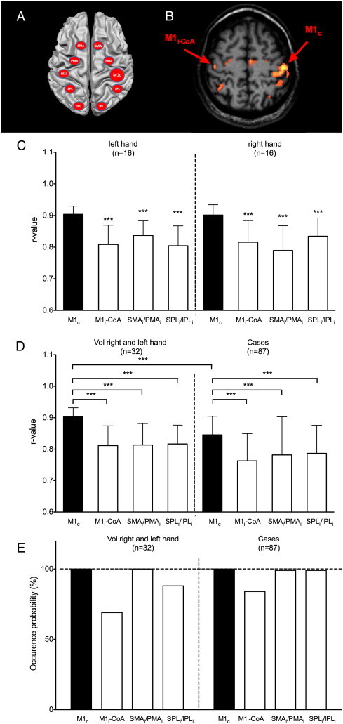
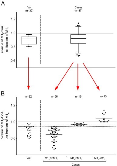
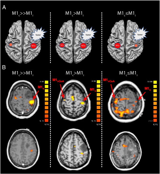
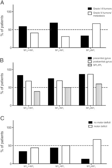
Hemispheric lateralization is a frequently encountered phenomenon of cortical function. It describes the functional specialization of a region on one side of the brain for a given task. It is well characterized in motor and sensory, as well as language systems and becomes more and more known for various cognitive domains. While in the adult healthy brain hemispheric lateralization is mostly set, pathological processes may lead to cortical reorganization. In these cases neuroplasticity of the corresponding region in the non-dominant hemisphere seems to play an important role. In a previous study we investigated language associated regions in right-handed patients with frontal and temporal tumors of the left hemisphere. We observed a marked change of language lateralization in these patients towards the non-dominant hemisphere as measured by functional MRI (Partovi et al., 2012). In the present study we evaluated activation and lateralization of cortical motor areas in patients with tumors of the central region. BOLD fMRI was performed during unilateral voluntary movements of the contralesional hand in 87 patients. Individual correlations of measured BOLD-signals with the model hemodynamic reference function were determined on a ROI basis in single subjects and compared to those of 16 healthy volunteers. In volunteers the strongest activation is usually found in the M1 hand representation contralateral to the movement, while a weaker homotopic co-activation is observed in ipsilateral M1 (Stippich et al., 2007a). In the patient group our results show significant changes of motor activations, ranging from a reduction of M1 lateralization to equalization of M1 activations or even inversion of M1 lateralization during contralesional movements. This study corroborates in a large patient group the idea that lesions affecting M1 may lead to functional reorganization of cortical motor systems and in particular equalize hemispheric lateralization. However, it is not yet clear whether these changes are only an epiphenomenon or indeed reflect an attempt of recovery of brain function.
Keywords: Functional MRI; Functional reorganization; Ipsilateral coactivation; M1; Plasticity.
Figures
Similar articles
-
Comparison of language cortex reorganization patterns between cerebral arteriovenous malformations and gliomas: a functional MRI study.J Neurosurg. 2015 May;122(5):996-1003. doi: 10.3171/2014.12.JNS14629. Epub 2015 Feb 6. J Neurosurg. 2015. PMID: 25658788
-
fMRI signal decreases in ipsilateral primary motor cortex during unilateral hand movements are related to duration and side of movement.Neuroimage. 2005 Feb 15;24(4):1080-7. doi: 10.1016/j.neuroimage.2004.10.003. Epub 2004 Nov 26. Neuroimage. 2005. PMID: 15670685 Clinical Trial.
-
Evaluating the Abnormality of Bilateral Motor Cortex Activity in Subacute Stroke Patients Executing a Unimanual Motor Task With Increasing Demand on Precision.Front Neurol. 2022 May 25;13:836716. doi: 10.3389/fneur.2022.836716. eCollection 2022. Front Neurol. 2022. PMID: 35693005 Free PMC article.
-
Integrated technology for evaluation of brain function and neural plasticity.Phys Med Rehabil Clin N Am. 2004 Feb;15(1):263-306. doi: 10.1016/s1047-9651(03)00124-4. Phys Med Rehabil Clin N Am. 2004. PMID: 15029909 Review.
-
Reorganization after pre- and perinatal brain lesions.J Anat. 2010 Oct;217(4):469-74. doi: 10.1111/j.1469-7580.2010.01262.x. J Anat. 2010. PMID: 20649910 Free PMC article. Review.
Cited by
-
On the bimanual integration of proprioceptive information.Exp Brain Res. 2015 Apr;233(4):1273-88. doi: 10.1007/s00221-015-4205-0. Epub 2015 Jan 25. Exp Brain Res. 2015. PMID: 25618007
-
Surgery of Motor Eloquent Glioblastoma Guided by TMS-Informed Tractography: Driving Resection Completeness Towards Prolonged Survival.Front Oncol. 2022 May 27;12:874631. doi: 10.3389/fonc.2022.874631. eCollection 2022. Front Oncol. 2022. PMID: 35692752 Free PMC article.
-
Transient effects of tumor location on the functional architecture at rest in glioblastoma patients: three longitudinal case studies.Radiat Oncol. 2016 Aug 17;11(1):107. doi: 10.1186/s13014-016-0683-x. Radiat Oncol. 2016. PMID: 27535235 Free PMC article.
-
[Functional neuroanatomy: sensorimotor system].Radiologe. 2013 Jul;53(7):584-91. doi: 10.1007/s00117-013-2483-8. Radiologe. 2013. PMID: 23784618 Review. German.
-
Comparative fMRI and MEG localization of cortical sensorimotor function: Bimodal mapping supports motor area reorganization in glioma patients.PLoS One. 2019 Mar 7;14(3):e0213371. doi: 10.1371/journal.pone.0213371. eCollection 2019. PLoS One. 2019. PMID: 30845241 Free PMC article.
References
-
- Blatow M. Clinical functional MRI of sensorimotor cortex using passive motor and sensory stimulation at 3 tesla. Journal of Magnetic Resonance Imaging. 2011;34(2):429–437. - PubMed
-
- Boroojerdi B., Diefenbach K., Ferbert A. Transcallosal inhibition in cortical and subcortical cerebral vascular lesions. Journal of Neurological Sciences. 1996;144(1–2):160–170. - PubMed
-
- Carpentier A.C. Patterns of functional magnetic resonance imaging activation in association with structural lesions in the rolandic region: a classification system. Journal of Neurosurgery. 2001;94:946–954. - PubMed
LinkOut - more resources
Full Text Sources
Other Literature Sources
