. 2013 Nov 12;110(46):18626-31.
doi: 10.1073/pnas.1311100110. Epub 2013 Oct 28.

Gata3/Ruvbl2 complex regulates T helper 2 cell proliferation via repression of Cdkn2c expression

Affiliations

Gata3/Ruvbl2 complex regulates T helper 2 cell proliferation via repression of Cdkn2c expression

Hiroyuki Hosokawa et al. Proc Natl Acad Sci U S A. .

Abstract

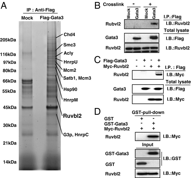
GATA-binding protein 3 (Gata3) controls the differentiation of naive CD4 T cells into T helper 2 (Th2) cells by induction of chromatin remodeling of the Th2 cytokine gene loci, direct transactivation of Il5 and Il13 genes, and inhibition of Ifng. Gata3 also facilitates Th2 cell proliferation via additional mechanisms that are far less well understood. We herein found that Gata3 associates with RuvB-like protein 2 (Ruvbl2) and represses the expression of a CDK inhibitor, cyclin-dependent kinase inhibitor 2c (Cdkn2c) to facilitate the proliferation of Th2 cells. Gata3 directly bound to the Cdkn2c locus in an Ruvbl2-dependent manner. The defect in the proliferation of Gata3-deficient Th2 cells is rescued by the knockdown of Cdkn2c, indicating that Cdkn2c is a key molecule involved in the Gata3-mediated induction of Th2 cell proliferation. Ruvbl2-knockdown Th2 cells showed decreased antigen-induced expansion and caused less airway inflammation in vivo. We therefore have identified a functional Gata3/Ruvbl2 complex that regulates the proliferation of differentiating Th2 cells through the repression of a CDK inhibitor, Cdkn2c.

Keywords: master transcription factor; polycomb group complex; transcriptional regulation.


Conflict of interest statement

The authors declare no conflict of interest.

Figures

Fig. 1.
Fig. 1.
Identification of Ruvbl2 as a molecule that interacts with Gata3 in Th2 cells. (A) Total extracts from 3xFlag–Gata3-expressing cross-linked D10G4.1 cells were subjected to affinity purification using a Flag mAb, followed by SDS/PAGE and SYPRO Ruby staining. Several specific polypeptides were identified by mass spectrometry as described in Materials and Methods. (B) The 3xFlag–Gata3-expressing D10G4.1 cells were treated with or without formaldehyde as indicated (Cross-link + or −) before extraction. The extracts were then immunoprecipitated (I.P.) with a Flag mAb, followed by immunoblotting (I.B.) with an Ruvbl2 Ab (Upper). Total lysates were also subjected to I.B. in parallel (Lower). (C) The 293T cells were transfected with expression plasmids encoding Flag-tagged Gata3 and Myc-tagged Ruvbl2. Two days later, the extracts were I.P. with a Flag mAb, followed by I.B. with a Myc mAb (Upper). The total lysates were also subjected to I.B. in parallel (Lower). (D) GST or GST–Gata3 proteins were bound to glutathione Sepharose 4B and incubated with purified Myc-tagged Ruvbl2. The bound proteins were subjected to I.B. with an anti-Myc mAb (Upper). The inputs represent 10% of the amount of protein used in the pull-down sample (Lower). Three (C) and two (B and D) independent experiments were performed, and similar results were obtained.
Fig. 2.
Fig. 2.
Ruvbl2 regulates the proliferation of Th2 cells. (A) A control or Ruvbl2 siRNA was transfected into naive CD4 T cells, and the cells were labeled with CFSE. Then, the cells were stimulated with an immobilized anti-TCRβ mAb and anti-CD28 mAb under Th2 conditions for 3 d. The cells were then restimulated and subjected to intracellular staining with an APC-conjugated anti–IL-4 mAb (Left). The percentages of the cells in the gates representing the number of cell divisions (nos. 1 to 7) are shown (Right). (B) Naive CD4 T cells from WT or Gata3-deficient mice were labeled with CFSE and cultured under Th2 conditions for 3 d. The cells were then restimulated and subjected to IL-4 staining (Left). The percentages of the cells in the gates are shown (Right). (C) A control or Ruvbl2 siRNA was transfected into naive CD4 T cells, and the cells were stimulated with an immobilized anti-TCRβ mAb and anti-CD28 mAb under Th2 conditions for 4 d (Left). Naive CD4 T cells from WT or Gata3-deficient mice were cultured under Th2 conditions for 5 d (Right). Representative intracellular staining profiles for BrdU are shown with the percentages of cells in the gate. Three independent experiments were performed and similar results were obtained (A, B, and C).
Fig. 3.
Fig. 3.
The expression of Cdkn2c controls the Gata3-dependent proliferation of Th2 cells. (A) Naive CD4 T cells from either WT or Gata3-deficient mice were cultured under Th1 or Th2 conditions for 5 d, and the expression levels of Cdkn2c mRNA in Th1 WT, Th2 WT, or Th2 Gata3 KO cells were determined by RT-qPCR. The relative expression (/Hprt) is shown with SDs. **P < 0.01 by Student t test. (B) Naive CD4 T cells were transfected with control or Ruvbl2 siRNA, and cultured under Th2 conditions for 4 d; then, the expression levels of Cdkn2c mRNA were determined by RT-qPCR. (C) Naive CD4 T cells from WT or Gata3-deficient mice were cultured under Th1 or Th2 conditions for 3 d. The binding of Gata3 to the Cdkn2c locus was determined by a ChIP assay with a qPCR analysis. The relative intensities (/input) are shown with SDs. (D) D10G4.1 cells were transfected with the indicated reporter constructs. Two days after transfection, the cells were assayed for luciferase activity. The data indicate the mean values of three independent experiments with SDs. (E) Naive CD4 T cells from WT or Gata3-deficient mice were cultured under Th2 conditions for 2 d and then infected with a retroviral vector encoding a control shRNA or shCdkn2c bicistronically with a GFP gene. Four days later, the retrovirus-infected GFP-expressing cells were purified, and the levels of Cdkn2c mRNA were measured by RT-qPCR. (F) The BrdU incorporation in a portion of the same cultured cells used in E was determined. (G) The same cultured cells used in E were labeled with Cell Proliferation Dye eFluor 670 on day 3 after stimulation. Two days later, cell division was assessed by FACS. Numbers represent mean fluorescent units. (H) The same cultured cells used in E were stimulated with immobilized TCRβ mAb and subjected to IL-4 staining, followed by FACS analysis. The percentages of IL-4-producing cells are shown. Four (A), three (B, C, E, and F), and two (D, G, and H) independent experiments were performed, and similar results were obtained.
Fig. 4.
Fig. 4.
Ruvbl2 is necessary for the recruitment of Gata3 at the Cdkn2c locus in developing Th2 cells. (A) Naive CD4 T cells from Gata3-deficient mice were stimulated under Th2 conditions for 2 d and then infected with a retroviral vector carrying WT or mutant (dTA) Gata3 cDNAs. Three days later, the retrovirus-infected GFP-expressing cells were purified, and the levels of mRNA of Cdkn2c were measured by RT-qPCR. **P < 0.01 by Student t test. (B) The binding of Gata3 WT or dTA to the Cdkn2c G3BS and Th2 cytokine loci (CGRE region) were determined by the ChIP assay with a qPCR analysis using the same cells shown in A. (C) A control or Ruvbl2 siRNA was transfected into naive CD4 T cells, and the cells were stimulated under Th1 or Th2 conditions for 3 d. The binding of Gata3 to the Cdkn2c locus was determined. (D) A schematic representation of the Cdkn2c locus. The locations of primers and exons are indicated. (E) Naive CD4 T cells from WT or Gata3-deficient mice were cultured under Th1 or Th2 conditions for 5 d. The status of H3-K27 Me3 and H2A-K119 Ub at the Cdkn2c and Hprt loci was determined by a ChIP assay, using specific primers to detect the indicated regions. **P < 0.01, *P < 0.05 by Student t test. (F) A control or Ruvbl2 siRNA was transfected into naive CD4 T cells, and the cells were stimulated under Th2 conditions for 4 d. The status of H3-K27 Me3 and H2A-K119 Ub at the Cdkn2c and Hprt promoters was determined by a ChIP assay. Four (A), three (B and C), and two (E and F) independent experiments were performed, and similar results were obtained.
Fig. 5.
Fig. 5.
Ruvbl2 regulates OVA-induced Th2 cell expansion and allergic airway inflammation in vivo. (A) A control or Ruvbl2 siRNA was transfected into naive CD4 T cells from DO11.10 Tg mice, and the cells were stimulated under Th2 conditions. On day 4, cells were i.v. transferred into BALB/c mice, and the mice were challenged with aerosolized OVA on days 5 and 7. Cells from lung were stained with antibodies against CD4 and KJ1.26 and assessed by FACS on days 5 (before inhalation) and 9 (after inhalation). A summary of the numbers of KJ1+ cells in the lung is presented (Right graph). Four mice per group were used. **P < 0.01 by Student t test. (B) The number of inflammatory cells in the bronchioalveolar lavage (BAL) fluid was counted. The absolute cell numbers of eosinophils (Eos.), neutrophils (Neu.), lymphocytes (Lym.) and macrophages (Mac.) are shown with SDs. Four mice per group were used. (C) The lungs were fixed and stained with hematoxylin/eosin (H&E) or with PAS. Representative staining patterns are shown. (Scale bar: 100 μm.) Two independent experiments were performed, and similar results were obtained (A, B, and C).

Similar articles

Cited by

References

    1. Zhu J, Yamane H, Paul WE. Differentiation of effector CD4 T cell populations. Annu Rev Immunol. 2010;28:445–489. - PMC - PubMed
    1. Reiner SL. Development in motion: Helper T cells at work. Cell. 2007;129(1):33–36. - PubMed
    1. Kurata H, Lee HJ, O’Garra A, Arai N. Ectopic expression of activated Stat6 induces the expression of Th2-specific cytokines and transcription factors in developing Th1 cells. Immunity. 1999;11(6):677–688. - PubMed
    1. Onodera A, et al. STAT6-mediated displacement of polycomb by trithorax complex establishes long-term maintenance of GATA3 expression in T helper type 2 cells. J Exp Med. 2010;207(11):2493–2506. - PMC - PubMed
    1. Yamashita M, et al. Ras-ERK MAPK cascade regulates GATA3 stability and Th2 differentiation through ubiquitin-proteasome pathway. J Biol Chem. 2005;280(33):29409–29419. - PubMed

Publication types

MeSH terms