Transgenic expression of pectin methylesterase inhibitors limits tobamovirus spread in tobacco and Arabidopsis
- PMID: 24127644
- PMCID: PMC6638747
- DOI: 10.1111/mpp.12090
Transgenic expression of pectin methylesterase inhibitors limits tobamovirus spread in tobacco and Arabidopsis
Erratum in
- Mol Plant Pathol. 2014 Aug;15(6):646
Abstract
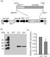
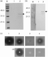
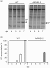
Plant infection by a virus is a complex process influenced by virus-encoded factors and host components which support replication and movement. Critical factors for a successful tobamovirus infection are the viral movement protein (MP) and the host pectin methylesterase (PME), an important plant counterpart that cooperates with MP to sustain viral spread. The activity of PME is modulated by endogenous protein inhibitors (pectin methylesterase inhibitors, PMEIs). PMEIs are targeted to the extracellular matrix and typically inhibit plant PMEs by forming a specific and stable stoichiometric 1:1 complex. PMEIs counteract the action of plant PMEs and therefore may affect plant susceptibility to virus. To test this hypothesis, we overexpressed genes encoding two well-characterized PMEIs in tobacco and Arabidopsis plants. Here, we report that, in tobacco plants constitutively expressing a PMEI from Actinidia chinensis (AcPMEI), systemic movement of Tobacco mosaic virus (TMV) is limited and viral symptoms are reduced. A delayed movement of Turnip vein clearing virus (TVCV) and a reduced susceptibility to the virus were also observed in Arabidopsis plants overexpressing AtPMEI-2. Our results provide evidence that PMEIs are able to limit tobamovirus movement and to reduce plant susceptibility to the virus.
© 2013 BSPP AND JOHN WILEY & SONS LTD.
Figures


Similar articles
-
How do pectin methylesterases and their inhibitors affect the spreading of tobamovirus?Plant Signal Behav. 2014;9(12):e972863. doi: 10.4161/15592316.2014.972863. Plant Signal Behav. 2014. PMID: 25482766 Free PMC article.
-
Interaction between the tobacco mosaic virus movement protein and host cell pectin methylesterases is required for viral cell-to-cell movement.EMBO J. 2000 Mar 1;19(5):913-20. doi: 10.1093/emboj/19.5.913. EMBO J. 2000. PMID: 10698933 Free PMC article.
-
The tobamovirus Turnip Vein Clearing Virus 30-kilodalton movement protein localizes to novel nuclear filaments to enhance virus infection.J Virol. 2013 Jun;87(11):6428-40. doi: 10.1128/JVI.03390-12. Epub 2013 Mar 27. J Virol. 2013. PMID: 23536678 Free PMC article.
-
Pectin methylesterase inhibitor.Biochim Biophys Acta. 2004 Feb 12;1696(2):245-52. doi: 10.1016/j.bbapap.2003.08.011. Biochim Biophys Acta. 2004. PMID: 14871665 Review.
-
The Multifaceted Role of Pectin Methylesterase Inhibitors (PMEIs).Int J Mol Sci. 2018 Sep 21;19(10):2878. doi: 10.3390/ijms19102878. Int J Mol Sci. 2018. PMID: 30248977 Free PMC article. Review.
Cited by
-
Cell-to-cell movement of viruses via plasmodesmata.J Plant Res. 2015 Jan;128(1):37-47. doi: 10.1007/s10265-014-0683-6. Epub 2014 Dec 21. J Plant Res. 2015. PMID: 25527904 Review.
-
Enzymatic fingerprinting reveals specific xyloglucan and pectin signatures in the cell wall purified with primary plasmodesmata.Front Plant Sci. 2022 Oct 25;13:1020506. doi: 10.3389/fpls.2022.1020506. eCollection 2022. Front Plant Sci. 2022. PMID: 36388604 Free PMC article.
-
Pectin Remodeling and Involvement of AtPME3 in the Parasitic Plant-Plant Interaction, Phelipanche ramosa-Arabidospis thaliana.Plants (Basel). 2024 Aug 5;13(15):2168. doi: 10.3390/plants13152168. Plants (Basel). 2024. PMID: 39124288 Free PMC article.
-
Overexpression of MePMEI1 in Arabidopsis enhances Pb tolerance.Front Plant Sci. 2022 Sep 16;13:996981. doi: 10.3389/fpls.2022.996981. eCollection 2022. Front Plant Sci. 2022. PMID: 36186034 Free PMC article.
-
Cell wall traits as potential resources to improve resistance of durum wheat against Fusarium graminearum.BMC Plant Biol. 2015 Jan 19;15:6. doi: 10.1186/s12870-014-0369-1. BMC Plant Biol. 2015. PMID: 25597920 Free PMC article.
References
-
- Andika, I.B. , Zheng, S.L. , Tan, Z.L. , Sun, L.Y. , Kondo, H. , Zhou, X.P. and Chen, J.P. (2013) Endoplasmic reticulum export and vesicle formation of the movement protein of Chinese wheat mosaic virus are regulated by two transmembrane domains and depend on the secretory pathway. Virology, 435, 493–503. - PubMed
-
- Ascencio‐Ibanez, J.T. , Sozzani, R. , Lee, T.J. , Chu, T.M. , Wolfinger, R.D. , Cella, R. and Hanley‐Bowdoin, L. (2008) Global analysis of Arabidopsis gene expression uncovers a complex array of changes impacting pathogen response and cell cycle during geminivirus infection. Plant Physiol. 148, 436–454. - PMC - PubMed
-
- Bradford, M.M. (1976) A rapid and sensitive method for the quantitation of microgram quantities of protein utilizing the principle of protein–dye binding. Anal. Biochem. 72, 248–254. - PubMed
Publication types
MeSH terms
Substances
Associated data
- Actions
- Actions
- Actions
LinkOut - more resources
Full Text Sources
Other Literature Sources
Research Materials

