. 2014;39(1):145-62.
doi: 10.3233/JAD-131238.

Experimental induction of type 2 diabetes in aging-accelerated mice triggered Alzheimer-like pathology and memory deficits

Affiliations

Experimental induction of type 2 diabetes in aging-accelerated mice triggered Alzheimer-like pathology and memory deficits

Jogender Mehla et al. J Alzheimers Dis. 2014.

Abstract

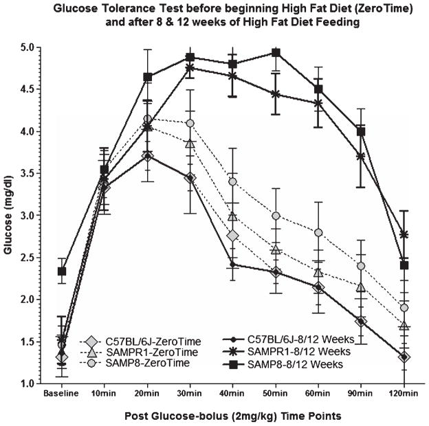
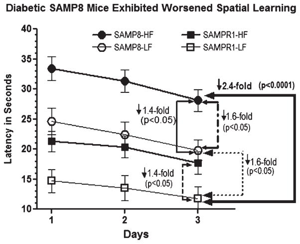
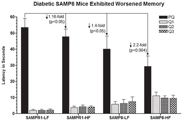
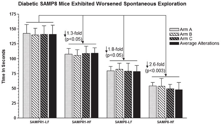
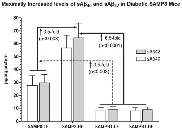
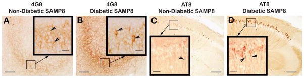
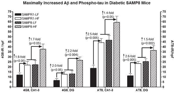
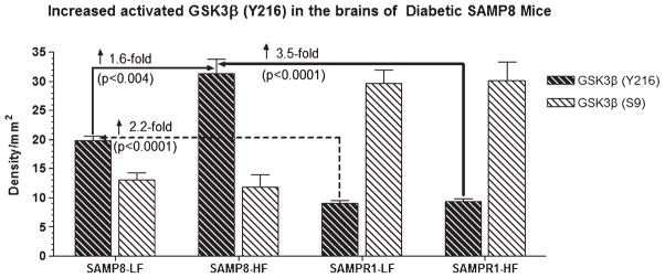
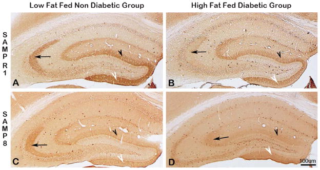
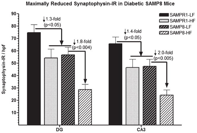
Alzheimer's disease (AD) is an age-dependent neurodegenerative disease constituting ~95% of late-onset non-familial/sporadic AD, and only ~5% accounting for early-onset familial AD. Availability of a pertinent model representing sporadic AD is essential for testing candidate therapies. Emerging evidence indicates a causal link between diabetes and AD. People with diabetes are >1.5-fold more likely to develop AD. Senescence-accelerated mouse model (SAMP8) of accelerated aging displays many features occurring early in AD. Given the role played by diabetes in the pre-disposition of AD, and the utility of SAMP8 non-transgenic mouse model of accelerated aging, we examined if high fat diet-induced experimental type 2 diabetes in SAMP8 mice will trigger pathological aging of the brain. Results showed that compared to non-diabetic SAMP8 mice, diabetic SAMP8 mice exhibited increased cerebral amyloid-β, dysregulated tau-phosphorylating glycogen synthase kinase 3β, reduced synaptophysin immunoreactivity, and displayed memory deficits, indicating Alzheimer-like changes. High fat diet-induced type 2 diabetic SAMP8 mice may represent the metabolic model of AD.

Keywords: Alzheimer's disease; amyloid-β; diabetes; glycogen synthase kinase-3β; learning and memory; pathological aging of the brain; senescence-accelerated mice; synaptophysin; tau.


Figures

Fig. 1
Fig. 1
Effects of high fat (HF) feeding on glucose tolerance test in SAMP8, SAMPR1, and C57BL/6J mice before beginning HF diet (Zero Time), and after 8/12 weeks of HF feeding, performed after overnight fasting at every 20 min up to 2 h post a single bolus glucose injection (2 mg/kg). Glucose tolerance test at Zero Time shows peaking of blood glucose levels between 20–30 min and quickly normalizing thereafter, suggestive of normal functioning of pancreas. On the other hand, glucose tolerance test after 8/12 weeks of HF diet showed that peaking of blood glucose levels for SAMP8 and SAMPR1 mice, but not for c57BL/6J mice, at 20–30 min, consistently maintained high up to 90 min with a gradual decline by 120 min, indicating failure of pancreas and development of type 2 diabetes, as opposed to normal functioning of pancreas in C57BL/6J mice.
Fig. 2
Fig. 2
Effect of high fat (HF) diet induced experimental T2DM on Morris water maze acquisition learning in SAMP8 and SAMPR1 mice, as measured by latency (Time in seconds required to reach the submerged platform). Data are presented as group means ± standard deviation (SD) derived from the average individual values (6 trials/animal/day for 3 days) for each group. Note that compared to low-fat (LF) fed non-diabetic SAMPR1 mice, spatial acquisition learning was observed to be gradually worsened from HF-fed diabetic SAMPR1 mice (1.4-fold) > LF-fed non-diabetic SAMP8 mice (1.6-fold) > HF-fed diabetic SAMP8 mice (2.4-fold). Data indicate the most significantly deteriorated spatial acquisition learning in diabetic SAMP8 mice.
Fig. 3
Fig. 3
Effect of high fat (HF) diet induced experimental T2DM on Morris water maze retention memory in SAMP8 and SAMPR1 mice, as measured by latency (Time in seconds required to explore quadrant of the pool that previously contained platform named PQ). Data are presented as group means ± standard deviation (SD) derived from the average individual values (a single probe trial/animal) for each group. Note that low-fat (LF) fed non-diabetic SAMPR1 mice spent maximum amount of time in PQ, indicating preserved memory of the previously learned location of the platform. By contrast, retention memory was observed to be gradually deteriorated from LF-fed non-diabetic SAMP8 mice > HF-fed diabetic SAMPR1 mice > HF-fed diabetic SAMP8 mice. Data indicate the most significantly deteriorated retention memory in diabetic SAMP8 mice.
Fig. 4
Fig. 4
Effect of high fat (HF) diet induced experimental T2DM on Y maze spontaneous exploration representing working reference memory in SAMP8 and SAMPR1 mice, as measured by latency (Time in seconds required to explore all arms with average alteration in all arms in Y maze). Data are presented as group means ± standard deviation (SD) derived from the average individual values (a single probe trial/animal) for each group. Note that low-fat (LF) fed non-diabetic SAMPR1 mice spent maximum amount of time in PQ, indicating preserved memory of the previously learned location of the platform. By contrast, retention memory was observed to be gradually deteriorated from HF-fed diabetic SAMPR1 mice > LF-fed non-diabetic SAMP8 mice > HF-fed diabetic SAMP8 mice. Data indicate the most significantly deteriorated retention memory in diabetic SAMP8 mice.
Fig. 5
Fig. 5
Effect of high fat (HF) diet induced experimental T2DM on cerebral levels of Tris-SDS soluble Aβ40 (sAβ40) and Tris-SDS soluble Aβ42 (sAβ42) in SAMP8 and SAMPR1 mice. Data are presented as group means ± standard deviation (SD) derived from the average individual values for each group. Note that SAMPR1 mice with or without HF diet did not differ significantly exhibiting base levels of cerebral sAβ40 and sAβ42, which were increased by ~3-fold in low-fat (LF) fed non-diabetic SAMP8 mice. HF-fed SAMP8 mice exhibited maximally increased levels of cerebral sAβ40 and sAβ42, both as compared to HF- and LF-fed SAMPR1 mice (p < 0.0001), and compared to LF-fed non-diabetic SAMP8 mice (p < 0.0003). Data indicate the most significantly increased cerebral sAβ40 and sAβ42 in diabetic SAMP8 mice.
Fig. 6
Fig. 6
Immunodistribution of 4G8 (A, B) and phospho-tau (AT8) (C, D) in the hippocampus of low-fat diet fed non-diabetic SAMP8 mice (A, C), and in the hippocampus of high-fat diet fed diabetic SAMP8 mice (B, D). Note faint 4G8 immunoreactivity within the perikarya of CA3 granule cells (Fig. 6A, inlet, arrowheads) of non-diabetic SAMP8 mice, indicating the presence of intraneuronal Aβ accumulated as a result of accelerated aging in absence of diabetes. Note stronger 4G8 immunoreaction within the perikarya of CA3 granule cells (Fig. 6B, inlet, arrowheads) of diabetic SAMP8 mice, indicating increased accumulation of intraneuronal Aβ than that of non-diabetic SAMP8 mice increased as a result of accelerated aging in presence of diabetes. Similarly, there was observed some evidence of tau phosphorylation within the CA1 hippocampal neurites in non-diabetic SAMP8 mice (Fig. 6C, inlet, arrowhead) merely due to accelerated aging, which was remarkably increased in the CA hippocampal CA1 of diabetic SAMP8 mice (Fig. 6D, inlet, arrowheads) as a result of accelerated aging compounded with diabetes. Scale bars in A-D = 100 μm; Scale bars in all inlets = 20 μm.
Fig. 7
Fig. 7
Effect of high fat diet induced experimental T2DM on the immunoreactivities (IR) of Aβ (4G8) and phospho-tau (AT8) in the CA1-3 and dentate gyrus (DG) hippocampal subfields of SAMPR1 and SAMP8 mice. Data are represented as group mean ± standard deviation (SD) derived from average individual values for each group. Note that both high fat fed diabetic SAMPR1 mice and low fat fed non-diabetic SAMP8 mice showed more or less similar pattern of 4G8-IR Aβ and AT8-IR phospho-tau within Ca1-3 and DG, which were 1.8–2.0-fold higher for 4G8-IR and 2.5–2.6-fold higher for AT8-IR than the low fat fed non-diabetic SAMPR1 mice. Feeding of high fat diet to SAMP8 mice resulted in 1.7–2.2-fold increase in 4G8-IR Aβ and 1.4–1.5-fold increase in AT8-IR phospho-tau within the respective hippocampal subfields. Data indicate that diabetic SAMP8 mice exhibited Alzheimer-like changes with regard to Aβ and phospho-tau.
Fig. 8
Fig. 8
Western blot of brain samples derived from SAMP8 or SAMPR1 mice fed with high fat (HF) or low fat (LF) diets showing the reactivity for glycogen synthase kinase-3β (GSK3β) phosphorylated at tyrosine 216 site-activated form of GSK3β (anti-Y216 GSK3β) and at serine 9 site-inactive form of GSK3β (anti-S9 GSK3β) (BioSource International). In general, density for activated GSK3β (Y216) is greater in the brains of SAMP8 mice than that of SAMPR1 mice. By contrast, density for inactive form of GSK3β (S9) is greater in the brains of SAMPR1 mice than that of SAMP8 mice. HF treatment did not affect the reaction for inactive GSK3β (S9) in the brains of SAMP8 mice. HF treatment increased the density for activated GSK3β (Y216) only in the brains of SAMP8 mice, but not in the brains of SAMPR1 mice, indicating that experimental induction of type 2diabetes in SAMP8 mice promotes abnormal phosphorylation of tau. Densitometric analysis showed that compared to LF-fed non-diabetic SAMP8 mice, the levels of GSK3β (Y216) increased by 1.6-fold (p < 0.004) in HF-fed diabetic SAMP8 mice. Compared to LF-or HF-fed non-diabetic or diabetic SAMPR1 mice, the levels of GSK3β (Y216) increased by 2.2-fold (p < 0.0001) in HF-fed diabetic SAMP8 mice.
Fig. 9
Fig. 9
Immunodistribution of synaptophysin (SYN) in the hippocampus of SAMPR1 (A, B, top panel), and SAMP8 (C, D, bottom panel); low-fat fed non-diabetic mice (A, C, left panel), and with high-fat fed diabetic mice (B, D, right panel). Note the strongest SYN immunoreactivity (IR) within the CA3 dendritic field (Fig. 9A, arrow), and within the supra-granular (Fig. 9A, black arrowhead) and infragranular (Fig. 9A, white arrowhead) molecular layers of DG of non-diabetic SAMPR1 mice. Note that experimental induction of diabetes in SAMPR1 mice resulted in the reduction of SYN immunoreactivity only in the CA3 dendritic field (Fig. 9B, black arrowhead), while preserving SYN immunoreactivity within the supra-granular (Fig. 9B, black arrowhead) and infragranular (Fig. 9B, white arrowhead) molecular layers of DG of diabetic SAMPR1 mice. On the other hand, in non-diabetic SAMP8 mice, SYN immunoreactivity in the CA3 dendritic field (Fig. 9C, black arrowhead) was preserved, but the SYN immunoreactivity within the supra-granular (Fig. 9C, black arrowhead) and infragranular (Fig. 9C, white arrowhead) molecular layers of DG was reduced. In the hippocampus of diabetic SAMP8 mice, SYN immunoreactivity in all CA3 (Fig. 9D, arrow) and respective DG subfields (Fig. 9D, black arrowhead and white arrowhead) were observed to be greatly reduced. Scale bar = 100 μm.
Fig. 10
Fig. 10
Effect of high fat (HF) diet induced experimental T2DM on the immunoreactivities (IR) of synaptophysin (SYN) in the CA3 and dentate gyrus (DG) hippocampal subfields of SAMPR1 and SAMP8 mice. Data are represented as group mean ± standard deviation (SD) derived from average individual values for each group. Note that both HF-fed diabetic SAMPR1 mice and low fat (LF) fed non-diabetic SAMP8 mice exhibited matching pattern of SYN-IR, which were 1.8–2.0-fold lower than the hippocampal SYN-IR observed in the LF-fed non-diabetic SAMPR1 mice. Feeding of HF diet to SAMP8 mice resulted in further reduction of SYN-IR by 1.8–2.0-fold in respective hippocampal of diabetic SAMP8 mice. Data indicate that diabetic SAMP8 mice showed Alzheimer-like synaptic deficits.

Similar articles

Cited by

References

    1. Finder VH. Alzheimer’s disease: A general introduction and pathomechanism. J Alzheimers Dis. 2010;22(Suppl 3):5–19. - PubMed
    1. Duyckaerts C, Delatour B, Potier MC. Classification and basic pathology of Alzheimer disease. Acta Neuropathol. 2009;118:5–36. - PubMed
    1. Bastos Leite AJ, Scheltens P, Barkhof F. Pathological aging of the brain: An overview. Top Magn Reson Imaging. 2004;15:369–389. - PubMed
    1. Craft S. The role of metabolic disorders in Alzheimer disease and vascular dementia: Two roads converged. Arch Neurol. 2009;66:300–305. - PMC - PubMed
    1. Luchsinger JA. Diabetes, related conditions, and dementia. J Neurol Sci. 2010;299:35–38. - PMC - PubMed

Publication types