

Substrate-based fragment identification for the development of selective, nonpeptidic inhibitors of striatal-enriched protein tyrosine phosphatase
- PMID: 24083656
- PMCID: PMC3875168
- DOI: 10.1021/jm401037h
Substrate-based fragment identification for the development of selective, nonpeptidic inhibitors of striatal-enriched protein tyrosine phosphatase
Abstract
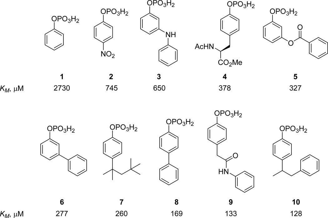
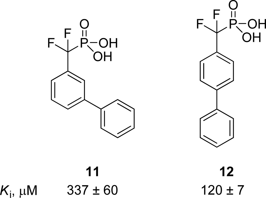
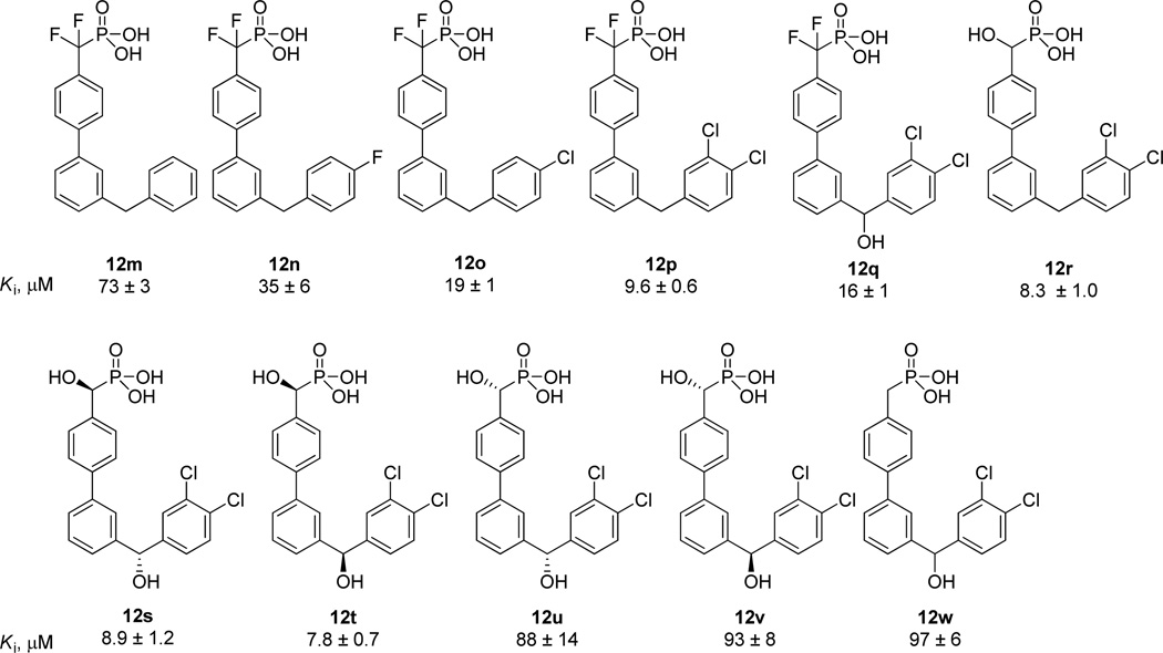
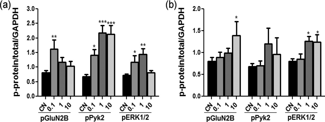
High levels of striatal-enriched protein tyrosine phosphatase (STEP) activity are observed in a number of neuropsychiatric disorders such as Alzheimer's disease. Overexpression of STEP results in the dephosphorylation and inactivation of many key neuronal signaling molecules, including ionotropic glutamate receptors. Moreover, genetically reducing STEP levels in AD mouse models significantly reversed cognitive deficits and decreased glutamate receptor internalization. These results support STEP as a potential target for drug discovery for the treatment of Alzheimer's disease. Herein, a substrate-based approach for the discovery and optimization of fragments called substrate activity screening (SAS) has been applied to the development of low molecular weight (<450 Da) and nonpeptidic, single-digit micromolar mechanism-based STEP inhibitors with greater than 20-fold selectivity across multiple tyrosine and dual specificity phosphatases. Significant levels of STEP inhibition in rat cortical neurons are also observed.
Figures
Similar articles
-
Inhibitor of the tyrosine phosphatase STEP reverses cognitive deficits in a mouse model of Alzheimer's disease.PLoS Biol. 2014 Aug 5;12(8):e1001923. doi: 10.1371/journal.pbio.1001923. eCollection 2014 Aug. PLoS Biol. 2014. PMID: 25093460 Free PMC article.
-
X-ray Characterization and Structure-Based Optimization of Striatal-Enriched Protein Tyrosine Phosphatase Inhibitors.J Med Chem. 2017 Nov 22;60(22):9299-9319. doi: 10.1021/acs.jmedchem.7b01292. Epub 2017 Nov 8. J Med Chem. 2017. PMID: 29116812 Free PMC article.
-
Abeta-mediated NMDA receptor endocytosis in Alzheimer's disease involves ubiquitination of the tyrosine phosphatase STEP61.J Neurosci. 2010 Apr 28;30(17):5948-57. doi: 10.1523/JNEUROSCI.0157-10.2010. J Neurosci. 2010. PMID: 20427654 Free PMC article.
-
Striatal-enriched Tyrosine Protein Phosphatase (STEP) in the Mechanisms of Depressive Disorders.Curr Protein Pept Sci. 2017 Aug 30;18(11):1152-1162. doi: 10.2174/1389203718666170710121532. Curr Protein Pept Sci. 2017. PMID: 28699511 Review.
-
An emerging role of STriatal-Enriched protein tyrosine Phosphatase in hyperexcitability-associated brain disorders.Neurobiol Dis. 2024 Oct 1;200:106641. doi: 10.1016/j.nbd.2024.106641. Epub 2024 Aug 17. Neurobiol Dis. 2024. PMID: 39159894 Review.
Cited by
-
Striatal-enriched protein tyrosine phosphatase controls responses to aversive stimuli: implication for ethanol drinking.PLoS One. 2015 May 20;10(5):e0127408. doi: 10.1371/journal.pone.0127408. eCollection 2015. PLoS One. 2015. PMID: 25992601 Free PMC article.
-
Disruption of striatal-enriched protein tyrosine phosphatase (STEP) function in neuropsychiatric disorders.Neurosci Res. 2014 Dec;89:1-9. doi: 10.1016/j.neures.2014.08.018. Epub 2014 Sep 10. Neurosci Res. 2014. PMID: 25218562 Free PMC article. Review.
-
Substrate deconstruction and the nonadditivity of enzyme recognition.J Am Chem Soc. 2014 May 21;136(20):7374-82. doi: 10.1021/ja501354q. Epub 2014 May 12. J Am Chem Soc. 2014. PMID: 24791931 Free PMC article.
-
Synthesis of benzopentathiepin analogs and their evaluation as inhibitors of the phosphatase STEP.Bioorg Med Chem Lett. 2015 Mar 1;25(5):1044-6. doi: 10.1016/j.bmcl.2015.01.020. Epub 2015 Jan 20. Bioorg Med Chem Lett. 2015. PMID: 25666825 Free PMC article.
-
Altered Intracellular Calcium Homeostasis Underlying Enhanced Glutamatergic Transmission in Striatal-Enriched Tyrosine Phosphatase (STEP) Knockout Mice.Mol Neurobiol. 2018 Oct;55(10):8084-8102. doi: 10.1007/s12035-018-0980-5. Epub 2018 Mar 5. Mol Neurobiol. 2018. PMID: 29508281
References
-
-
For a general overview of synaptic plasticity in neuropsychiatric disorders, see: Lüscher C, Issac JT. The synapse: center stage for many brain diseases. J. Physiol. 2009;587:727–729. Palop JJ, Chin J, Mucke L. A network dysfunction perspective on neurodegenerative diseases. Nature. 2006;443:768–773.
-
-
- Stephan KE, Friston KJ, Frith CD. Dysconnection in Schizophrenia: from abnormal synaptic plasticity to failures of self-monitoring. Schizophrenia Bull. 2009;35:509–527. - PMC - PubMed
- Stephan KE, Baldeweg T, Friston KJ. Synaptic plasticity and dysconnection in Schizophrenia. Biol. Psychiatry. 2006;59:929–939. - PubMed
-
- Duman RS. Pathophysiology of depression: the concept of synaptic plasticity. Eur. Psychiatry. 2002;17:306–310. - PubMed
-
- Garner CC, Wetmore DZ. Synaptic pathology of Down syndrome. Adv. Exp. Med. Biol. 2012;970:451–468. - PubMed
- Huttenlocher PR. Dendritic and synaptic pathology in mental retardation. Pediatr. Neurol. 1991;7:79–85. - PubMed
- O’Donnell WT, Warren ST. A decade of molecular studies of Fragile X syndrome. Annu. Rev. Neurosci. 2002;25:315–338. - PubMed
Publication types
MeSH terms
Substances
Grants and funding
LinkOut - more resources
Full Text Sources
Other Literature Sources
Chemical Information
