. 2013 Nov 1;288(44):32050-63.
doi: 10.1074/jbc.M113.504365. Epub 2013 Sep 19.

Reduction of synaptojanin 1 accelerates Aβ clearance and attenuates cognitive deterioration in an Alzheimer mouse model

Affiliations

Reduction of synaptojanin 1 accelerates Aβ clearance and attenuates cognitive deterioration in an Alzheimer mouse model

Li Zhu et al. J Biol Chem. .

Abstract

Recent studies link synaptojanin 1 (synj1), the main phosphoinositol (4,5)-biphosphate phosphatase (PI(4,5)P2-degrading enzyme) in the brain and synapses, to Alzheimer disease. Here we report a novel mechanism by which synj1 reversely regulates cellular clearance of amyloid-β (Aβ). Genetic down-regulation of synj1 reduces both extracellular and intracellular Aβ levels in N2a cells stably expressing the Swedish mutant of amyloid precursor protein (APP). Moreover, synj1 haploinsufficiency in an Alzheimer disease transgenic mouse model expressing the Swedish mutant APP and the presenilin-1 mutant ΔE9 reduces amyloid plaque load, as well as Aβ40 and Aβ42 levels in hippocampus of 9-month-old animals. Reduced expression of synj1 attenuates cognitive deficits in these transgenic mice. However, reduction of synj1 does not affect levels of full-length APP and the C-terminal fragment, suggesting that Aβ generation by β- and γ-secretase cleavage is not affected. Instead, synj1 knockdown increases Aβ uptake and cellular degradation through accelerated delivery to lysosomes. These effects are partially dependent upon elevated PI(4,5)P2 with synj1 down-regulation. In summary, our data suggest a novel mechanism by which reduction of a PI(4,5)P2-degrading enzyme, synj1, improves amyloid-induced neuropathology and behavior deficits through accelerating cellular Aβ clearance.

Keywords: Alzheimer Disease; Amyloid; Clearance; Endosomal/Lysosomal Degradation; Intracellular Trafficking; PIP2; Protein Degradation; Synaptojanin 1; Transgenic Mice.


Figures

FIGURE 1.
FIGURE 1.
Knockdown of synj1 reduces Aβ in N2a Swedish mutant APP cells. A, N2a Swedish mutant APP cells were treated with synj1 or control siRNA duplex for 4–5 days before analysis. Levels of media and intracellular Aβ, as well as βCTF, were analyzed by immunoprecipitation with 4G8 followed by immunoblotting with 6E10. Levels of synj1 and β-actin were detected from cell lysates. Treatment with DAPT was included as a control. B, protein levels were normalized to actin and expressed as percentages of control. The data were collected in duplicate or triplicate from three independent experiments. Significant reductions (**, p < 0.001) of Aβ in media and lysate and reduction of synj1 protein levels were observed upon synj1 knockdown, as compared with control. Levels of Aβ40 and Aβ42 in the media were determined by sandwich ELISA analysis (**, p < 0.001). WB, Western blotting; ctrl, control.
FIGURE 2.
FIGURE 2.
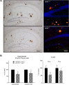
synj1 haploinsufficiency in AD transgenic mice decreases hippocampal amyloid plaque load. A, extracellular amyloid plaque deposits in hippocampal regions of transgenic mice expressing Swedish mutant APP/PS1ΔE9 (9 months old) with synj1+/+ (upper panels) or synj1+/− (lower panels) genetic background. Left panels, immunohistochemical staining of amyloid plaques with anti-amyloid antibody AB2454. Right panels, overlay fluorescent staining of amyloid plaques in dentate gyrus of hippocampus with 6E10 (red), LCO (green), and DAPI (blue). B, the total amyloid plaque load in hippocampus, as well as in the dentate gyrus (DG) subregion of animals, was determined by stereological quantification of brain sections through the whole hippocampus, and results were expressed as percentages of control (n = 3/group). Statistically significant reductions (*, p < 0.05) in amyloid plaque load were observed in synj1+/− mice, as compared with controls. Levels of Aβ40 and Aβ42 in the hippocampal brain lysates are determined by sandwich ELISA analysis (*, p < 0.05).
FIGURE 3.
FIGURE 3.
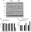
Several AD-related proteins remain unchanged with synj1 haploinsufficiency in AD transgenic mice. A, protein levels of holoAPP, C99/βCTF, PS1, BACE1, and synj1 in the hippocampal brain lysates of APP/PS1+/− synj1+/+ and APP/PS1+/− synj1+/− mice were determined by Western blot. B, results were normalized to actin and expressed as percentages of control. Reduction in synj1 protein levels by 60.2 ± 9.2% (*, p < 0.05) was observed in synj1 haploinsufficiency mice, as compared with control. C, the inositol lipid levels from hippocampal tissue of APP/PS1+/− synj1+/+ (n = 5) and APP/PS1+/− synj1+/− (n = 5) mice were determined by HPLC combined with suppressed conductivity.
FIGURE 4.
FIGURE 4.
synj1 haploinsufficiency in AD transgenic mice attenuates learning and memory deficits. A, novel object recognition test; APP/PS1+/−/synj1+/+ APP/PS1+/−/synj1+/−, APP/PS1−/−/synj1+/+, and APP/PS1−/−/synj1+/− (n = 5/group). The novel object discrimination index is expressed as percentages of time exploring novel object = (time spent exploring novel object × 100)/total exploration time. B, Y maze test is used for analysis of spontaneous alteration: APP/PS1+/−/synj1+/+ (n = 5), APP/PS1+/−/synj1+/− (n = 6), APP/PS1−/−/synj1+/+ (n = 8), and APP/PS1−/−/synj1+/− (n = 9). The percentage of spontaneous alternation (% of SAP) = the number of triads × 100/(the number of total arm entries-2). *, p < 0.05; **, p < 0.001.
FIGURE 5.
FIGURE 5.
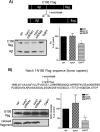
synj1 haploinsufficiency does not affect γ-secretase activity in vitro. A, C100-FLAG and Aβ generated by control or synj1 siRNA-treated membrane fractions were analyzed by immunoblot with 6E10. B, alternatively, N100-FLAG was analyzed by the in vitro γ-secretase assay. The Notch cleavage specific antibody Val1744 was used to detect the Notch cleavage fragment. The addition of DAPT to control membrane fractions was included as a control (69.3 ± 2.3% and 38.2 ± 16% of controls, respectively; lane 4). The results represent three independent experiments, and the data are presented as means ± S.D., *, p < 0.05. synj1 knockdown did not affect γ-secretase cleavage of C100 to generate Aβ (101 ± 11.1% of controls, n = 6, p = 0.74) or N100 to generate its cleavage product (recognized by Val1744 antibody only; 129.9 ± 27.4, n = 5, p = 0.11). ctrl, control.
FIGURE 6.
FIGURE 6.
Reduction of synj1 accelerates Aβ uptake into cells. A, biotin-Aβ42 internalized into the cells after 1 h of incubation was analyzed by immunoprecipitation of lysates with streptavidin beads. The amount of Aβ42 taken up by the cells was increased in N2a cells with synj1 siRNA knockdown (middle panel) and primary neurons (right panel) derived from embryonic day 17 synj1−/− animals (125.2 ± 13.4% of controls, n = 3, p = 0.03; 119.6 ± 9.0% of synj1 WT neurons, n = 4, p = 0.039, respectively). B, N2a cells transfected with control siRNA or synj1 siRNA were incubated with Alexa488-Aβ42 (500 nm) for 1 h before analyzed by fluorescence confocal microscopy. Representative cells are shown. Upper panels, control siRNA-treated cells. Lower panels, synj1 siRNA-treated cells. Left panels, internalized Alexa488-Aβ42. Scale bar, 10 μm. Middle panels, overlay view of Alexa488-Aβ (green)/Rab5 (red) colocalization. DAPI for nuclear staining (blue). Scale bar, 10 μm. Right panels, enlarged view of colocalization of internalized Alexa488-Aβ42 and Rab5+ early endosomes. Scale bar, 5 μm. C, the increased amount of Aβ42 taken up by N2a cells with synj1 siRNA knockdown was unchanged with treatment of a PIP2-depleting reagent, m-3-FBS (149.6 ± 8.0% of controls in m-3-FBS-treated versus 144.3 ± 24.3% of controls in o-3-FBS-treated conditions, n = 3, p = 0.79). An inactive analog was used as a control (o-3-FBS). ctrl, control.
FIGURE 7.
FIGURE 7.
Reduction of synj1 increases cellular Aβ degradation. A, left panel, immunoblot analysis of Aβ turnover rate in the presence of CHX (50 μg/ml) in N2a cells transfected with either control or synj1 siRNA. Right panel, densitometric analysis of Aβ accumulation in the presence of CHX at 0, 30, 60, and 90 min. B, left panel, immunoblot analysis of holoAPP turnover rate in the presence of CHX (50 μg/ml) in N2a cells transfected with either control or synj1 siRNA. Right panel, densitometric analysis of holoAPP accumulation in the presence of CHX at 0, 60, 120, and 180 min. The results represent three independent experiments, and the data are presented as percentages of controls at time point 0; means ± S.E. *, p < 0.05. ctrl, control.
FIGURE 8.
FIGURE 8.
Delivery of internalized Aβ42 to LAMP1+ lysosomes increases in synj1−/− primary neurons. Embryonic cortical neurons (top panels, synj1+/+; middle panels, synj1+/−; bottom panels, synj1−/−) were incubated with Alexa555-Aβ42 (500 nm) for 24 h. Cells were then fixed and stained for a lysosomal marker LAMP1 (red) before analyzed by fluorescence confocal microscopy. Representative neurons are shown. A, left panels, internalized Alexa555-Aβ42 shown in red fluorescence. Right panels, a lysosomal marker LAMP1 shown in green fluorescence. Scale bar, 10 μm. B, enlarged view of colocalization of internalized Alexa555-Aβ42 with LAMP1+ lysosomes at cell body (left panels) and processes (right panels). Yellow, overlay view of Alexa555-Aβ/LAMP1 colocalization. Scale bar, 5 μm.
FIGURE 9.
FIGURE 9.
Reduction of synj1 increases Aβ degradation in lysosomes partially through elevated PIP2 levels. A, N2a cells transfected with control or synj1 siRNA were incubated in the presence or absence of lysosomal inhibitors for 24 h. Media and lysate Aβ were quantified by IP/Western blot. The left panel shows one typical example of Western blot analysis. Right panel, data are plotted as percentages of controls, means ± S.D. (n = 3). *, p < 0.05. B, the amounts of degraded Aβ were calculated by subtracting the values of secreted or cell-associated Aβ in the absence of inhibitors from the values in the presence of inhibitors. The data are plotted as percentages of controls, means ± S.D. (n = 3). *, p < 0.05. C, N2a cells transfected with control or synj1 siRNA were incubated with a PIP2-depleting reagent m-3-FBS or its inactive analog o-3-FBS, in the presence or absence of lysosomal inhibitors for 24 h. Lysate Aβ were quantified by IP/Western blot. The data are plotted as percentages of controls, means ± S.D. (n = 3). *, p < 0.05. ctrl, control; DMSO, dimethyl sulfoxide; inh, inhibitor; N.S., not significant.
FIGURE 10.
FIGURE 10.
Model for synj1-regulated intracellular Aβ trafficking and lysosomal degradation. In WT cells, secreted Aβ is internalized into early endosomes, followed by either recycling back to plasma membrane through recycling endosomes or delivered to late endosomes/lysosomes for degradation. With synj1 knockdown (KD), Aβ is more rapidly internalized to the early endosomes and delivered to the late endosomal/lysosomal pathway for degradation. As a consequence, the amount of secreted and cellular Aβ is decreased with synj1 reduction. The increased Aβ uptake and internalization to early endosomes with synj1 reduction are independent upon elevated PIP2, whereas the effects of accelerated Aβ degradation in lysosomes by synj1 reduction are PIP2-dependent.

Similar articles

Cited by

References

    1. Barthet G., Georgakopoulos A., Robakis N. K. (2012) Cellular mechanisms of r-secretase substrate selection, processing and toxicity. Prog. Neurobiol. 98, 166–175 - PMC - PubMed
    1. Zhang H., Ma Q., Zhang Y. W., Xu H. (2012) Proteolytic processing of Alzheimer's β-amyloid precursor protein. J. Neurochem. 120, 9–21 - PMC - PubMed
    1. Mawuenyega K. G., Sigurdson W., Ovod V., Munsell L., Kasten T., Morris J. C., Yarasheski K. E., Bateman R. J. (2010) Decreased clearance of CNS β-amyloid in Alzheimer's disease. Science 330, 1774. - PMC - PubMed
    1. Cremona O., Di Paolo G., Wenk M. R., Lüthi A., Kim W. T., Takei K., Daniell L., Nemoto Y., Shears S. B., Flavell R. A., McCormick D. A., De Camilli P. (1999) Essential role of phosphoinositide metabolism in synaptic vesicle recycling. Cell 99, 179–188 - PubMed
    1. Voronov S. V., Frere S. G., Giovedi S., Pollina E. A., Borel C., Zhang H., Schmidt C., Akeson E. C., Wenk M. R., Cimasoni L., Arancio O., Davisson M. T., Antonarakis S. E., Gardiner K., De Camilli P., Di Paolo G. (2008) Synaptojanin 1-linked phosphoinositide dyshomeostasis and cognitive deficits in mouse models of Down's syndrome. Proc. Natl. Acad. Sci. U.S.A. 105, 9415–9420 - PMC - PubMed

Publication types

MeSH terms