

Tissue-resident macrophages
- PMID: 24048120
- PMCID: PMC4045180
- DOI: 10.1038/ni.2705
Tissue-resident macrophages
Abstract
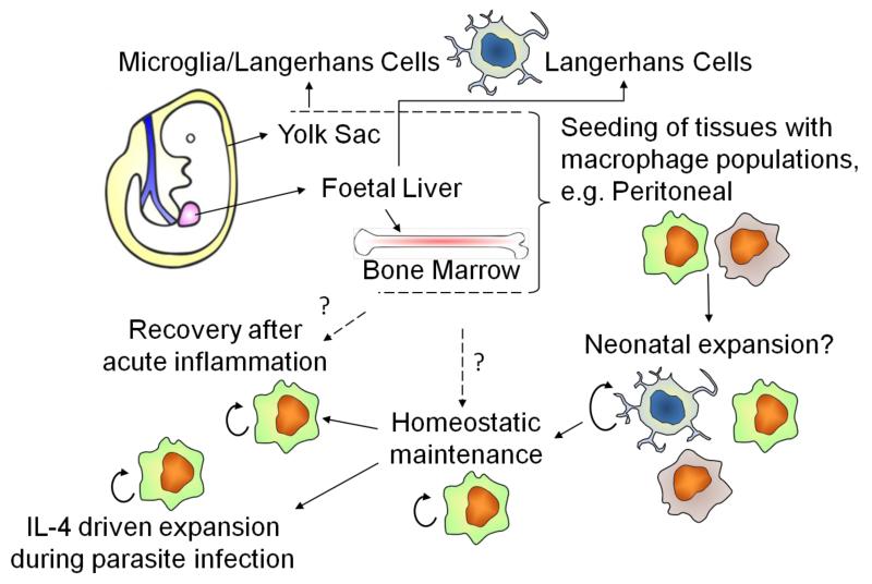
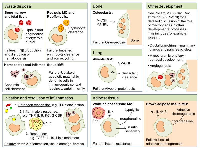
Tissue-resident macrophages are a heterogeneous population of immune cells that fulfill tissue-specific and niche-specific functions. These range from dedicated homeostatic functions, such as clearance of cellular debris and iron processing, to central roles in tissue immune surveillance, response to infection and the resolution of inflammation. Recent studies highlight marked heterogeneity in the origins of tissue macrophages that arise from hematopoietic versus self-renewing embryo-derived populations. We discuss the tissue niche-specific factors that dictate cell phenotype, the definition of which will allow new strategies to promote the restoration of tissue homeostasis. Understanding the mechanisms that dictate tissue macrophage heterogeneity should explain why simplified models of macrophage activation do not explain the extent of heterogeneity seen in vivo.
Figures
Similar articles
-
Establishment and Maintenance of the Macrophage Niche.Immunity. 2020 Mar 17;52(3):434-451. doi: 10.1016/j.immuni.2020.02.015. Immunity. 2020. PMID: 32187515 Review.
-
Macrophages in wound healing: activation and plasticity.Immunol Cell Biol. 2019 Mar;97(3):258-267. doi: 10.1111/imcb.12236. Epub 2019 Feb 11. Immunol Cell Biol. 2019. PMID: 30746824 Free PMC article. Review.
-
Determinants of Resident Tissue Macrophage Identity and Function.Immunity. 2020 Jun 16;52(6):957-970. doi: 10.1016/j.immuni.2020.05.014. Immunity. 2020. PMID: 32553181 Review.
-
Protective and pathogenic functions of macrophage subsets.Nat Rev Immunol. 2011 Oct 14;11(11):723-37. doi: 10.1038/nri3073. Nat Rev Immunol. 2011. PMID: 21997792 Free PMC article. Review.
-
Macrophage Related Chronic Inflammation in Non-Healing Wounds.Front Immunol. 2021 Jun 16;12:681710. doi: 10.3389/fimmu.2021.681710. eCollection 2021. Front Immunol. 2021. PMID: 34220830 Free PMC article. Review.
Cited by
-
Deciphering the role of CD47 in cancer immunotherapy.J Adv Res. 2024 Sep;63:129-158. doi: 10.1016/j.jare.2023.10.009. Epub 2023 Oct 28. J Adv Res. 2024. PMID: 39167629 Free PMC article. Review.
-
Elevated Endomyocardial Biopsy Macrophage-Related Markers in Intractable Myocardial Diseases.Inflammation. 2015 Dec;38(6):2288-99. doi: 10.1007/s10753-015-0214-1. Inflammation. 2015. PMID: 26205770
-
Macrophage LAMTOR1 Deficiency Prevents Dietary Obesity and Insulin Resistance Through Inflammation-Induced Energy Expenditure.Front Cell Dev Biol. 2021 May 20;9:672032. doi: 10.3389/fcell.2021.672032. eCollection 2021. Front Cell Dev Biol. 2021. PMID: 34095141 Free PMC article.
-
Macrophages, Chronic Inflammation, and Insulin Resistance.Cells. 2022 Sep 26;11(19):3001. doi: 10.3390/cells11193001. Cells. 2022. PMID: 36230963 Free PMC article. Review.
-
Imaging liver biology in vivo using conventional confocal microscopy.Nat Protoc. 2015 Feb;10(2):258-68. doi: 10.1038/nprot.2015.006. Epub 2015 Jan 8. Nat Protoc. 2015. PMID: 25569332
References
-
- Metchnikoff E. Leçons sur la pathologie comparée de l’inflammation. Paris, Masson. 1892
-
- Aschoff L. Das reticuloendotheliale System. Erg. Inn. Med. Kinderheilk. 1924;26
-
- Sabin FR, Doan CA, Cunningham RS. Discrimination of two types of phagocytic cells in the connective tissues by the supravital technique. Contrib. Embryol. (Am) 1925;16:125–162.
-
- Daems WT, Brederoo P. The Fine Structure and Peroxidase Activity of Resident and Exudate Peritoneal Macrophages in the Guinea Pig. The Reticuloendothelial System and Immune Phenomena: Advances in Experimental Medicine and Biology. 1971;15:19–31.
Publication types
MeSH terms
Grants and funding
LinkOut - more resources
Full Text Sources
Other Literature Sources
