

Review
doi: 10.1038/nrg3513.
The interplay between cell signalling and mechanics in developmental processes
Affiliations
- PMID: 24045690
- PMCID: PMC4056017
- DOI: 10.1038/nrg3513
Item in Clipboard
Review
The interplay between cell signalling and mechanics in developmental processes
Nat Rev Genet.
2013 Oct.
Abstract
Force production and the propagation of stress and strain within embryos and organisms are crucial physical processes that direct morphogenesis. In addition, there is mounting evidence that biomechanical cues created by these processes guide cell behaviours and cell fates. In this Review we discuss key roles for biomechanics during development to directly shape tissues, to provide positional information for cell fate decisions and to enable robust programmes of development. Several recently identified molecular mechanisms suggest how cells and tissues might coordinate their responses to biomechanical cues. Finally, we outline long-term challenges in integrating biomechanics with genetic analysis of developing embryos.
Figures
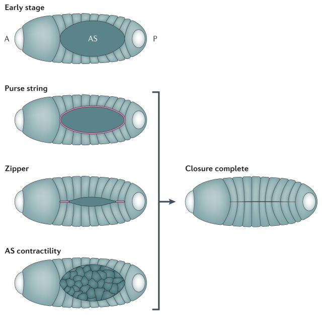
This cartoon shows the process of Drosophila dorsal closure and illustrates the individual mechanical components which help complete closure. In the early stage embryo, the amnioserosa (AS) cells are located in the “hole” which prospective epidermal cells will cover. The embryo is already polarized with anterior (A), where the head will form, and posterior (P) axes. Closure is driven to completion by three distinct cellular processes that contribute to global tissue movements: 1) The actomyosin purse string is shown in pink and is located at the edge of the epithelial cells and AS cells. 2) As epidermal cells meet at the anterior and posterior ends of the AS they fuse and create a zipper , shown in pink, that progressively seals the epidermal sheet over the AS. .3) Cells in the AS, shown in pink, undergo cycles of actomyosin contractions (AS contractility ) to narrow their exposed apical faces and draw the epidermal margins of the AS together.
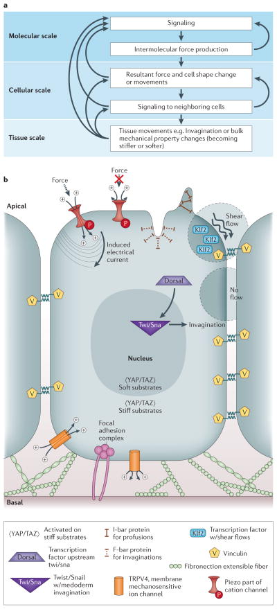
(A) This flow chart shows the intricacies involved with connecting molecular, cellular, and tissue scale behaviors and mechanisms. At the molecular scale, there is molecular signaling which causes intermolecular force production. This force production then feeds back into more molecular signaling. From the molecular scale, there are resultant forces and cell shape changes/movements on the cellular scale, which induce signaling into neighboring cells. The cellular scale can then feedback into molecular scale dynamics or result in tissue scale movements or bulk mechanical property changes. Isolating any portion of this intricate feedback loop is extremely difficult without considering all upstream and downstream effects. (B) Different molecules involved in sensing and signaling to force are shown. Vinculin is located inside the cell at sites of focal adhesions and is also a candidate for sensing forces during development. Fibronectin is an extensible ECM fiber that may sense tension within the tissue. TRPV4 is a membrane mechanosensitive ion channel, believed to open based on tension in the cell membrane. Piezo is a protein part of a cation channel which induces an electrical current based on force. I-bar protein is located at protrusions of the cell membrane and F-bar protein is located at invaginations of the cell membrane. KLF2 is a transcription factor which becomes transcribed when the cell experiences shear flow. YAP/TAZ is activated when the cell is on stiff substrates. Dorsal is a transcription factor upstream of Twist/Snail which is responsible for mesoderm invagination.
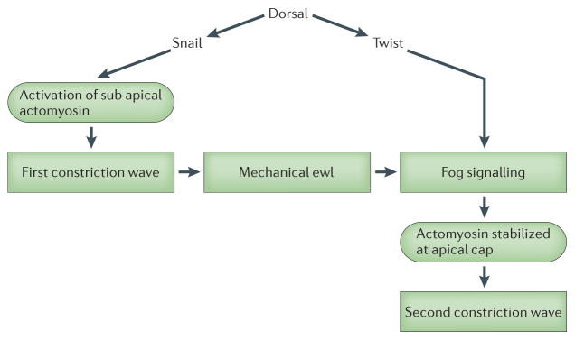
A pathway depicting genetic control of ventral furrow and anterior midgut invagination and a role in that network for mechanical cues. Snail expression is responsible for the mechanical cues produced by the first phase of myosin II contraction. Strain within the epithelium may redirect actomyosin to the apical cap and activate fog signaling which act during the second, Twist-dependent phase to stabilize actomyosin contractions and drive epithelial folding. Furrowing and invagination in Twist mutants can be rescued by exogenously applied strain either by laser ablation or by physical indentation. In the case of invagination-rescued anterior midgut differentiation, larval development was also reported to be rescued.
Similar articles
-
Embryo mechanics: balancing force production with elastic resistance during morphogenesis.Curr Top Dev Biol. 2011;95:215-41. doi: 10.1016/B978-0-12-385065-2.00007-4. Curr Top Dev Biol. 2011. PMID: 21501753 Review.
-
Mechanical control of tissue and organ development.Development. 2010 May;137(9):1407-20. doi: 10.1242/dev.024166. Development. 2010. PMID: 20388652 Free PMC article. Review.
-
[The irruption of mechanics in the chemistry of life].Med Sci (Paris). 2018 Nov;34(11):963-971. doi: 10.1051/medsci/2018241. Epub 2018 Dec 10. Med Sci (Paris). 2018. PMID: 30526840 Review. French.
-
Interplay between mechanics and signalling in regulating cell fate.Nat Rev Mol Cell Biol. 2022 Jul;23(7):465-480. doi: 10.1038/s41580-022-00472-z. Epub 2022 Apr 1. Nat Rev Mol Cell Biol. 2022. PMID: 35365816 Review.
-
Mechanical design in embryos: mechanical signalling, robustness and developmental defects.Philos Trans R Soc Lond B Biol Sci. 2017 May 19;372(1720):20150516. doi: 10.1098/rstb.2015.0516. Philos Trans R Soc Lond B Biol Sci. 2017. PMID: 28348252 Free PMC article. Review.
Cited by
-
Hydraulic fracture during epithelial stretching.Nat Mater. 2015 Mar;14(3):343-51. doi: 10.1038/nmat4206. Epub 2015 Feb 9. Nat Mater. 2015. PMID: 25664452 Free PMC article.
-
Brillouin microscopy.Nat Rev Methods Primers. 2024;4:8. doi: 10.1038/s43586-023-00286-z. Epub 2024 Feb 1. Nat Rev Methods Primers. 2024. PMID: 39391288 Free PMC article.
-
The Cell as Matter: Connecting Molecular Biology to Cellular Functions.Matter. 2021 Jun 2;4(6):1863-1891. doi: 10.1016/j.matt.2021.03.013. Matter. 2021. PMID: 35495565 Free PMC article.
-
Viscoelastic biomarker for differentiation of benign and malignant breast lesion in ultra- low frequency range.Sci Rep. 2019 Apr 5;9(1):5737. doi: 10.1038/s41598-019-41885-9. Sci Rep. 2019. PMID: 30952880 Free PMC article.
-
Socket Array Irregularities and Wing Membrane Distortions at the Eyespot Foci of Butterfly Wings Suggest Mechanical Signals for Color Pattern Determination.Insects. 2024 Jul 16;15(7):535. doi: 10.3390/insects15070535. Insects. 2024. PMID: 39057268 Free PMC article.
References
-
- His W. On the principles of animal morphology. Proceedings of Royal Society of Edinburgh. 1888;15:287–298.
-
- Rhumbler L. Zur mechanik des gastrulations vorganges insbesondere der invagination. Archiv Fur Entwicklungs mechanic. 1902;14:401–476.
-
- Morgan TH. Experimental Embryology. Columbia University Press; 1927.
-
- Lewis WH. Mechanics of Invagination. Anatomical Record. 1947;97:139–56. - PubMed
-
- Howard J. Mechanics of Motor Proteins and the Cytoskeleton. Sinauer Associates; Sunderland, MA: 2001. p. 367.
Publication types
MeSH terms
Grants and funding
LinkOut - more resources
Full Text Sources
Other Literature Sources
