. 2014 Mar 15;75(6):508-16.
doi: 10.1016/j.biopsych.2013.07.033. Epub 2013 Sep 13.

Late adolescent expression of GluN2B transmission in the prefrontal cortex is input-specific and requires postsynaptic protein kinase A and D1 dopamine receptor signaling

Affiliations

Late adolescent expression of GluN2B transmission in the prefrontal cortex is input-specific and requires postsynaptic protein kinase A and D1 dopamine receptor signaling

Eden Flores-Barrera et al. Biol Psychiatry. .

Abstract

Background: Refinement of mature cognitive functions, such as working memory and decision making, typically takes place during adolescence. The acquisition of these functions is linked to the protracted development of the prefrontal cortex (PFC) and dopamine facilitation of glutamatergic transmission. However, the mechanisms that support these changes during adolescence remain elusive.

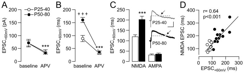
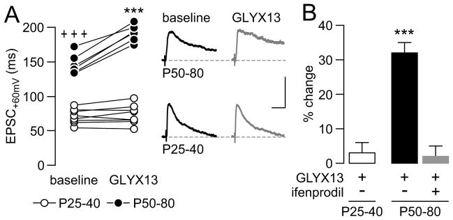
Methods: Electrophysiological recordings (in vitro and in vivo) combined with pharmacologic manipulations were employed to determine how N-methyl-D-aspartate transmission in the medial PFC changes during the adolescent transition to adulthood. The relative contribution of GluN2B transmission and its modulation by postsynaptic protein kinase A and D1 receptor signaling were determined in two distinct age groups of rats: postnatal day (P)25 to P40 and P50 to P80.

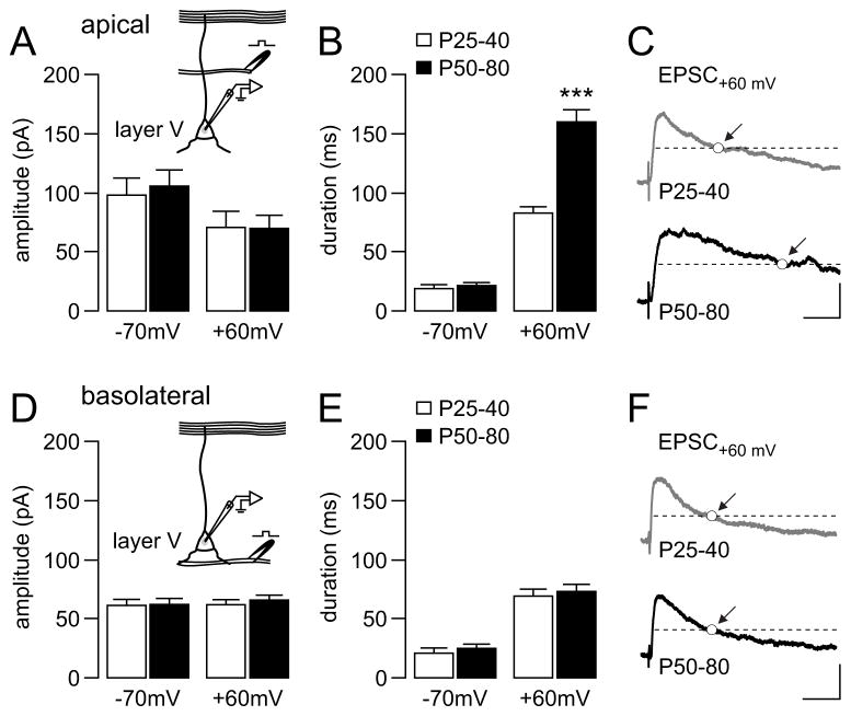
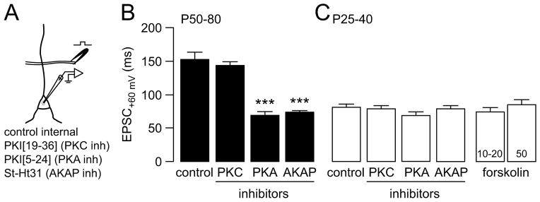
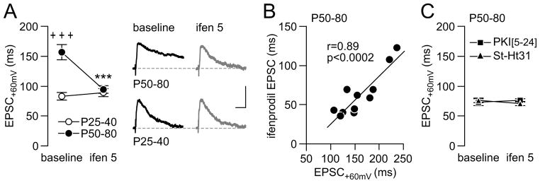
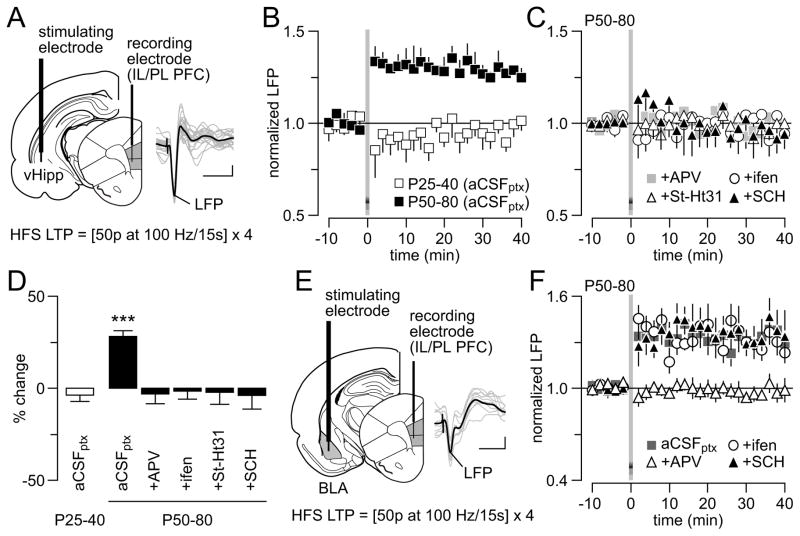
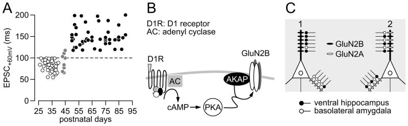
Results: We found that only N-methyl-D-aspartate receptor transmission onto the apical dendrite of layer V pyramidal neurons undergoes late adolescent remodeling due to a functional emergence of GluN2B function after P40. Both protein kinase A and dopamine D1 receptor signaling are required for the functional expression of GluN2B transmission and to sustain PFC plasticity in response to ventral hippocampal, but not basolateral amygdala, inputs.

Conclusions: Thus, the late adolescent acquisition of GluN2B function provides a mechanism for dopamine D1-mediated regulation of PFC responses in an input-specific manner.

Keywords: Adolescence; NMDA; amygdala; dopamine; hippocampus; signaling.


Figures

Figure 1
Figure 1
(A) Electrical stimulation of glutamatergic synaptic transmission onto the apical dendrite of layer V medial PFC pyramidal neuron revealed no apparent age differences in the amplitude of EPSC−70mV and EPSC+60mV (n=10, P25–40; n=14, P50–80). (B) However, the duration of apical stimulation-evoked EPSC+60mV (duration to half amplitude) in the P50–80 age group was significantly longer to that recorded in the P25–40 group (***p<0.0005, unpaired t-test). (C) Average traces of apical stimulation-evoked synaptic response illustrating the age-dependent increase in EPSC duration recorded at the +60 mV holding potential (calibration bars: 50 pA, 50 ms). (DE) Bar graph summarizing the results obtained from the basolateral stimulation revealed similar EPSC kinetics across cells recorded from both age groups (n=7, P25–40; n=14, P50–80). (F) Average traces of basolateral-evoked EPSC recorded at the +60 mV holding potential (calibration bars: 50 pA, 50 ms).
Figure 2
Figure 2
(A) Bath application of the NMDA receptor antagonist APV (50 μM, 10 min) significantly reduced the amplitude of apical-evoked EPSC+60mV in all cells tested from both P25–40 (n=10) and P50–80 (n=14) age groups (***p<0.001 vs. baseline, paired t-test; main effect of treatment p<0.0006, F(1,46) = 13.65; no significant age x treatment interaction, two-way ANOVA). (B) Similarly, APV markedly reduced the duration of apical-evoked EPSC+60mV in both age groups (+++p<0.0005 vs. P25–40, ***p<0.0005 vs. baseline, Fisher LSD post-hoc test; significant age x treatment interaction p<0.0005, F(1,46) = 24.92, two-way ANOVA). Note that the late-adolescent increase in EPSC+60mV duration is not longer detectable following bath application of APV. (C) Analyses of the APV-sensitive component digital subtraction of control vs. APV treatment revealed that the prolonged EPSC+60mV duration recorded in the P50–80 PFC is due to a facilitation of the NMDA-mediated EPSC (***p<0.0005 vs. P25–40, unpaired t-test). Insets are example traces showing the prolonged EPSCNMDA obtained in the P50–80 PFC (calibration bars: 15 pA, 100 ms). (D) Further analyses revealed a significant positive correlation between the duration of the pre-APV EPSC+60mV and the EPSCNMDA component (P25–40: white circles; P50–80: black circles).
Figure 3
Figure 3
(A) Diagram summarizing the experimental design used to assess the contribution of postsynaptic PKA and PKC signaling in mediating the late-adolescent facilitation of NMDA-mediated transmission onto medial PFC layer V pyramidal neuron apical dendrite. (B) In the P50–80 age group, the inclusion of PKI[19–36] (PKC inhibitor, 10 μM; n=7) failed to alter the characteristic prolonged EPSC+60mV duration obtained in response to apical stimulation (control internal solution, n=11). However, the inclusion of the PKA inhibitor PKI[5–24] (n=12) or the AKAP inhibitor St-Ht31 (n=10) into the recording electrode markedly reduced the duration of apical-evoked EPSC+60mV (***p<0.0005 vs. control, Fisher LSD post-hoc test after significant one-way ANOVA, F(3,36) = 31.61, p<0.0005), resembling that observed in the P25–40 medial PFC. (C) In contrast, the inclusion of PKI[19–36] (10 μM; n=7), PKI[5–24] (20 μM; n=10) or St-Ht31 (10 μM; n=7) into the recording electrode did not affect the duration of apical-evoked EPSC+60mV in the P25–40 age group (control internal solution, n=13). In addition, bath application of forskolin failed to increased the duration of the evoked EPSC+60mV to the P50–80 age group level (10–20 μM, n=7; 50 μM, n=6).
Figure 4
Figure 4
(A) Age-dependent effect of the selective GluN2B antagonist ifenprodil on apical stimulation-evoked EPSC+60mV duration recorded from layer V medial PFC pyramidal neuron (age x treatment interaction, p<0.0005, F(1,44) = 17.59, two-way ANOVA). Bath application of ifenprodil (ifen 5 μM; 10 min) markedly reduced the duration of apical-evoked EPSC+60mV only in the P50–80 age group (n=11; ***p<0.0001 vs. baseline, +++p<0.0005 vs. P25–40, Fisher LSD post-hoc test). In contrast, ifenprodil failed to further reduce the duration of apical-evoked EPSC+60mV in the P25–40 PFC (n=13, p=0.95, Fisher LSD post-hoc test). Insets are traces of apical-evoked responses illustrating the age-dependent effect of ifenprodil on EPSC+60mV duration (calibration bars: 50 pA/50 ms). (B) Further analyses revealed a significant positive correlation between EPSC+60mV duration (pre-ifenprodil) and the ifenprodil/GluN2B-sensitive component of the evoked response. (C) The inclusion of the PKA inhibitor PKI[5–24] (20 μM, n=9) or AKAP inhibitor St-Ht31 (5 μM, n=8) into the recording electrode (10–15 min) was sufficient to occlude the effect of ifenprodil on EPSC+60mV duration.
Figure 5
Figure 5
(A) Age-dependent effect of the D1 receptor antagonist SCH23390 (SCH) on apical stimulation-evoked EPSC+60mV in layer V pyramidal neuron of the medial PFC (age x treatment interaction, p<0.0001, F(1,20) = 105.8, two-way ANOVA). Bath application of SCH23390 (10 μM; 10–15 min) markedly reduced EPSC+60mV duration in all pyramidal neurons recorded from the P50–80 PFC (n=6; ***p<0.0005 vs. baseline, +++p<0.0005 vs. P25–40, Fisher LSD post-hoc test). In contrast, SCH23390 failed to reduce the duration of the evoked EPSC+60mV in the P25–40 age group (n=6, p=0.97, Fisher LSD post-hoc test). Inset traces are examples of apical-evoked EPSC+60mV illustrating the age-dependent effect of SCH23390 (calibration bars: 50 pA/50 ms). (B) Bar graph summarizing the percent change (% change) to baseline of the results shown in A. Overall, SCH23390 reduced the duration of apical-evoked EPSC+60mV by ~40% in the P50–80 age group whereas a non-significant ~5% change was observed in the P25–40 PFC. Notably, SCH23390 failed to further reduce EPSC+60mV duration in the P50–80 age group when recordings were conducted from slices pre-incubated with ifenprodil (5 μM, n=6; ***p<0.0005 vs. P25–40 or P50–80 + ifenprodil, Fisher LSD post-hoc test after significant one-way ANOVA, p<0.0001, F(2,15) = 128.9).
Figure 6
Figure 6
(A) Age-dependent effect of GLYX13 on apical-evoked EPSC+60mV duration recorded from layer V medial PFC pyramidal neurons (age x treatment interaction, p<0.0005, F(1,24) = 18.98, two-way ANOVA). Bath application of GLYX13 (1 μM; 10 min) significantly increased the duration of the evoked EPSC+60mV only in prefrontal pyramidal neurons recorded from the P50–80 age group (n=6; ***p<0.0005 vs. baseline, +++p<0.0005 vs. P25–40, Fisher LSD post-hoc test). All neurons recorded from the P25–40 PFC remained unchanged following bath application of GLYX13 (n=8, p=0.97, Fisher LSD post-hoc test). Insets are example traces of apical-evoked responses illustrating the age-dependent effect of GLYX13 on EPSC+60mV duration (calibration bars: 50 pA/50 ms). (B) Bar graph summarizing the results shown in A as percent change (% change) to baseline. Overall, GLYX13 increased the duration of apical-evoked EPSC+60mV by ~30% in the P50–80 age group whereas a non-significant ~5% change was observed in the P25–40 PFC. Note that the facilitatory effect of GLYX13 observed in the P50–80 age group failed to occur in brain slices pre-incubated with ifenprodil (5 μM, n=5; ***p<0.0005 vs. P25–40 or P50–80 + ifenprodil, Fisher LSD post-hoc test after significant one-way ANOVA, p<0.0001, F(2,16) = 30.2).
Figure 7
Figure 7
(A) Diagram illustrating the recording arrangement used to study ventral hippocampal high-frequency stimulation (HFS)-induced plasticity in the medial PFC by means of local field potential (LFP) recordings in vivo (IL: infralimbic; PL: prelimbic; calibration bars: 2 μV/50 ms). To pharmacologically isolate glutamatergic transmission, all recordings were conducted following local prefrontal microinfusion of aCSF-containing picrotoxin (aCSFptx; see details in Methods and Materials). (B) In the P50–80 age group (aCSFptx, n=7), ventral hippocampal HFS typically elicited a sustained facilitation of the evoked LFP response in the medial PFC. Such LTP of the evoked LFP response was not observed in the medial PFC of P25–40 rats (aCSFptx, n=6). (C) Summary of the effects of ventral hippocampal HFS following single local prefrontal microinfusion of aCSFptx-containing APV (50 μM, n=6), ifenprodil (5 μM, n=6), St-Ht31 (10 μM, n=6) or SCH23390 (10 μM, n=6). Note that the characteristic LFP-LTP observed in the P50–80 PFC is not longer present after prefrontal removal of NMDA-GluN2B transmission, PKA signaling and D1 receptor tone. (D) Bar graph summarizing the statistical analyses of the results shown in C (average from the last 20 min; ***p<0.0005 vs. P25–40 or any treatment group, Fisher LSD post-hoc test after significant one-way ANOVA, p<0.0005, F(5,31) = 5.86). (E) Diagram illustrating the recording arrangement used to study basolateral amygdala (BLA)-induced plasticity in the medial PFC by means of LFP recordings in vivo (calibration bars: 2 μV/50 ms). As in A, all prefrontal LFP recordings were conducted following local microinfusion of aCSFptx into the PFC. (F) BLA-HFS also elicited LTP in the PFC of P50–80 rats (aCSFptx, n=6), a form plasticity that was blocked by the NMDA receptor antagonist APV (50 μM, n=5). However, prefrontal infusions of ifenprodil (5 μM, n=5) or SCH23390 (10 μM, n=5) failed to block BLA-induced LTP in the medial PFC.
Figure 8
Figure 8
(A) Summary of the baseline EPSC+60mV duration of all layer V pyramidal neurons recorded from the medial PFC included in the present study. Additional data obtained from the P41–49 age gap were included to highlight that the gain of long-lasting NR2B transmission begins to emerge ~P45. Note that all PFC cells recorded after P50 exhibit EPSC+60mV duration >100 ms (dashed line). (B) Simplified diagram illustrating how dopamine D1 receptor signaling and AKAP-PKA interacts to facilitate (directly or indirectly) GluN2B transmission in the apical dendrite of layer V PFC pyramidal neurons. (C) Two plausible scenarios for explaining the GluN2B-dependent input-specific LTP observed in the adult PFC following HFS of the ventral hippocampus. Model 1 suggests that ventral hippocampal inputs to the PFC may synapse preferentially onto the apical dendrite whereas afferents from the basolateral amygdala may synapse preferentially onto the basolateral dendrites. Model 2 proposes a convergence of inputs originating from both structures onto the apical dendrite of layer V pyramidal neurons. However, a functional segregation of these two afferents is determined by the emergence of GluN2B transmission preferentially at hippocampal synapses.

Similar articles

Cited by

References

    1. Best JR, Miller PH. A developmental perspective on executive function. Child Dev. 2010;81:1641–1660. - PMC - PubMed
    1. Luna B, Garver KE, Urban TA, Lazar NA, Sweeney JA. Maturation of cognitive processes from late childhood to adulthood. Child Dev. 2004;75:1357–1372. - PubMed
    1. Ishikawa A, Nakamura S. Convergence and interaction of hippocampal and amygdalar projections within the prefrontal cortex in the rat. J Neurosci. 2003;23:9987–9995. - PMC - PubMed
    1. Phillips ML, Drevets WC, Rauch SL, Lane R. Neurobiology of emotion perception I: The neural basis of normal emotion perception. Biol Psychiatry. 2003;54:504–514. - PubMed
    1. Tseng KY, O’Donnell P. Post-pubertal emergence of prefrontal cortical up states induced by D1-NMDA co-activation. Cereb Cortex. 2005;15:49–57. - PubMed

Publication types

MeSH terms

Substances