. 2014 Mar 6;426(5):1061-76.
doi: 10.1016/j.jmb.2013.09.007. Epub 2013 Sep 13.

The plant host can affect the encapsidation of brome mosaic virus (BMV) RNA: BMV virions are surprisingly heterogeneous

Affiliations

The plant host can affect the encapsidation of brome mosaic virus (BMV) RNA: BMV virions are surprisingly heterogeneous

Peng Ni et al. J Mol Biol. .

Abstract

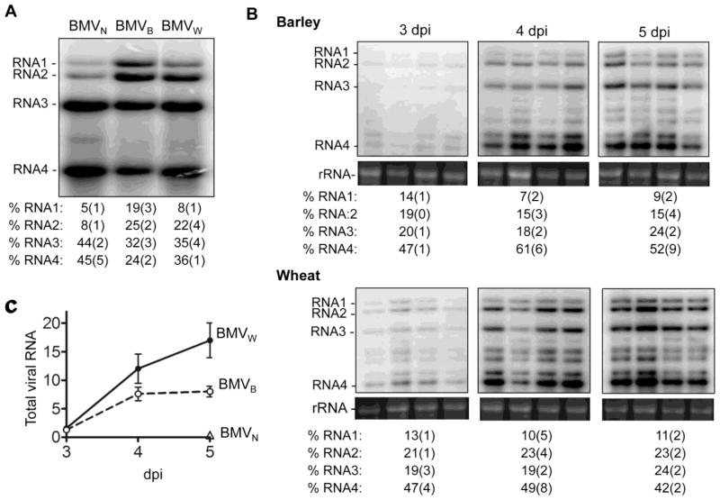
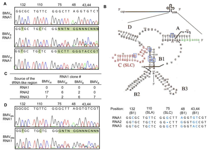
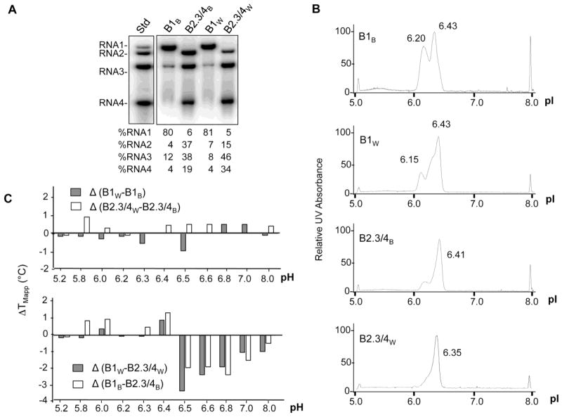
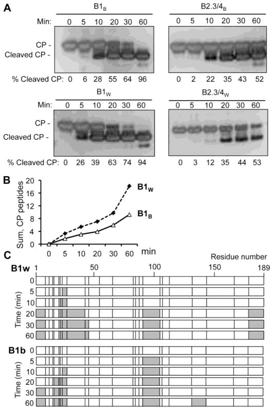
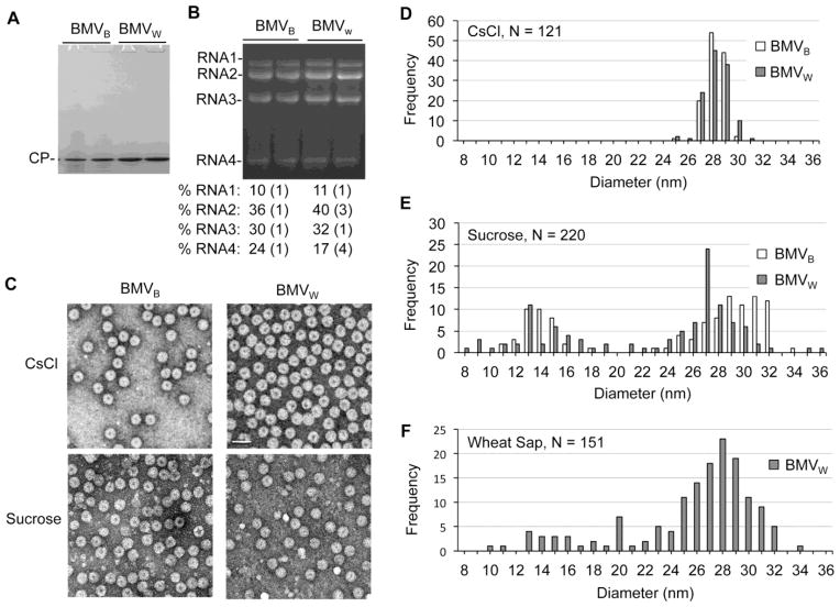
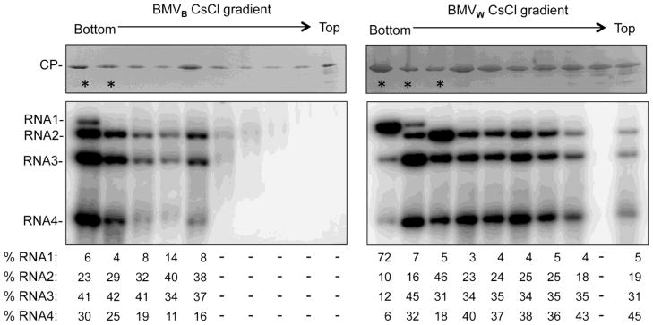
Brome mosaic virus (BMV) packages its genomic and subgenomic RNAs into three separate viral particles. BMV purified from barley, wheat, and tobacco have distinct relative abundances of the encapsidated RNAs. We seek to identify the basis for the host-dependent differences in viral RNA encapsidation. Sequencing of the viral RNAs revealed recombination events in the 3' untranslated region of RNA1 of BMV purified from barley and wheat, but not from tobacco. However, the relative amounts of the BMV RNAs that accumulated in barley and wheat are similar and RNA accumulation is not sufficient to account for the difference in RNA encapsidation. Virions purified from barley and wheat were found to differ in their isoelectric points, resistance to proteolysis, and contacts between the capsid residues and the RNA. Mass spectrometric analyses revealed that virions from the three hosts had different post-translational modifications that should impact the physiochemical properties of the virions. Another major source of variation in RNA encapsidation was due to the purification of BMV particles to homogeneity. Highly enriched BMV present in lysates had a surprising range of sizes, buoyant densities, and distinct relative amounts of encapsidated RNAs. These results show that the encapsidated BMV RNAs reflect a combination of host effects on the physiochemical properties of the viral capsids and the enrichment of a subset of virions. The previously unexpected heterogeneity in BMV should influence the timing of the infection and also the host innate immune responses.

Keywords: Brome mosaic virus; RNA encapsidation; capsid–RNA interaction; coat protein; virion polymorphism.


Figures

Fig. 1
Fig. 1. The encapsidation and replication of BMV RNA in different plant hosts
A) The relative abundances of the four viral RNAs isolated from the CsCl banded BMVW, BMVB, BMVN. The RNA was detected by a riboprobe that anneals to the common 3′tRNA-like region. The quantification below the image were from four independent experiments. All quantifications show the percentage of the four viral RNAs in individual sample with errors in the brackets. B). Time course of viral RNA accumulation in barley and wheat. 5 μg total RNA isolated from the wheat and barley leaves at the indicated time were separated by gel electrophoresis and blotted for BMV RNA by the riboprobe. rRNA served as a loading control. The relative amounts of the four viral RNA accumulated from the four independent samples are summarized under the Northern blot images. All quantifications show the percentage of the four viral RNAs in individual sample with errors in the brackets. C) Graph summarizing the accumulation of BMV RNAs in barley, wheat and N. benthamiana over time. Quantification used summation of the intensity of all four viral RNAs in quadruplicates at each time point.
Fig. 2
Fig. 2. RNA recombination occurred within the tRNA-like structure of RNA1 in BMVW and BMVB
A) Representative sequencing chromatograms of the cDNA population derived from the tRNA-like structure in RNA1 for BMVW, BMVB and BMVN. Residues shaded in green identified mismatches to the sequences present in the Agrobacterium constructs used to start BMV infection. The numbers indicate the position of the nucleotides, with the 3′-most nucleotide being residue 1. B) Schematic of the common tRNA-like structure and the sequences of the diagnostic sites. The schematic adapted from Felden et al. is modeled after the tRNA-like region in RNA3. The name of each stem-loop structure is in bold and the core promoter for minus-strand RNA synthesis, SLC, is in red. The nucleotides affected by the recombination in RNA1 are boxed. In the lower panel, the SNPs (nt 43, 44, 132) from the three BMV RNAs are colored blue with their positions shown above. In addition to the three SNPs, substitutions at nt 48 and 110 in RNA1 and a deletion at nt 75 in RNA2 are present in the BMV cDNA in the Agrobacterium. These five positions are used to map the intersegment crossovers in the RNA1 recombinants. C) The number of clones obtained from the RNA1 cDNA library of BMVW or BMVB that contained sequences mapped to the tRNA-like structure in RNA1, RNA2 or RNA3. Two independent preparations of BMVw and BMVB were used to generate the results. D) Representative sequencing chromatograms of the cDNA population derived from the tRNA-like structure in the RNA1 after BMVW and BMVN were passed through N. benthamiana. The progeny virions are denoted as BMVWN and BMVNN respectively.
Fig. 3
Fig. 3. Enrichment of the B1 and B2.3/4 virions from wheat or barley and characterization of their physiochemical properties
A) Northern blot demonstrating the enrichment of the subsets of the three BMV virions. The proportion of each RNA in the purified virions is below the blot image. B) Isoelectric focusing chromatographs of the subsets of the BMV virions isolated from barley and wheat. The peaks near pI of 5.0 and 8.0 are pI standards added to each sample. C) The difference (ΔTmApp) in apparent melting temperature of the capsid for the subsets of the BMV virions isolated from barley and wheat as a function of pH. The upper panel compares subsets of BMV enriched for the same RNAs but isolated from different hosts. The lower panel compares the subsets of BMV isolated from the same host but enriched for different RNAs.
Fig. 4
Fig. 4. BMVW and BMVB are differentially sensitive to protease digestion
A) Partial trypsin digestion of B1 and B2.3/4 particle from BMVW and BMVB. The SDS-PAGE gel was stained with silver. The signal in the cleaved CP and the full length CP were used to calculate the percentage of the cleaved product. B) Quantification of the trypsin cleaved CP peptides by mass-spectrometry. All MALDI-TOF spectra of the CP fragments generated over time in partial trypsin digestion were normalized to the intensity of the bradykinin fragment that was added to the sample prior to the sample processing. The intensities of the CP fragments were summed as a function of digestion time. C) Locations of the CP tryptic peptide fragments released from B1B and B1W. The locations of each arginine or lysine are denoted by a vertical line in the schematics. The proteolytic fragments of the CP detected by mass spectrometry are colored grey.
Fig. 5
Fig. 5. BMV capsid-RNA interacting residues
A) Capsid peptide that contact the encapsidated RNA. The peptides associated with RNA1 or RNA2, 3, and 4 in an RCAP assay (summarized in Table 2) are highlighted in red and blue, respectively, for both BMVN and BMVB. A cross-section of the capsid is shown to allow viewing of the CPs from the internal cavity of the BMV virion. B) The RNA sequences that contact the capsid. The normalized coverage of BMV RNAs by the CP-associated RNA fragments in a CLIP-seq assay is shown. The normalized coverage was obtained by dividing the actual coverage at each nucleotide position by the average coverage of the corresponding RNA to account for different depth of sequencing. The traces for BMV isolated from N. benthamiana, wheat or barley were colored blue, red and green, respectively. Note that there was nearly complete overlap of the results.
Fig. 6
Fig. 6. The BMV capsid contains post-translational modifications
A) Mass spectrometry spectra showing modified forms of the BMV CP from virions purified from barley, wheat and N. benthamiana. Below each spectrum are summaries of the potential modifications predicted to exist on the three CPs labeled within each spectrum. Abbreviations: Met: methionine, Acet: acetylation, Met Ox: methionine oxidation, Hydrox: hydroxylation, ΔMethyl: removal of the N-terminal methionine. B) Comparison of lysine acetylation in tryptic fragments of the CPs from BMVB and BMVW. The mass of the observed peptides, the theoretical mass, and the mass differences are shown. The residue most likely to be modified due to predictions are shown in color and underlined. C) The location of the acetylated lysine residues in a CP subunit. The colors of labeled residues correspond to those in panel B.
Fig. 7
Fig. 7. Variation in the diameters of the BMV particles
A) The CP is the most abundant protein from BMVW and BMVB purified by sucrose density cushions. 2 μg of total protein determined by optical density was loaded per lane in the SDS-PAGE. The gel was stained with Coomassie blue. B) Relative abundances of the virion RNAs from BMVW and BMVB enriched with sucrose cushions. RNAs was visualized by staining with ethidium bromide. The quantification of the relative amount of the RNAs were from three independent experiments. C) Negative-stained electron micrograph of BMVW and BMVB purified from CsCl gradients or the sucrose cushion. D) The size distribution of CsCl purified BMVW and BMVB. The outer diameters of the viral particles were measured from multiple micrographs to ensure random sampling. E) The size distribution of the BMVW and BMVB particles enriched through the sucrose cushion. G) The size distribution of BMVW for clarified wheat sap.
Fig. 8
Fig. 8. BMV virions exhibit a range of densities in CsCl density gradients
BMVW or BMVB enriched by sedimentation through a sucrose cushion were further centrifuged in a 45% cesium chloride gradient for 20 h at 50,000 g. Fractions were then collected following puncturing the bottom of the tube. The stars denote the fractions that appear in the opalescent white band. The upper images show the presence of the CP in each fraction. The SDS-PAGE gel was stained with Commassie blue. The lower images show the detection of viral RNAs by Northern blot. The quantification show the relative amount of the four viral RNAs in each fraction. Approximately equal amount of virions or viral RNAs from each fraction were used for the visualization, except for the fractions that contained significantly less viral RNAs.

Similar articles

Cited by

References

    1. Levine B, Hardwick JM, Griffin DE. Persistence of alphaviruses in vertebrate hosts. Trends Microbiol. 1994;2:25–28. - PubMed
    1. Hayes EB, Komar N, Nasci RS, Montgomery SP, O’Leary DR, Campbell GL. Epidemiology and transmission dynamics of West Nile virus disease. Emerg Infect Dis. 2005;11:1167–1173. - PMC - PubMed
    1. Rao AL. Genome packaging by spherical plant RNA viruses. Annu Rev Phytopathol. 2006;44:61–87. - PubMed
    1. Lane LC. The bromoviruses. Adv Virus Res. 1974;19:151–220. - PubMed
    1. Rao A, Grantham G. Biological significance of the seven amino-terminal basic residues of brome mosaic virus coat protein. Virology. 1995;211:42–52. - PubMed

Publication types

MeSH terms

LinkOut - more resources