Role of HGF in epithelial-stromal cell interactions during progression from benign breast disease to ductal carcinoma in situ
- PMID: 24025166
- PMCID: PMC3978616
- DOI: 10.1186/bcr3476
Role of HGF in epithelial-stromal cell interactions during progression from benign breast disease to ductal carcinoma in situ
Abstract
Introduction: Basal-like and luminal breast cancers have distinct stromal-epithelial interactions, which play a role in progression to invasive cancer. However, little is known about how stromal-epithelial interactions evolve in benign and pre-invasive lesions.
Methods: To study epithelial-stromal interactions in basal-like breast cancer progression, we cocultured reduction mammoplasty fibroblasts with the isogenic MCF10 series of cell lines (representing benign/normal, atypical hyperplasia, and ductal carcinoma in situ). We used gene expression microarrays to identify pathways induced by coculture in premalignant cells (MCF10DCIS) compared with normal and benign cells (MCF10A and MCF10AT1). Relevant pathways were then evaluated in vivo for associations with basal-like subtype and were targeted in vitro to evaluate effects on morphogenesis.
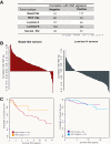
Results: Our results show that premalignant MCF10DCIS cells express characteristic gene expression patterns of invasive basal-like microenvironments. Furthermore, while hepatocyte growth factor (HGF) secretion is upregulated (relative to normal, MCF10A levels) when fibroblasts are cocultured with either atypical (MCF10AT1) or premalignant (MCF10DCIS) cells, only MCF10DCIS cells upregulated the HGF receptor MET. In three-dimensional cultures, upregulation of HGF/MET in MCF10DCIS cells induced morphological changes suggestive of invasive potential, and these changes were reversed by antibody-based blocking of HGF signaling. These results are relevant to in vivo progression because high expression of a novel MCF10DCIS-derived HGF signature was correlated with the basallike subtype, with approximately 86% of basal-like cancers highly expressing the HGF signature, and because high expression of HGF signature was associated with poor survival.
Conclusions: Coordinated and complementary changes in HGF/MET expression occur in epithelium and stroma during progression of pre-invasive basal-like lesions. These results suggest that targeting stroma-derived HGF signaling in early carcinogenesis may block progression of basal-like precursor lesions.
Figures


Similar articles
-
Epithelial p53 Status Modifies Stromal-Epithelial Interactions During Basal-Like Breast Carcinogenesis.J Mammary Gland Biol Neoplasia. 2021 Jun;26(2):89-99. doi: 10.1007/s10911-020-09477-w. Epub 2021 Jan 13. J Mammary Gland Biol Neoplasia. 2021. PMID: 33439408 Free PMC article.
-
Dynamic stromal-epithelial interactions during progression of MCF10DCIS.com xenografts.Int J Cancer. 2007 May 15;120(10):2127-34. doi: 10.1002/ijc.22572. Int J Cancer. 2007. PMID: 17266026
-
Induction of hepatocyte growth factor in fibroblasts by tumor-derived factors affects invasive growth of tumor cells: in vitro analysis of tumor-stromal interactions.Cancer Res. 1997 Aug 1;57(15):3305-13. Cancer Res. 1997. PMID: 9242465
-
Gene expression analysis of in vitro cocultures to study interactions between breast epithelium and stroma.J Biomed Biotechnol. 2011;2011:520987. doi: 10.1155/2011/520987. Epub 2011 Dec 13. J Biomed Biotechnol. 2011. PMID: 22203785 Free PMC article. Review.
-
Roles of the HGF/Met signaling in head and neck squamous cell carcinoma: Focus on tumor immunity (Review).Oncol Rep. 2020 Dec;44(6):2337-2344. doi: 10.3892/or.2020.7799. Epub 2020 Oct 9. Oncol Rep. 2020. PMID: 33125120 Review.
Cited by
-
Three-dimensional modelling identifies novel genetic dependencies associated with breast cancer progression in the isogenic MCF10 model.J Pathol. 2016 Nov;240(3):315-328. doi: 10.1002/path.4778. J Pathol. 2016. PMID: 27512948 Free PMC article.
-
TGFβ1 regulates HGF-induced cell migration and hepatocyte growth factor receptor MET expression via C-ets-1 and miR-128-3p in basal-like breast cancer.Mol Oncol. 2018 Sep;12(9):1447-1463. doi: 10.1002/1878-0261.12355. Epub 2018 Jul 30. Mol Oncol. 2018. PMID: 30004628 Free PMC article.
-
Recurrence analysis on prostate cancer patients with Gleason score 7 using integrated histopathology whole-slide images and genomic data through deep neural networks.J Med Imaging (Bellingham). 2018 Oct;5(4):047501. doi: 10.1117/1.JMI.5.4.047501. Epub 2018 Nov 15. J Med Imaging (Bellingham). 2018. PMID: 30840742 Free PMC article.
-
Paper-based Transwell assays: an inexpensive alternative to study cellular invasion.Analyst. 2018 Dec 17;144(1):206-211. doi: 10.1039/c8an01157e. Analyst. 2018. PMID: 30328422 Free PMC article.
-
Weight Loss Reversed Obesity-Induced HGF/c-Met Pathway and Basal-Like Breast Cancer Progression.Front Oncol. 2014 Jul 8;4:175. doi: 10.3389/fonc.2014.00175. eCollection 2014. Front Oncol. 2014. PMID: 25072025 Free PMC article.
References
-
- Krtolica A, Campisi J. Integrating epithelial cancer, aging stroma and cellular senescence. Adv Gerontol. 2003;15:109–116. - PubMed
-
- Pirone JR, D'Arcy M, Stewart DA, Hines WC, Johnson M, Gould MN, Yaswen P, Jerry DJ, Smith Schneider S, Troester MA. Age-associated gene expression in normal breast tissue mirrors qualitative age-at-incidence patterns for breast cancer. Cancer Epidemiol Biomarkers Prev. 2012;15:1735–1744. doi: 10.1158/1055-9965.EPI-12-0451. - DOI - PMC - PubMed
Publication types
MeSH terms
Substances
Grants and funding
LinkOut - more resources
Full Text Sources
Other Literature Sources
Medical
Molecular Biology Databases
Miscellaneous

