

Near-infrared light-sensitive liposomes for the enhanced photothermal tumor treatment by the combination with chemotherapy
- PMID: 24022681
- PMCID: PMC4126419
- DOI: 10.1007/s11095-013-1180-7
Near-infrared light-sensitive liposomes for the enhanced photothermal tumor treatment by the combination with chemotherapy
Abstract
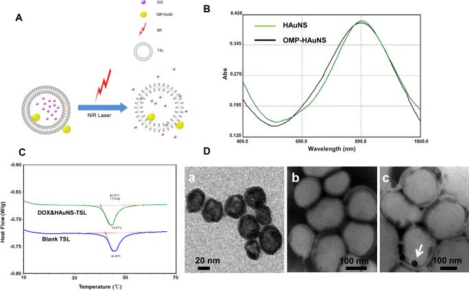
Purpose: To develop a near-infrared (NIR) light-sensitive liposome, which contains hollow gold nanospheres (HAuNS) and doxorubicin (DOX), and evaluate their potential utility for enhancing antitumor activity and controlling drug release.
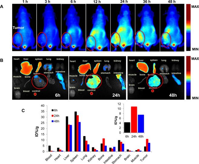
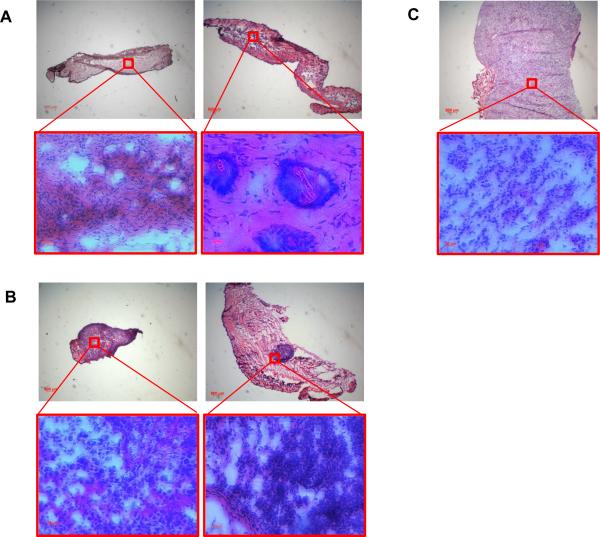
Methods: The liposomes (DOX&HAuNS-TSL) were designed based on a thermal sensitive liposome (TSL) formulation, and hydrophobically modified HAuNS were attached onto the membrane of the liposomes. The behavior of DOX release from the liposomes was investigated by the dialysis, diffusion in agarose gel and cellular uptake of the drug. The biodistribution of DOX&HAuNS-TSL was assessed by i.v. injection in tumor-bearing nude mice. Antitumor efficacy was evaluated both histologically using excised tissue and intuitively by measuring the tumor size and weight.
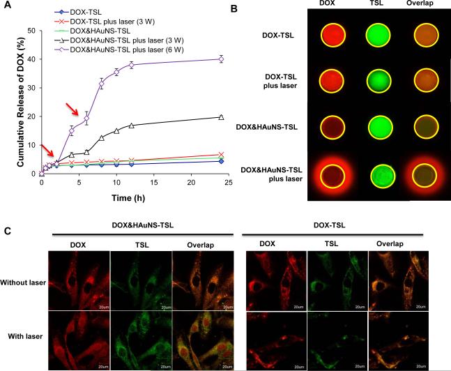
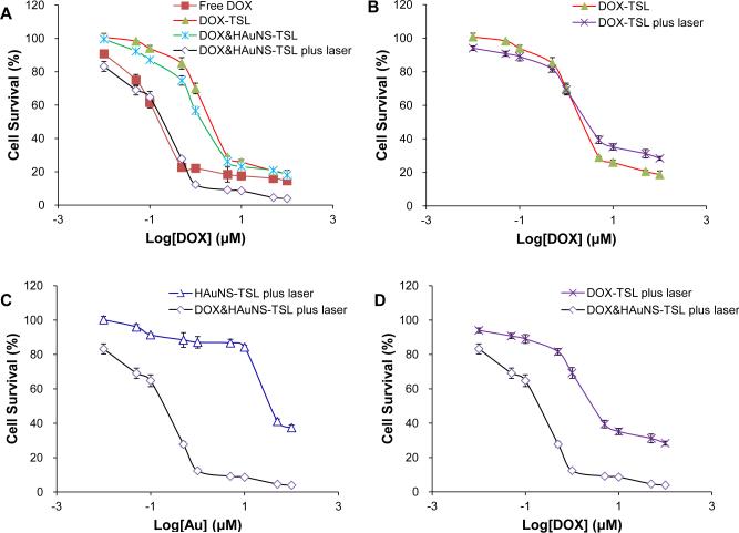
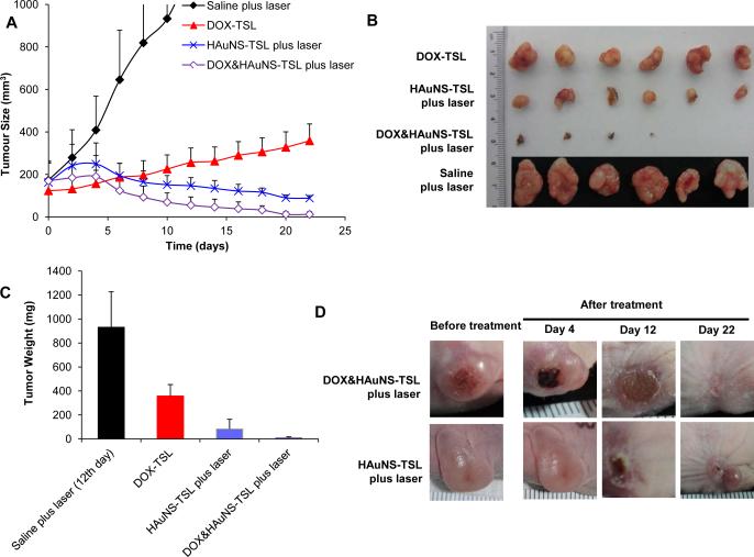
Results: Rapid and repetitive DOX release from the liposomes (DOX&HAuNS-TSL), could be readily achieved upon NIR laser irradiation. The treatment of tumor cells with DOX&HAuNS-TSL followed by NIR laser irradiation showed significantly greater cytotoxicity than the treatment with DOX&HAuNS-TSL alone, DOX-TSL alone (chemotherapy alone) and HAuNS-TSL plus NIR laser irradiation (Photothermal ablation, PTA, alone). In vivo antitumor study indicated that the combination of simultaneous photothermal and chemotherapeutic effect mediated by DOX&HAuNS-TSL plus NIR laser presented a significantly higher antitumor efficacy than the PTA alone mediated by HAuNS-TSL plus NIR laser irradiation.
Conclusions: Our study could be as the valuable reference and direction for the clinical application of PTA in tumor therapy.
Figures
Similar articles
-
Photothermal-chemotherapy with doxorubicin-loaded hollow gold nanospheres: A platform for near-infrared light-trigged drug release.J Control Release. 2012 Mar 10;158(2):319-28. doi: 10.1016/j.jconrel.2011.10.028. Epub 2011 Oct 28. J Control Release. 2012. PMID: 22063003 Free PMC article.
-
Effective photothermal chemotherapy using doxorubicin-loaded gold nanospheres that target EphB4 receptors in tumors.Cancer Res. 2012 Sep 15;72(18):4777-86. doi: 10.1158/0008-5472.CAN-12-1003. Epub 2012 Aug 3. Cancer Res. 2012. PMID: 22865457 Free PMC article.
-
Gold cluster-labeled thermosensitive liposmes enhance triggered drug release in the tumor microenvironment by a photothermal effect.J Control Release. 2015 Oct 28;216:132-9. doi: 10.1016/j.jconrel.2015.08.002. Epub 2015 Aug 4. J Control Release. 2015. PMID: 26247553
-
Chemophototherapeutic Ablation of Doxorubicin-Resistant Human Ovarian Tumor Cells.Photochem Photobiol. 2023 Mar;99(2):844-849. doi: 10.1111/php.13677. Epub 2022 Aug 2. Photochem Photobiol. 2023. PMID: 35842741 Free PMC article. Review.
-
Review of the Delivery Kinetics of Thermosensitive Liposomes.Cancers (Basel). 2023 Jan 7;15(2):398. doi: 10.3390/cancers15020398. Cancers (Basel). 2023. PMID: 36672347 Free PMC article. Review.
Cited by
-
Recent Advancements in Stimuli Responsive Drug Delivery Platforms for Active and Passive Cancer Targeting.Cancers (Basel). 2021 Feb 7;13(4):670. doi: 10.3390/cancers13040670. Cancers (Basel). 2021. PMID: 33562376 Free PMC article. Review.
-
Stimuli-responsive liposomes for drug delivery.Wiley Interdiscip Rev Nanomed Nanobiotechnol. 2017 Sep;9(5):10.1002/wnan.1450. doi: 10.1002/wnan.1450. Epub 2017 Feb 15. Wiley Interdiscip Rev Nanomed Nanobiotechnol. 2017. PMID: 28198148 Free PMC article. Review.
-
Design and Application of Near-Infrared Nanomaterial-Liposome Hybrid Nanocarriers for Cancer Photothermal Therapy.Pharmaceutics. 2021 Dec 3;13(12):2070. doi: 10.3390/pharmaceutics13122070. Pharmaceutics. 2021. PMID: 34959351 Free PMC article. Review.
-
Photoswitchable anticancer activity via trans-cis isomerization of a combretastatin A-4 analog.Org Biomol Chem. 2016 Jan 7;14(1):40-9. doi: 10.1039/c5ob02005k. Epub 2015 Oct 27. Org Biomol Chem. 2016. PMID: 26503632 Free PMC article.
-
Synergic effect of doxorubicin release and two-photon irradiation of Mn2+-doped Prussian blue nanoparticles on cancer therapy.RSC Adv. 2020 Jan 15;10(5):2646-2649. doi: 10.1039/c9ra09133e. eCollection 2020 Jan 14. RSC Adv. 2020. PMID: 35496092 Free PMC article.
References
-
- Loo C, Lowery A, Halas N, West J, Drezek R. Immunotargeted nanoshells for integrated cancer imaging and therapy. Nano Lett. 2005;5:709–711. - PubMed
-
- Schwartz JA, Shetty AM, Price RE, Stafford RJ, Wang JC, Uthamanthil RK, Pham K, McNichols RJ, Coleman CL, Payne JD. Feasibility study of particle-assisted laser ablation of brain tumors in orthotopic canine model. Cancer Res. 2009;69:1659–1667. - PubMed
-
- Abdulla-Al-Mamun M, Kusumoto Y, Mihata A, Islam MS, Ahmmad B. Plasmon-induced photothermal cell-killing effect of gold colloidal nanoparticles on epithelial carcinoma cells. Photochemical & Photobiological Sciences. 2009;8:1125–1129. - PubMed
Publication types
MeSH terms
Substances
Grants and funding
LinkOut - more resources
Full Text Sources
Other Literature Sources
Miscellaneous
