. 2013 Nov;24(21):3420-34.
doi: 10.1091/mbc.E13-05-0236. Epub 2013 Sep 4.

Identification and characterization of multiple novel Rab-myosin Va interactions

Affiliations

Identification and characterization of multiple novel Rab-myosin Va interactions

Andrew J Lindsay et al. Mol Biol Cell. 2013 Nov.

Abstract

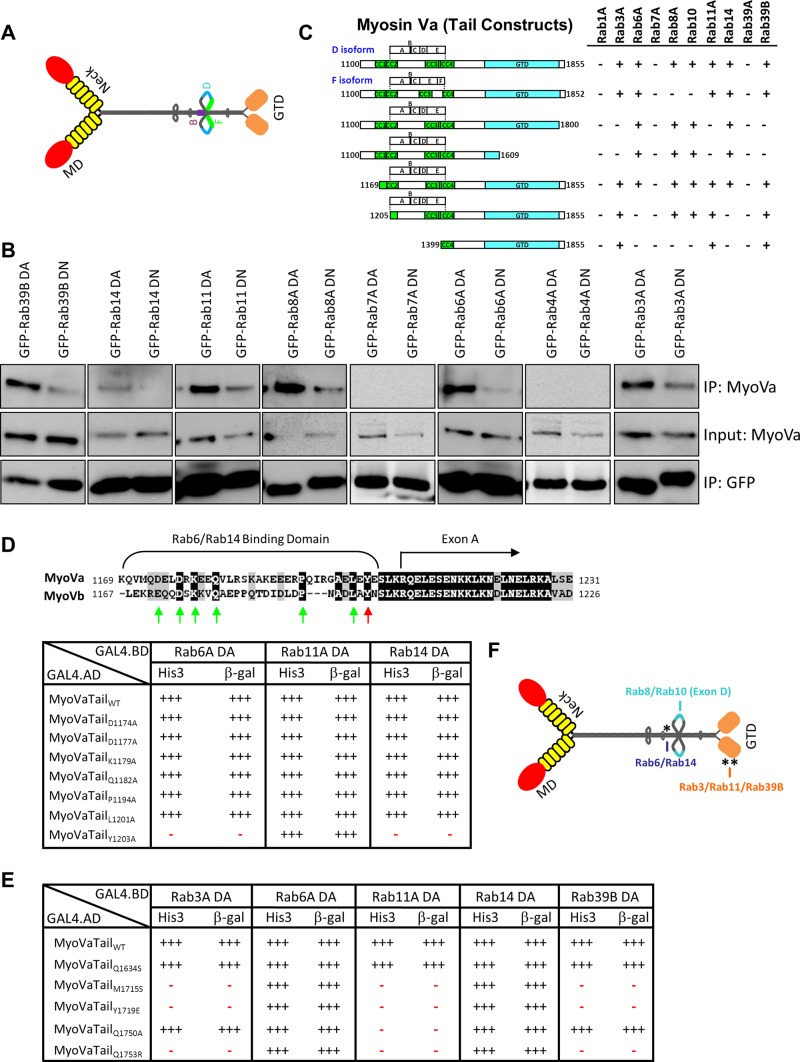
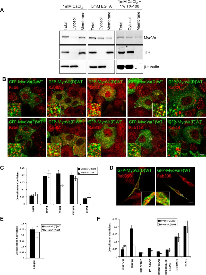
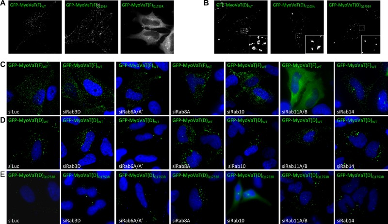
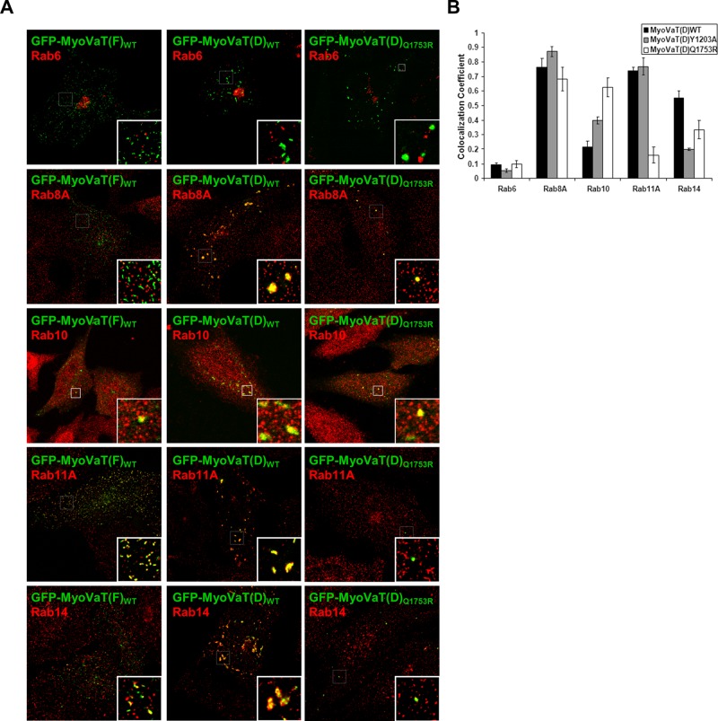
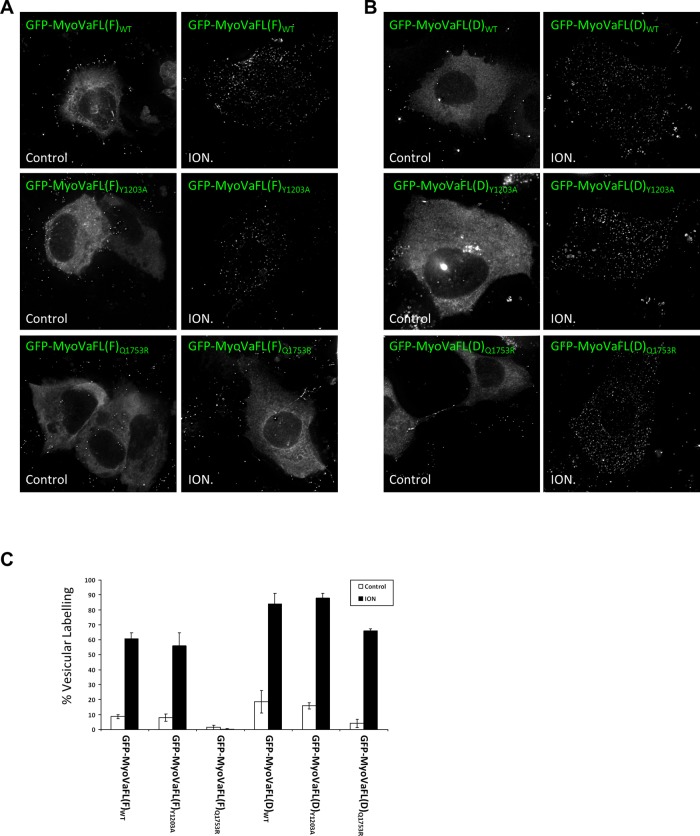
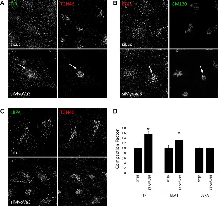
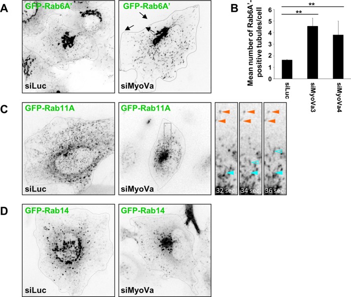
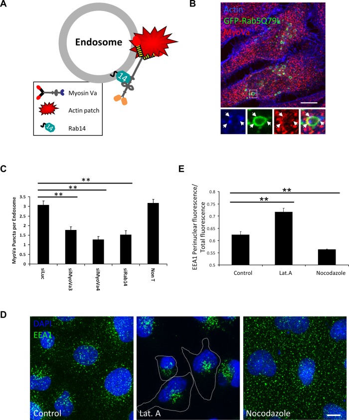
Myosin Va is a widely expressed actin-based motor protein that binds members of the Rab GTPase family (3A, 8A, 10, 11A, 27A) and is implicated in many intracellular trafficking processes. To our knowledge, myosin Va has not been tested in a systematic screen for interactions with the entire Rab GTPase family. To that end, we report a yeast two-hybrid screen of all human Rabs for myosin Va-binding ability and reveal 10 novel interactions (3B, 3C, 3D, 6A, 6A', 6B, 11B, 14, 25, 39B), which include interactions with three new Rab subfamilies (Rab6, Rab14, Rab39B). Of interest, myosin Va interacts with only a subset of the Rabs associated with the endocytic recycling and post-Golgi secretory systems. We demonstrate that myosin Va has three distinct Rab-binding domains on disparate regions of the motor (central stalk, an alternatively spliced exon, and the globular tail). Although the total pool of myosin Va is shared by several Rabs, Rab10 and Rab11 appear to be the major determinants of its recruitment to intracellular membranes. We also present evidence that myosin Va is necessary for maintaining a peripheral distribution of Rab11- and Rab14-positive endosomes.


Figures

FIGURE 1:
FIGURE 1:
Myosin Va interacts with multiple Rab GTPases. (A) Schematic diagram depicting the extended “active” structure of myosin Va. The location of the alternatively spliced exons B, D, and F are indicated. GTD, globular-tail domain; MD, motor domain. (B) HeLa cells expressing dominant-active (DA) or dominant-negative (DN) mutants of the indicated Rab GTPases fused to GFP were lysed, and the fusion proteins were immunoprecipitated with an anti-GFP antibody. The ability of myosin Va to form a complex with these Rabs was tested by Western blot analysis using anti-myosin Va antibody. Input represents 10% of the starting material used in all conditions. (C) Regions of the myosin Va tail tested using the yeast two-hybrid technique. The constitutively active mutant of each Rab GTPase was used. CC, coiled coil; GTD, globular tail domain. (D) Identification of a critical amino acid for binding to Rab6 and Rab14. ClustalW alignment of the Rab6/Rab14-binding regions of human myosin Va and myosin Vb. Arrows indicate the conserved amino acids that were mutated to alanine, and the red arrow indicates tyrosine 1203, which, when mutated, abolishes the Rab6 and Rab14 interaction. The table indicates the strength of interaction of each mutant with constitutively active Rab6A, Rab11A, and Rab14 using the yeast two-hybrid HIS3 and LacZ reporter assays. (E) Identification of critical amino acids that mediate binding to Rab3A, Rab11A, and Rab39B. The amino acids in myosin Va that correspond to those identified by Lipatova et al. (2008) that mediate the binding of Myo2 to Ypt31/32 were mutated and the mutants tested for interaction with the constitutively active mutants of the indicated Rab GTPases by yeast two-hybrid assay. (F) Schematic diagram of myosin Va indicating the identified Rab-binding domains. Asterisk indicates the location of the Y1203A mutation; double asterisks indicate the location of the Q1753R mutation.
FIGURE 2:
FIGURE 2:
Characterization of the intracellular localization of myosin Va. (A) A431 (epidermoid carcinoma) cells were separated into cytosol and membrane fractions in the presence of CaCl2, EGTA, or CaCl2 and 1% TX-100. The presence of myosin Va in these fractions was determined by Western blot analyses using anti–myosin Va antibody. The distribution of TfR and β-tubulin was used to determine the accuracy of the fractionation procedure. (B) A431 cells stably expressing GFP-MyoVaFL(D)WT or GFP-MyoVaFL(F)(WT) were treated with 5 μM ionomycin for 4 min before fixation, permeabilization, and labeling with antibodies to the indicated endogenous Rab GTPases. Insets contain zoomed images of the boxed regions. (C, E, F) Quantitative analysis of the colocalization coefficients of each GFP–myosin Va splice variant with the indicated Rab GTPase or organelle marker (mean ± SEM; n = 15–20 cells). (D) SH-SY5Y cells expressing the indicated GFP–myosin Va splice variant treated with 5 μM ionomycin for 5 min before fixation and labeling with an antibody that detects endogenous Rab39B. Each immunofluorescence image is a single confocal section. Bar, 10 μm.
FIGURE 3:
FIGURE 3:
Myosin Va is recruited to membranes by Rab10 and Rab11. HeLa cells expressing GFP-MyoVaTail(F)WT (A) or GFP-MyoVaTail(D)WT (B) or their mutants were fixed and imaged by fluorescence deconvolution microscopy. HeLa cells were transfected for 72 h with control siRNA or siRNA targeting the indicated Rab GTPase, and GFP-MyoVaTail(F)WT (C), GFP-MyoVaTail(D)WT (D), or GFP-MyoVaTail(D)Q1753R (E) was transfected into these cells for the final 18 h. The cells were fixed and the nuclei labeled with DAPI and imaged by fluorescence deconvolution microscopy. (See Figure S5D for Western blots analyzing the knockdown efficiency of the siRNA duplexes.)
FIGURE 4:
FIGURE 4:
Expression of the myosin Va tail induces the aggregation of Rab-positive vesicles. (A) HeLa cells expressing the indicated myosin Va tail fused to GFP were fixed and labeled with antibodies that detect endogenous Rab GTPases. Insets show zoomed images of the boxed areas. (B) Quantitative analysis of the colocalization coefficients of each of the GFP–myosin Va tail constructs in HeLa cells with the indicated Rab GTPase (mean ± SEM; n = 20–40 cells).
FIGURE 5:
FIGURE 5:
Calcium is required for membrane binding of myosin Va. GFP-MyoVaFL(F)WT (A) or GFP-MyoVaFL(D)WT (B) and their Y1203A or Q1753R mutants were treated with 5 μM ionomycin, or solvent alone, for 4 min at 37°C. The cells were then fixed immediately and processed for fluorescence microscopy. (C) Percentage of cells displaying vesicular labeling of the GFP–myosin Va fusion proteins for each condition (mean ± SEM; n = 150 cells from three independent experiments).
FIGURE 6:
FIGURE 6:
Myosin Va depletion induces perinuclear clustering of endocytic recycling pathway membrane compartments. HeLa cells transfected with control siRNA or myosin Va siRNA for 72 h were fixed, permeabilized, and colabeled with antibodies to the TfR and TGN46 (A), EEA1 and GM130 (B), or LBPA and TGN46 (C). (D) Quantification of the fluorescence intensity of TfR, EEA1, or LBPA in the pericentrosomal region relative to the total cellular fluorescence intensity and expressed as a “compaction factor.” Data were normalized to the control (mean ± SEM; n = 30–50 cells). *p <10−4, Student's t test.
FIGURE 7:
FIGURE 7:
Myosin Va depletion affects Rab motility and organelle distribution. (A) A HeLa cell line stably expressing GFP-Rab6A′ was transfected with control or myosin Va siRNA for 72 h. Two-minute time-lapse movies were recorded on a spinning disk confocal microscope. Representative frames from Supplemental Movie S1. Black arrows indicate tubules emerging from the Golgi complex. (B) Quantification of the number of Rab6A′-positive tubules connected to the Golgi for each condition (mean ± SEM; n = 11–15 cells). Two independent siRNAs targeting myosin Va were used. **p < 10−3. (C) HeLa cells were transfected with control or myosin Va siRNA for 72 h and plasmid DNA encoding GFP-Rab11A for the final 18 h. Two-minute time-lapse movies were recorded on a spinning disk confocal microscope. Representative frames from Supplemental Movie S2. Insets depict zoomed images of the boxed region. Indicated are examples of immobile vesicles (orange arrowheads) and rapidly moving vesicles (blue arrowheads). Closed arrowheads indicate the position of the vesicle in the first frame (recorded at 32 s), and the open arrowheads indicate the position of the same vesicle in the given frame. (D) HeLa cells transfected with control or myosin Va siRNA for 72 h and plasmid DNA encoding GFP-Rab14 for the final 18 h. Two-minute time-lapse movies were recorded on a spinning disk confocal microscope. Representative frames from Supplemental Movie S3.
FIGURE 8:
FIGURE 8:
Endosomes are tethered to the actin cytoskeleton by Rab14 and myosin Va. (A) Model depicting the tethering of endosomes to the actin cytoskeleton by myosin Va, which is associated with the endosomal membrane via a direct interaction with Rab14. (B) HeLa cells expressing GFP-Rab5Q79L for ∼24 h were fixed and labeled with coumarin–phalloidin and an antibody to myosin Va. Inset contains a zoomed image of the indicated enlarged endosome. (C) Quantification of the number of endogenous myosin Va puncta on GFP-Rab5AQ79L enlarged endosomes in cells transfected with the indicated siRNA for 72 h (mean ± SEM; n ≥ 60 endosomes per condition). **p <10−3. (D) A431 cells treated with solvent alone, 0.5 μM latrunculin A, or 20 μM nocodazole for 1 h at 37°C before fixation and labeling with anti-EEA1 (green) and DAPI (blue). (E) The fluorescence intensity in the perinuclear region of the cell was expressed as a ratio of the total cellular fluorescence using the Radial Profile plug-in in ImageJ (mean ± SEM; n ≥ 100 cells per condition). **p <10−3.

Similar articles

Cited by

References

    1. Chen W, Feng Y, Chen D, Wandinger-Ness A. Rab11 is required for trans-Golgi network-to-plasma membrane transport and a preferential target for GDP dissociation inhibitor. Mol Biol Cell. 1998;9:3241–3257. - PMC - PubMed
    1. Darchen F, Goud B. Multiple aspects of Rab protein action in the secretory pathway: focus on Rab3 and Rab6. Biochimie. 2000;82:375–384. - PubMed
    1. Delevoye C, et al. AP-1 and KIF13A coordinate endosomal sorting and positioning during melanosome biogenesis. J Cell Biol. 2009;187:247–264. - PMC - PubMed
    1. Desnos C, et al. Rab27A and its effector MyRIP link secretory granules to F-actin and control their motion towards release sites. J Cell Biol. 2003;163:559–570. - PMC - PubMed
    1. English AR, Voeltz GK. Rab10 GTPase regulates ER dynamics and morphology. Nat Cell Biol. 2013;15:169–178. - PMC - PubMed

Publication types

MeSH terms

LinkOut - more resources