. 2013 Nov 1;383(1):90-105.
doi: 10.1016/j.ydbio.2013.08.015. Epub 2013 Aug 29.

Sost and its paralog Sostdc1 coordinate digit number in a Gli3-dependent manner

Affiliations

Sost and its paralog Sostdc1 coordinate digit number in a Gli3-dependent manner

Nicole M Collette et al. Dev Biol. .

Abstract

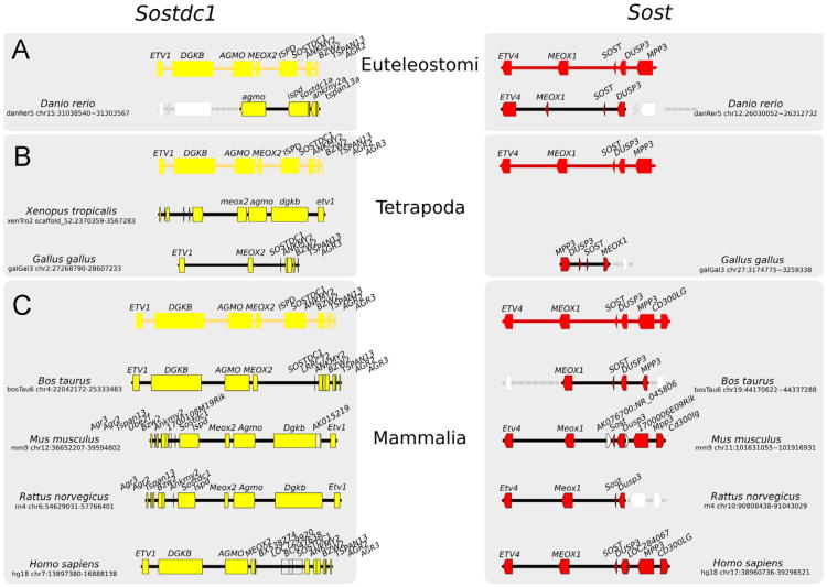
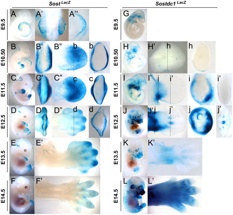
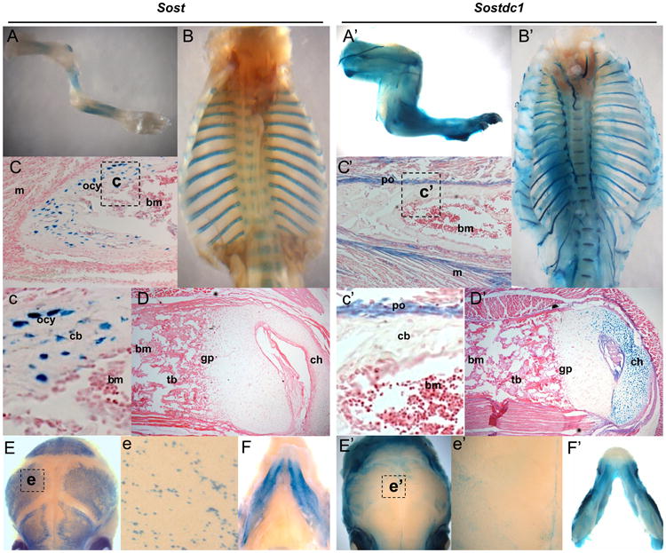
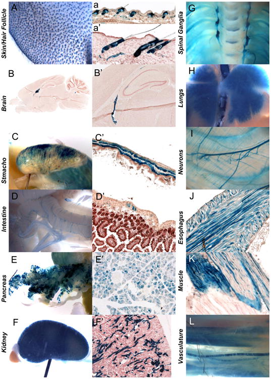
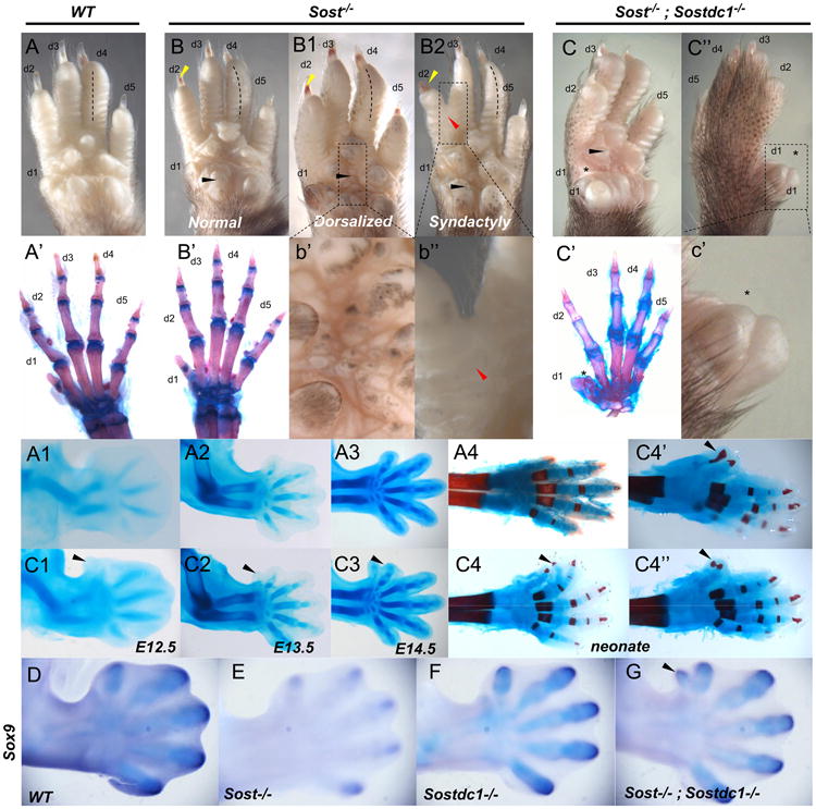
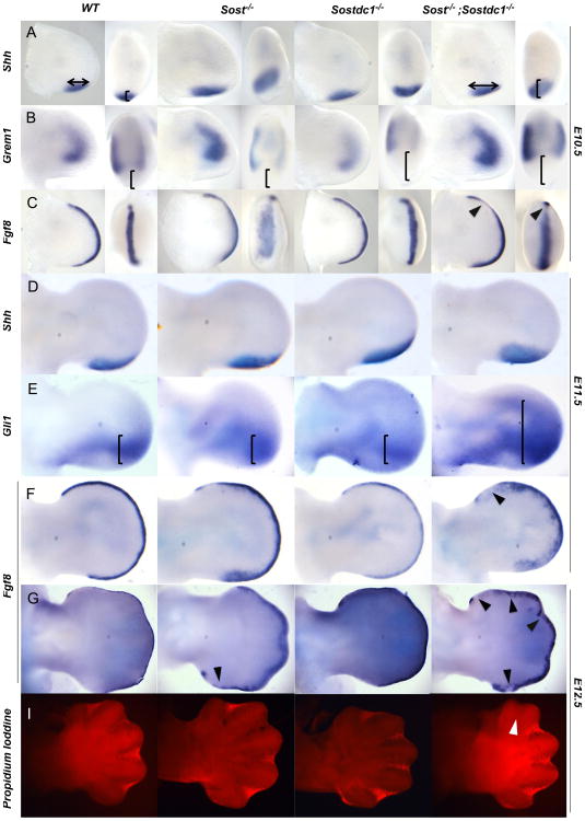
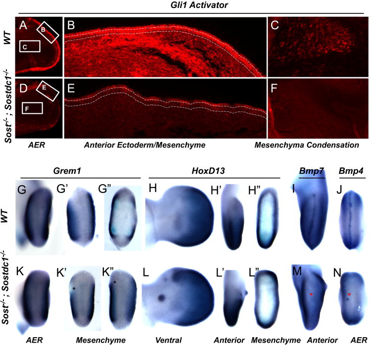
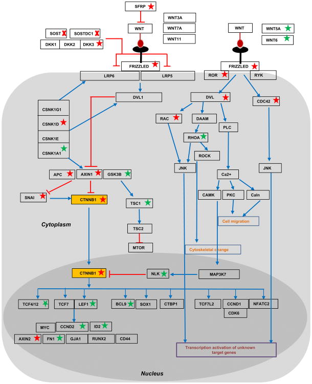
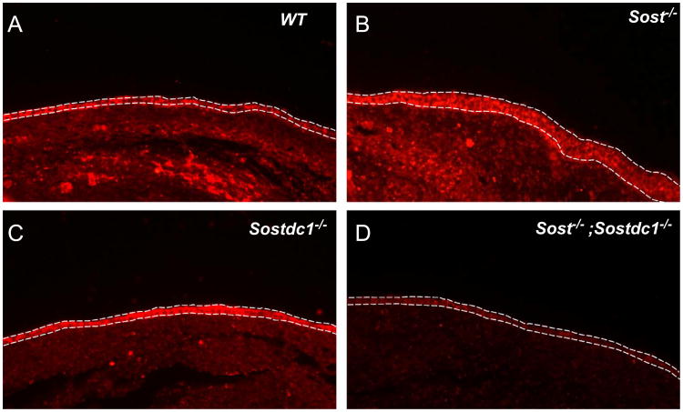
WNT signaling is critical in most aspects of skeletal development and homeostasis, and antagonists of WNT signaling are emerging as key regulatory proteins with great promise as therapeutic agents for bone disorders. Here we show that Sost and its paralog Sostdc1 emerged through ancestral genome duplication and their expression patterns have diverged to delineate non-overlapping domains in most organ systems including musculoskeletal, cardiovascular, nervous, digestive, reproductive and respiratory. In the developing limb, Sost and Sostdc1 display dynamic expression patterns with Sost being restricted to the distal ectoderm and Sostdc1 to the proximal ectoderm and the mesenchyme. While Sostdc1(-/-) mice lack any obvious limb or skeletal defects, Sost(-/-) mice recapitulate the hand defects described for Sclerosteosis patients. However, elevated WNT signaling in Sost(-/-); Sostdc1(-/-) mice causes misregulation of SHH signaling, ectopic activation of Sox9 in the digit 1 field and preaxial polydactyly in a Gli1- and Gli3-dependent manner. In addition, we show that the syndactyly documented in Sclerosteosis is present in both Sost(-/-) and Sost(-/-); Sostdc1(-/-) mice, and is driven by misregulation of Fgf8 in the AER, a region lacking Sost and Sostdc1 expression. This study highlights the complexity of WNT signaling in skeletal biology and disease and emphasizes how redundant mechanism and non-cell autonomous effects can synergize to unveil new intricate phenotypes caused by elevated WNT signaling.

Keywords: Limb formation; Polydactyly; Sclerostin; Shh; Sost; Sostdc1; WNT signaling; syndactyly.


Figures

Fig. 1
Fig. 1
Sost–Sostdc1 evolutionary relationship. An overview of SOSTDC1 (left) and SOST (right) evolution, created by tracking the SOSTDC1 and SOST gene loci through representative vertebrate genomes (not to scale) from (A) Euteleostomi, (B) Tetrapoda and (C) Mammals clades. Predicted orthologs (where annotation is not available) are shown with gray dotted lines. Genes are shown as arrowheads, with their gene symbols above. Genes that are not conserved/likely to be poorly annotated are represented in white. The direction of the arrowhead indicates the relative transcriptional orientation and the relevant genome coordinates indicated on the left and right respectively.
Fig. 2
Fig. 2
Sost and Sostdc1 expression during limb development visualized by LacZ expression. Sost (A–F) and Sostdc1 (G–L) expressions were examined in a time-course panel of E9.5–E14.5 heterozygous embryos referred to as SostLacZ and Sostdc1LacZ. At E9.5 in SostLacZ embryos, a dorsal view of the whole embryo (A′) and of the forelimb (A″) shows expression in the emerging limb bud while no limb expression is detected in Sostdc1LacZ (G). For E10.5 to E12.5 embryos (B–D and H–J), AER views (B′–D′), dorsal limb views (B″–D″ and H′–J′) and transverse section views (b–d; h–j; and i′, j′) are provided. For E13.5 and E14.5 embryos (E–F; K–L), dorsal limb views (E′–F′; K′–L′) are provided.
Fig. 3
Fig. 3
Sost and Sostdc1 expression in the neonatal skeleton. Sost expression marked cells in the appendicular (A) and axial (B, E) skeleton, while Sostdc1 was more broadly expressed in the limbs (A′) and rib cage (B′) to encompass connective tissue, muscle, cartilage and neurons. Sectioned long bones revealed Sost expression primarily in the osteocytes of cortical bone (C and c), but no obvious Sost expression was detected in the articular cartilage (D). Sostdc1 however was not detected in the mineralized bone; it was expressed in the periosteum (C and c′), the immediately adjacent muscles (C′) and the periarticular chondrocytes in the condyle (D′). Both Sost and Sostdc1 were also detected in the skull (E and E′) and mandible (F and F′); Sost expression was localized to osteoblasts and osteocytes in wholemount calvaria (e), while Sostdc1 was present in the connective tissue over the calvarial bones (e′); and m muscle; ocy osteocytes; bm bone marrow; po periosteum; cb cortical bone; gp growth plate; and ch chondrocytes.
Fig. 4
Fig. 4
Sostdc1 expression in adult tissues. Sostdc1 expression was examined in wholemount and sectioned LacZ stained tissues, and was detected in the skin and hair follicles (A, a, and a′). A highly specialized region in the brain was positive for Sostdc1 (B and B′). Smooth muscles of the stomach (C and C′), intestine (D and D′) and esophagus (J), and skeletal muscle (K) expressed Sostdc1. Sostdc1 was also robustly expressed in the pancreas (E and E′) and kidney (F and F′) and in the nervous system Sostdc1 stained spinal ganglia (G) and the lungs (H). Neurons (I) and vasculature was also positive for Sostdc1 (L).
Fig. 5
Fig. 5
Limb defects in Sost–/– and Sost–/–; Sostdc1–/– mice. Compared to adult WT autopods (A), Sost–/– autopods (B, B1, and B2, insets at b′ and b″) displayed pigmentation on the ventral side (B1 and b′), digit 2–3 syndactyly (B2 and b″; red arrow), nail dysplasia (B, B1 and B2; yellow arrows) and radial deviation of digits, primarily observed for digit 4 (B, B1 and B2, dotted lines). Ventral pigmentation was also observed in Sost–/–; Sostdc1–/– autopods. Unlike WT and Sost–/– autopods that had normal digit patterning (A′–B′), Sost–/–; Sostdc1–/– digit 1 was thicker (C, C″ and c′; asterisk) and skeletal preparation indicated the presence of extra bones (C′) in digit 1. A time course skeletal preparation examination revealed that an ectopic digit 1 was distinguishable as a tissue projection as early as E12.5 (A1-3 vs. C1-3); and the neonate Sost–/–; Sostdc1–/– limbs displayed a range of extra digits (C4 and C4″) associated with ectopic projections primarily from digit 1 (C4′ and C4″) and in rare occasions from digit 2 (C4; black arrow). Sox9 in situ hybridization on E13 embryos revealed an ectopic digit 1 field in Sost–/–; Sostdc1–/– autopods. d digit.
Fig. 6
Fig. 6
Altered SHH and FGF signaling causes polydactyly and syndactyly. Shh domain was expanded along the anterior–posterior and proximal–distal axis in Sost–/– and Sost–/–; Sostdc1–/– relative to WT and Sostdc1–/– limb buds at E10.5 (A, arrows, bracket) and E1 1.5 (D). Downstream of Shh, Gli1 expression was dramatically expanded in Sost–/–; Sostdc1–/– E11.5 limb buds relative to all other genotypes (E, brackets). Grem1 expression was absent in Shh positive region, and in Sostdc1–/– and Sost–/–; Sostdc1–/– limbs the Grem1 domain was reduced on the posterior side (B, brackets). Fgf8 AER expression domain was expanded and disorganized in Sost–/– and Sost–/–; Sostdc1–/– limbs, and on the anterior side of the Sost–/–; Sostdc1–/– limbs Fgf8 expression was reduced in all time points examined (B and F–G, arrows). A reduction in interdigital apoptosis was also detected on the anterior side of Sost–/–; Sostdc1–/– limb buds at E12.5 (I, arrow).
Fig. 7
Fig. 7
Gli3, Grem1, HoxD13, Bmp4 and Bmp7 expression is affected in Sost–/–; Sostdc1–/– E11.5 limbs. Consistent with a reduction in mRNA expression of Gli3, Gli3 activator protein expression was dramatically reduced in the ectoderm of Sost–/–; Sostdc1–/– E11.5 limbs (A and D). Higher magnification images of the anterior region of the limb showed a dramatic reduction in Gli3 both in the ectoderm (marked by dashed lines) and the underlying mesenchyme (B and E). Similarly, the pre-chondrocytes in the cartilage condensation stained positive for Gli3 in the WT limbs, but had little expression in the double knockouts (F). Grem1 expression was reduced in the anterior mesenchyme in double knockout (K′–K″) relative to WT limbs (G′–G″). Asterisks indicate region of lost anterior expression. HoxD13 was ectopically up-regulated in the anterior mesenchyme in the regions corresponding to digit 1 (L, L′, and L″; green arrows) and on the ventral side of the autopod in an ectodermal nubbin (L′ green arrow). Both Bmp4 and Bmp7 expression was absent from the AER (M and N; red asterisks). Views are indicated at the bottom of the figure.
Fig. 8
Fig. 8
Transcriptional changes in the WNT signaling pathway of E11.5 Sost–/–; Sostdc1–/– limbs. Genes found to be transcriptionally up- or down- regulated by more than 2-fold in Sost–/–;Sostdc1–/– limb buds are marked by a green or red star, respectively. Red arrows mark inhibitory relationships, and blue arrows mark other relationships such as transcriptional up-regulation or protein stabilization. β-catenin (CTNNB1) was found to be down-regulated (yellow box).
Fig. 9
Fig. 9
Activated β-catenin is dramatically reduced in Sost–/–; Sostdc1–/– limbs. Lack of Sost up-regulates WNT signaling as evidenced by increased staining for activated β-catenin in the ectoderm (marked by dashed lines) and underlining mesenchyme (B). Sost–/– ectoderm also appears thicker than all other genotypes; lack of Sostdc1 has little effect on ectodermal β-catenin (C), but causes a slight reduction in the mesenchyme; removing both Sost and Sostdc1 dramatically reduces both ectodermal and mesenchymal activated β-catenin protein (D).

Similar articles

Cited by

References

    1. Ahn Y, Sanderson BW, Klein OD, Krumlauf R. Inhibition of Wnt signaling by Wise (Sostdc1) and negative feedback from Shh controls tooth number and patterning. Development. 2010;137:3221–3231. - PMC - PubMed
    1. Akiyama H, Stadler HS, Martin JF, Ishii TM, Beachy PA, Nakamura T, de Crombrugghe B. Misexpression of Sox9 in mouse limb bud mesenchyme induces polydactyly and rescues hypodactyly mice. Matrix Biol: J Int Soc Matrix Biol. 2007;26:224–233. - PubMed
    1. Altenhoff AM, Dessimoz C. Phylogenetic and functional assessment of orthologs inference projects and methods. PLoS Comput Biol. 2009;5:e1000262. - PMC - PubMed
    1. Balemans W, Ebeling M, Patel N, Van Hul E, Olson P, Dioszegi M, Lacza C, Wuyts W, Van Den Ende J, Willems P, Paes-Alves AF, Hill S, Bueno M, Ramos FJ, Tacconi P, Dikkers FG, Stratakis C, Lindpaintner K, Vickery B, Foernzler D, Van Hul W. Increased bone density in sclerosteosis is due to the deficiency of a novel secreted protein (SOST) Hum Mol Genet. 2001;10:537–543. - PubMed
    1. Bandyopadhyay A, Tsuji K, Cox K, Harfe BD, Rosen V, Tabin CJ. Genetic analysis of the roles of BMP2, BMP4, and BMP7 in limb patterning and skeletogenesis. PLoS Genet. 2006;2:e216. - PMC - PubMed

Publication types

MeSH terms