Alteration of cancer stem cell-like phenotype by histone deacetylase inhibitors in squamous cell carcinoma of the head and neck
- PMID: 23992541
- PMCID: PMC7654248
- DOI: 10.1111/cas.12271
Alteration of cancer stem cell-like phenotype by histone deacetylase inhibitors in squamous cell carcinoma of the head and neck
Abstract
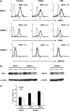
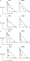
Recent progression in the understanding of stem cell biology has greatly facilitated the identification and characterization of cancer stem cells (CSCs). Moreover, evidence has accumulated indicating that conventional cancer treatments are potentially ineffective against CSCs. Histone deacetylase inhibitors (HDACi) have multiple biologic effects consequent to alterations in the patterns of acetylation of histones and are a promising new group of anticancer agents. In this study, we investigated the effects of two HDACi, suberoylanilide hydroxamic acid (SAHA) and trichostatin A (TSA), on two CD44+ cancer stem-like cell lines from squamous cell carcinoma of the head and neck (SCCHN) cultured in serum-free medium containing epidermal growth factor and basic fibroblast growth factor. Histone deacetylase inhibitors inhibited the growth of SCCHN cell lines in a dose-dependent manner as measured by MTS assays. Moreover, HDACi induced cell cycle arrest and apoptosis in these SCCHN cell lines. Interestingly, the expression of cancer stem cell markers, CD44 and ABCG2, on SCCHN cell lines was decreased by HDACi treatment. In addition, HDACi decreased mRNA expression levels of stemness-related genes and suppressed the epithelial-mesencymal transition phenotype of CSCs. As expected, the combination of HDACi and chemotherapeutic agents, including cisplatin and docetaxel, had a synergistic effect on SCCHN cell lines. Taken together, our data indicate that HDACi not only inhibit the growth of SCCHN cell lines by inducing apoptosis and cell cycle arrest, but also alter the cancer stem cell phenotype in SCCHN, raising the possibility that HDACi may have therapeutic potential for cancer stem cells of SCCHN.
© 2013 Japanese Cancer Association.
Figures


Similar articles
-
Expansion and characterization of cancer stem-like cells in squamous cell carcinoma of the head and neck.Oral Oncol. 2009 Jul;45(7):633-9. doi: 10.1016/j.oraloncology.2008.10.003. Epub 2008 Nov 21. Oral Oncol. 2009. PMID: 19027347
-
Carfilzomib and oprozomib synergize with histone deacetylase inhibitors in head and neck squamous cell carcinoma models of acquired resistance to proteasome inhibitors.Cancer Biol Ther. 2014 Sep;15(9):1142-52. doi: 10.4161/cbt.29452. Epub 2014 Jun 10. Cancer Biol Ther. 2014. PMID: 24915039 Free PMC article.
-
The histone deacetylase inhibitor trichostatin a decreases lymphangiogenesis by inducing apoptosis and cell cycle arrest via p21-dependent pathways.BMC Cancer. 2016 Sep 30;16(1):763. doi: 10.1186/s12885-016-2807-y. BMC Cancer. 2016. PMID: 27716272 Free PMC article.
-
Histone deacetylase inhibitors in cancer therapy.Curr Opin Oncol. 2008 Nov;20(6):639-49. doi: 10.1097/CCO.0b013e3283127095. Curr Opin Oncol. 2008. PMID: 18841045 Free PMC article. Review.
-
Histone deacetylase inhibitors: potential targets responsible for their anti-cancer effect.Invest New Drugs. 2010 Dec;28 Suppl 1(Suppl 1):S3-20. doi: 10.1007/s10637-010-9596-y. Epub 2010 Dec 14. Invest New Drugs. 2010. PMID: 21161327 Free PMC article. Review.
Cited by
-
Sensitization of chemo-resistant human chronic myeloid leukemia stem-like cells to Hsp90 inhibitor by SIRT1 inhibition.Int J Biol Sci. 2015 Jun 11;11(8):923-34. doi: 10.7150/ijbs.10896. eCollection 2015. Int J Biol Sci. 2015. PMID: 26157347 Free PMC article.
-
Lurbinectedin (PM01183), a selective inhibitor of active transcription, effectively eliminates both cancer cells and cancer stem cells in preclinical models of uterine cervical cancer.Invest New Drugs. 2019 Oct;37(5):818-827. doi: 10.1007/s10637-018-0686-6. Epub 2018 Oct 30. Invest New Drugs. 2019. PMID: 30374654
-
Targeting Head and Neck Cancer Stem Cells: Current Advances and Future Challenges.J Dent Res. 2015 Nov;94(11):1516-23. doi: 10.1177/0022034515601960. Epub 2015 Aug 25. J Dent Res. 2015. PMID: 26307039 Free PMC article. Review.
-
HPV+ve/-ve oral-tongue cancer stem cells: A potential target for relapse-free therapy.Transl Oncol. 2021 Jan;14(1):100919. doi: 10.1016/j.tranon.2020.100919. Epub 2020 Oct 24. Transl Oncol. 2021. PMID: 33129107 Free PMC article. Review.
-
Identification of a cancer stem cell-specific function for the histone deacetylases, HDAC1 and HDAC7, in breast and ovarian cancer.Oncogene. 2017 Mar 23;36(12):1707-1720. doi: 10.1038/onc.2016.337. Epub 2016 Oct 3. Oncogene. 2017. PMID: 27694895 Free PMC article.
References
-
- Clevers H. The cancer stem cell: premises, promises and challenges. Nat Med 2011; 17: 313–9. - PubMed
-
- Shackleton M, Quintana E, Fearon ER, Morrison SJ. Heterogeneity in cancer: cancer stem cells versus clonal evolution. Cell 2009; 138: 822–9. - PubMed
-
- Collins AT, Berry PA, Hyde C, Stower MJ, Maitland NJ. Prospective identification of tumorigenic prostate cancer stem cells. Cancer Res 2005; 65: 10. - PubMed
Publication types
MeSH terms
Substances
LinkOut - more resources
Full Text Sources
Other Literature Sources
Research Materials
Miscellaneous

