. 2013 Aug 19;8(8):e72281.
doi: 10.1371/journal.pone.0072281. eCollection 2013.

Human glioblastoma multiforme: p53 reactivation by a novel MDM2 inhibitor

Affiliations

Human glioblastoma multiforme: p53 reactivation by a novel MDM2 inhibitor

Barbara Costa et al. PLoS One. .

Abstract

Cancer development and chemo-resistance are often due to impaired functioning of the p53 tumor suppressor through genetic mutation or sequestration by other proteins. In glioblastoma multiforme (GBM), p53 availability is frequently reduced because it binds to the Murine Double Minute-2 (MDM2) oncoprotein, which accumulates at high concentrations in tumor cells. The use of MDM2 inhibitors that interfere with the binding of p53 and MDM2 has become a valid approach to inhibit cell growth in a number of cancers; however little is known about the efficacy of these inhibitors in GBM. We report that a new small-molecule inhibitor of MDM2 with a spirooxoindolepyrrolidine core structure, named ISA27, effectively reactivated p53 function and inhibited human GBM cell growth in vitro by inducing cell cycle arrest and apoptosis. In immunoincompetent BALB/c nude mice bearing a human GBM xenograft, the administration of ISA27 in vivo activated p53, inhibited cell proliferation and induced apoptosis in tumor tissue. Significantly, ISA27 was non-toxic in an in vitro normal human cell model and an in vivo mouse model. ISA27 administration in combination with temozolomide (TMZ) produced a synergistic inhibitory effect on GBM cell viability in vitro, suggesting the possibility of lowering the dose of TMZ used in the treatment of GBM. In conclusion, our data show that ISA27 releases the powerful antitumor capacities of p53 in GBM cells. The use of this MDM2 inhibitor could become a novel therapy for the treatment of GBM patients.


Conflict of interest statement

Competing Interests: The authors have declared that no competing interests exist.

Figures

Figure 1
Figure 1. ISA27 inhibits the growth/survival of GBM cell lines expressing wild-type p53.
GBM cells with wild-type p53 (U87MG, U343MG) were exposed to a range of ISA27 or Nutlin-3 concentrations for 24 h. After incubation, GBM cell viability was determined by MTS assay. A and B) Effect of ISA27 on the growth/survival of GBM cell lines expressing wild-type p53: curves for U87MG and U343MG cell samples were generated using a sigmoidal dose-response curve model (GraphPad Prism 4 software) from which the IC50 values were derived. ISA27-treated cells (▾), Nutlin-3-treated cells (▪); C) Effect of ISA27 on the growth/survival of the GBM T98G cell line expressing mutant p53: T98G cells were incubated with increasing concentrations of ISA27 or Nutlin-3 (ranging from 1 µM to 25 µM), and cell viability was measured after 24 h by MTS assay. ISA27-treated cells (), Nutlin-3-treated cells (). The results are expressed as the percentages of cell viability with respect to the vehicle-treated sample for each cell line; the viability of the vehicle-treated sample was arbitrarily set at 100%. Points, means of three independent experiments performed in duplicate; bars, SEM.
Figure 2
Figure 2. ISA27 increases p53 protein levels in GBM cell lines.
GBM cells (U87MG, U343MG) were treated with ISA27 or Nutlin-3 for the indicated incubation times, and protein levels of p53 were analysed in whole-cell lysates by Western blot. One representative Western blot is presented (upper panel) for each cell line. The blots show that the antibody to p53 (FL-393; Santa Cruz Biotechnology) recognised a single specific band at approximately 55 kDa, corresponding to the molecular weight of the p53 protein; β-actin is used as the loading control. Densitometric analyses of Western blots (lower panel) demonstrated that ISA27 induced a significant enhancement of p53 protein levels in U87MG or U343MG cells with a maximal effect at the 8 h incubation time.
Figure 3
Figure 3. ISA27 induces the stabilisation of p53 protein levels by decreasing the interaction of p53 with MDM2 in GBM cells.
A and B) Effect of ISA27 on p53 gene expression: U87MG and U343MG cells were treated with ISA27 or Nutlin-3 for 6, 12 and 24 h. After incubation, the relative quantification of p53 mRNA was performed by real-time RT-PCR. In U87MG cells, p53 mRNA levels did not change within 12 h of cell treatment; in U343 MG cells, p53 mRNA levels did not change during any of the incubation times. 3C) ISA27 sustains p53 protein expression in the presence of CHX: U87MG cells were incubated with 50 µM CHX alone or CHX in combination with ISA27 or Nutlin-3 for 0.5, 1, 2, 4 and 6 h. After incubation, cell lysates were used for Western blot analysis. One representative Western blot is presented for each cell treatment. The inhibition of protein synthesis by treatment with CHX alone blocked p53 protein expression after a 4-h treatment. ISA27 extended the stability of p53 in the presence of CHX. 3D) ISA27 reduces MDM2-p53 complex formation: U87MG cells were incubated with ISA27 or Nutlin-3 for 8 h followed by immunoprecipitation using an anti-MDM2 antibody. The MDM2-p53 complex and the relative input of the proteins were detected by immunoblot; a significant reduction in MDM2-p53 complex formation was shown in U87MG cells following 8 h of treatment with ISA27.
Figure 4
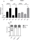
Figure 4. ISA27 restores p53 function in GBM cells.
4A) ISA27 induces p53 target gene expression: Real-time PCR analyses showed a statistically significant increase in MDM2 and p21 mRNA levels following short-term ISA27 treatment in U87MG cells. 4B) ISA27 induces cell cycle arrest: Cell cycle analysis (24 h) revealed a statistically significant increase in the G1 fraction and a nearly complete depletion of the S-phase population in short-term-treated U87MG cells.
Figure 5
Figure 5. Long-term ISA27 treatment of U87MG cells.
U87MG cells were treated with 2.5 µM ISA27 or 10 µM Nutlin-3 for 24, 48, 72, 96 and 120 h. The figure shows the viable cells that were counted by Trypan blue exclusion at the indicated times. The percentage of ISA27- () or Nutlin-3- () treated viable cells was calculated with respect to untreated viable cells at the indicated incubation times. Points, means of three independent experiments performed in duplicate; bars, SEM.
Figure 6
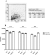
Figure 6. Lack of ISA27 toxicity in an in vitro human cell model (lymphomonocytes).
6A) Scatter cytogram analysis of cell populations from peripheral blood: Scatter cytogram analysis showing cell separation by size and granularity (G2 = lymphocytes; G3 = monocytes). One representative experiment is presented. 6B) Time-response of human lymphomonocyte viability following ISA27 treatment: lymphomonocytes were treated with 2.5 µM ISA27 or 10 µM Nutlin-3 for 12, 24 and 48 h. The figure shows the viable cells and dead cells counted by Trypan blue exclusion at the indicated times. Data represent the means of three independent experiments performed in duplicate; bars, SEM. No significant differences were observed between MDM2 inhibitor-treated and untreated-control viable or dead cells at each incubation time.
Figure 7
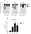
Figure 7. Long-term ISA27 treatment induces cell cycle arrest and a persistent increase in p21 mRNA levels in U87MG cells.
7A) Flow cytometric cell cycle profiling: the figure shows the percentage of untreated and MDM2 inhibitor-treated U87MG cells in G1-, S- and G2/M-phase. 7B) p21 mRNA evaluation: ISA27 treatment resulted in a statistically significant increase in p21 mRNA levels at 24, 48 and 72 h.
Figure 8
Figure 8. ISA27 induces U87MG cell senescence.
8A) Representative images of SA-β-Gal-expressing cells: The panel shows the SA-β-Gal-expressing MDM2 inhibitor-treated and untreated cells at 72 h. 8B) Percentage of SA-β-Gal-expressing cells: The number of SA-β-Gal-expressing cells was determined with respect to the total cells in each sample (untreated cells, ISA27- and Nutlin-3-treated cells). The percentage of SA-β-Gal-expressing MDM2 inhibitor-treated cells was then calculated with respect to the untreated cells, for which an arbitrary value of 100% was assigned.
Figure 9
Figure 9. U87MG cells did not recover normal growth upon removal of ISA27.
U87MG cells were cultured in the presence of 2.5 µM ISA27 or 10 µM Nutlin-3. After 4 days of MDM2 inhibitor treatment, cell culture medium was replaced with fresh culture medium without the MDM2 inhibitor. The figure shows the number of viable cells that were counted by Trypan blue exclusion after 4 days of MDM2 inhibitor treatment and 1, 2 and 3 days after MDM2 inhibitor removal. Data represent the means of three independent experiments performed in duplicate; bars, SEM.
Figure 10
Figure 10. ISA27 induces the dissipation of the mitochondrial membrane potential.
10A) Representative dot plots of untreated and MDM2 inhibitor-treated cells: After ISA27 treatment, mitochondrial depolarisation is visible by a decrease and an increase in fluorescence in the FL-2 and FL-1 channels, respectively. After Nutlin-3 treatment, mitochondrial depolarisation is not visible; CCCP is the positive control. 10B and C) Time-course analysis of ΔΨm dissipation: Histograms show the mean values of cell percentages either in the UR (polarised mitochondria) or LR (depolarised mitochondria) quadrant of the ΔΨm analysis plots derived from three independent experiments.
Figure 11
Figure 11. ISA27 induces an increase in PUMA mRNA levels, mitochondrial cytochrome c release, and DNA fragmentation.
11A) Relative quantification of PUMA mRNA: ISA27 induced a statistically significant increase in PUMA mRNA levels at 24 and 48 h. Nutlin-3 treatment resulted in a statistically significant increase at 72 h. 11B) Evaluation of cytosolic cytochrome c content: ISA27-treated cells showed a 25% increase in cytochrome c levels in the cytosolic fraction. Nutlin-3-treated cells did not give statistically significant results. 11C) Evaluation of DNA content: Frequency histograms from a representative experiment are shown. ISA27-treated cells showed a significant increase in the percentage of nuclei with hypodiploid DNA content at 72 h compared with control cells. In contrast, Nutlin-3 did not induce significant nuclear DNA fragmentation.
Figure 12
Figure 12. siRNA against p21 abrogates ISA27-mediated cell growth inhibition.
A) Evaluation of p21 levels after transient transfection of U87MG cells with p21 siRNA: Western blotting and real-time RT-PCR analyses demonstrated decreased p21 levels in U87MG cells after 24 h transient transfection. B) Evaluation of ISA27 effect on cell viability. p21 siRNA samples were exposed to a range of ISA27 concentrations for 24 h. After incubation, U87MG cell viability was determined by MTS assay. The IC50 value was 3.6±0.5 µM for random siRNA ISA27 sample. Transfection of U87MG cells with p21 siRNA rendered ISA27 much less effective to inhibit cell viability. C) Evaluation of ISA27 effect on cell cycle in p21 siRNA sample: random siRNA and p21siRNA samples were exposed to a fixed dose of ISA27 (5 µM) or DMSO for 24 h. The ISA27-treated random siRNA sample showed the increase of G1 phase compared to random siRNA sample. The comparison of cell cycle phases between p21 siRNA and ISA27-treated p21 siRNA sample did not show statistically significant differences.
Figure 13
Figure 13. siRNA against p21 made ISA27 unable to induce ΔΨm dissipation and PE.
Random siRNA and p21siRNA samples were exposed to a fixed dose of ISA27 (5 µM) or DMSO for 24 h. A) Evaluation of ISA27 effect on the ΔΨm in p21 siRNA samples. The ISA27-treated random siRNA sample showed dissipation of ΔΨm compared to random siRNA sample. The comparison of ΔΨm between p21 siRNA and ISA27-treated p21 siRNA sample did not show statistically significant differences. Upper panel shows representative dot plots of untreated and ISA27-treated random or p21siRNA samples. B) Evaluation of ISA27 effect on PE in p21 siRNA samples. The ISA27-treated random siRNA sample showed a statistical significant increase of early and late apoptosis compared to random siRNA sample. The comparison of early and late apoptosis between p21 siRNA and ISA27-treated p21 siRNA sample did not show statistically significant differences.
Figure 14
Figure 14. In vivo effects of ISA27 on GBM growth.
After inoculation of U87MG cells into BALB/c nude mice, tumor growth was measured twice a week (for 14 days of treatment) with a caliper. Both intra-peritoneal (5 mg/kg) and intra-tumoral (3 mg/kg) treatments with ISA27 delayed tumor growth compared with controls. As shown in the graph, the intra-tumoral treatment was more effective for inhibition of tumor growth. The figure also shows a representative image of tumors at the end of the treatment. All values are presented as the mean of the values observed in mice from the same group (5 mice/group). Two-way ANOVA was performed to compare the different parameters among the different groups. A significance level of P<0.05 was assumed for all statistical evaluations. Statistics were computed using GraphPad Prism software.
Figure 15
Figure 15. ISA27 inhibits cell proliferation and induces apoptosis in tumor tissues.
14 days from starting treatment, tumors were harvested from mice of different ISA27 treatment groups (IP = intraperitoneal treatment; IT = intratumor treatment) and processed for histological and Western blotting analysis. A) Histological analysis: apoptosis and cell proliferation were evaluated by analysis of cleaved caspase 3 and PCNA levels by immunohistochemistry in paraffin embedded sections of tumors. Upper panel shows representative images of immunohistochemistry analysis. ISA27-treated tumors show increased cleaved caspase 3 levels and reduced cell proliferation. Cleaved caspase 3 and PCNA levels in tumors were quantified from digital images. Results are shown in graph as percent of cleaved caspase 3 and PCNA expression respect to controls (*P<0.05 vs control). B) Western blotting analysis: apoptosis and cell proliferation were evaluated by analysis of cleaved caspase 3 and phosphorylated histone H3 (pH3) levels by Western blotting in whole lysates of tumors using specific antibodies. Densitometric analysis shows that ISA27 induced a significant increase in cleaved-caspase-3 levels and a significant reduction in pH3 levels (*P<0.05 vs control).
Figure 16
Figure 16. ISA27 activates p53 in vivo.
Tumors were harvested from mice of different ISA27 treatment groups (IP = intraperitoneal treatment; IT = intratumor treatment), processed to obtain cell lysates for Western blot and total RNA for real-time RT-PCR analysis. A) Western blot analysis showed up-regulation of p53 and p53 transcriptional target p21 in tumors from ISA27 IT group and up-regulation of p53 in tumors from ISA27 IP group. In this group, the increase of p21 was near to significance. B) Real-time RT-PCR analysis showed a statistically significant increase in MDM2, p21 and PUMA mRNA levels in tumors from ISA27 IT and IP groups.
Figure 17
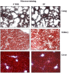
Figure 17. ISA27 is not toxic to normal tissues.
Two weeks from the start of the treatment, mice were sacrificed, and internal organs were removed for histological analysis. Masson trichrome staining was performed on liver, kidney and lung tissues from treated and control mice. No morphological differences were found in the treated mice compared with the controls.
Figure 18
Figure 18. Synergistic effect of ISA27 and temozolomide on the survival/growth of GBM cells.
Isobologram 2-D showing the interactions between TMZ and ISA27 in MTS viability tests performed in U87MG cells treated for 72 h with TMZ and/or ISA27. The IC50 values for TMZ and ISA27 are shown on the X- and Y-axes, respectively. The isobole of additivity is shown as a solid line drawn between the aforementioned IC50 values of TMZ and ISA27 and connects the X- and Y-axes. The open points (○) on the additivity line depict the theoretical IC50,add values for total dose expressed as the proportion of TMZ and ISA27 that produced a 50% effect. The solid points (•) depict the experimental IC50,mix values for total dose expressed as the proportion of TMZ and ISA27 that produced a 50% effect. The experimental IC50,mix values of the mixture of ISA27 and TMZ for the fixed-ratio combinations of 1∶80, 1∶150 and 1∶1,000 were found to be significantly below the theoretical isoboles of additivity, indicating super-additive (synergy) interactions.

Similar articles

Cited by

References

    1. Liu J, Ma Q, Zhang M, Wang X, Zhang D, et al. (2012) Alterations of TP53 are associated with a poor outcome for patients with hepatocellular carcinoma: Evidence from a systematic review and meta-analysis. Eur J Cancer 48: 2328–2338. - PMC - PubMed
    1. Furnari FB, Fenton T, Bachoo RM, Mukasa A, Stommel JM, et al. (2007) Malignant astrocytic glioma: genetics, biology, and paths to treatment. Genes Dev 21: 2683–2710. - PubMed
    1. Ohgaki H, Dessen P, Jourde B, Horstmann S, Nishikawa T, et al. (2004) Genetic pathways to glioblastoma: a population-based study. Cancer Res 64: 6892–6899. - PubMed
    1. Conrad CA, Milosavljevic VP, Yung WK (1995) Advances in chemotherapy for brain tumors. Neurol Clin 13: 795–812. - PubMed
    1. Levin VA, Leibel SA, Gutin PH (2001) Neoplasms of the central nervous system. In: De Vita Jr VT, Hellman S, Rosenberg SA, editors. Cancer principles of oncology. Philadelphia: Lippincott-Raven. 2100–2161.

Publication types

MeSH terms

Substances

Grants and funding

Funding for this study was provided by the Italian Ministry of University and Scientific Research (PRIN2009-prot. 20098SJX4F). The funders had no role in study design, data collection and analysis, decision to publish, or preparation of the manuscript.