

Redox-sensitive glycogen synthase kinase 3β-directed control of mitochondrial permeability transition: rheostatic regulation of acute kidney injury
- PMID: 23973862
- PMCID: PMC3859848
- DOI: 10.1016/j.freeradbiomed.2013.08.169
Redox-sensitive glycogen synthase kinase 3β-directed control of mitochondrial permeability transition: rheostatic regulation of acute kidney injury
Abstract
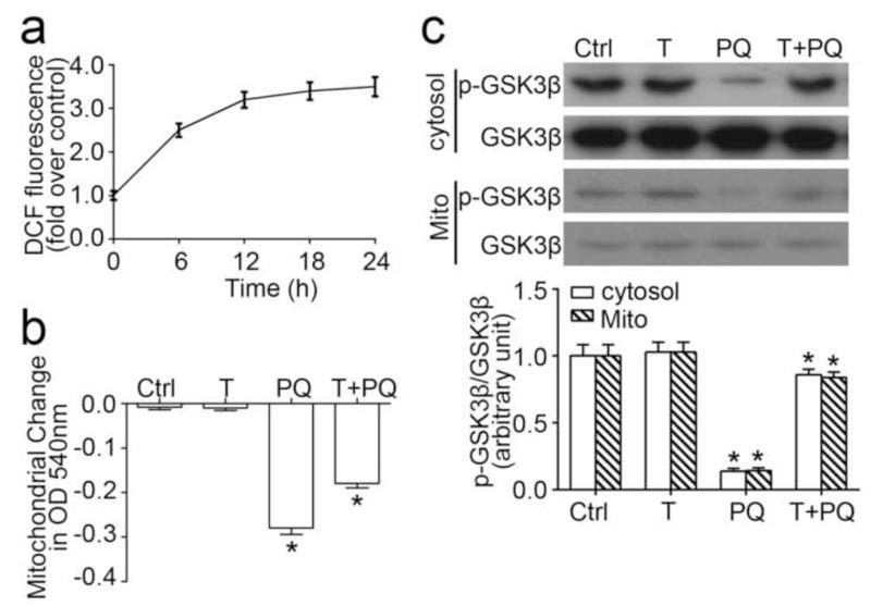
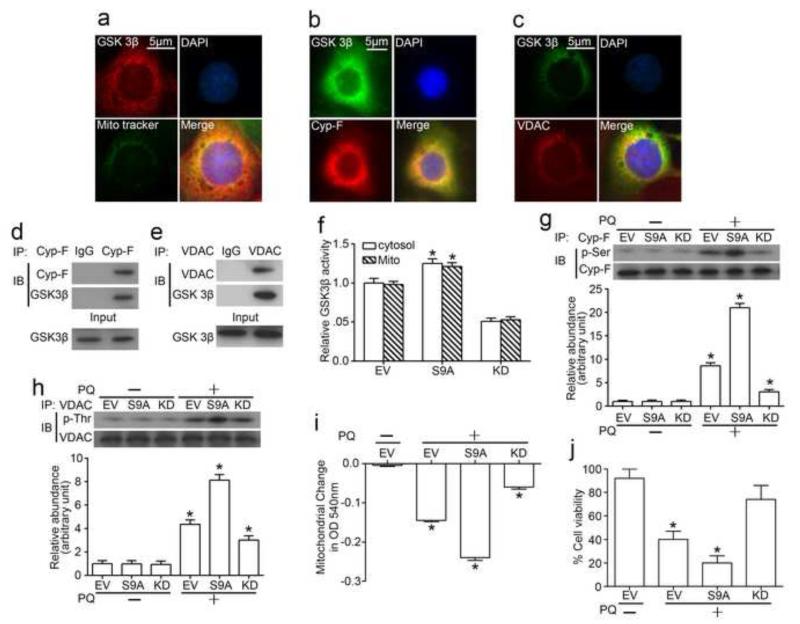
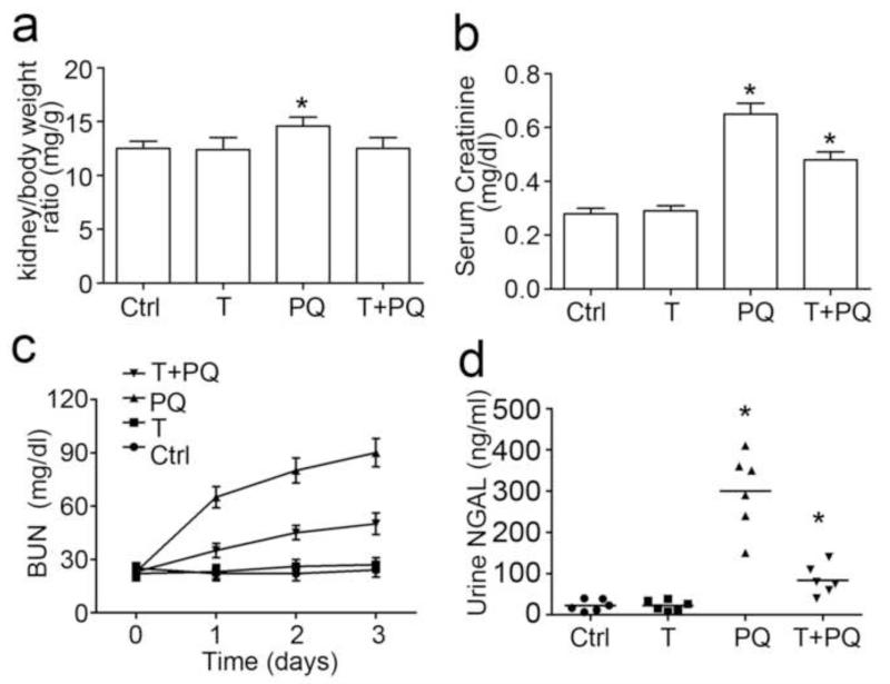
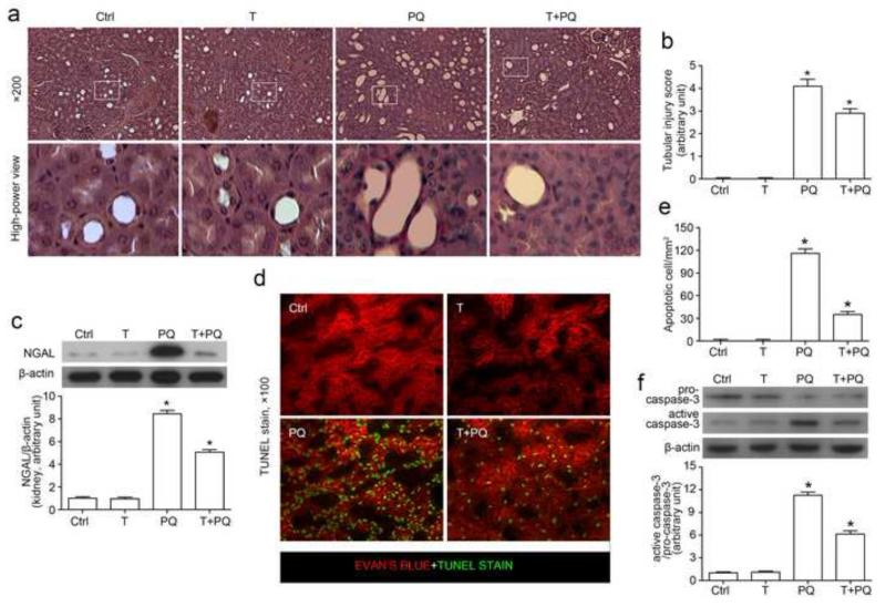
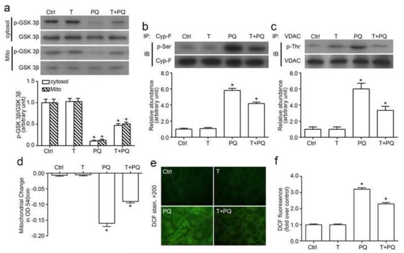
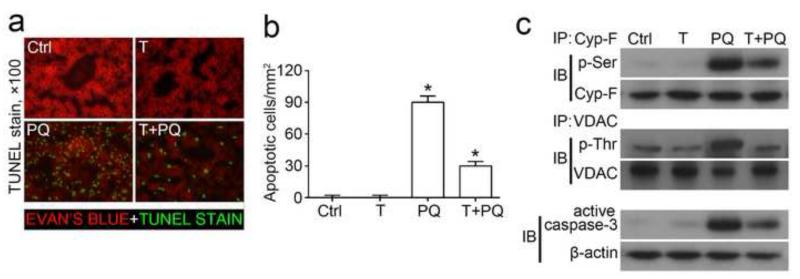
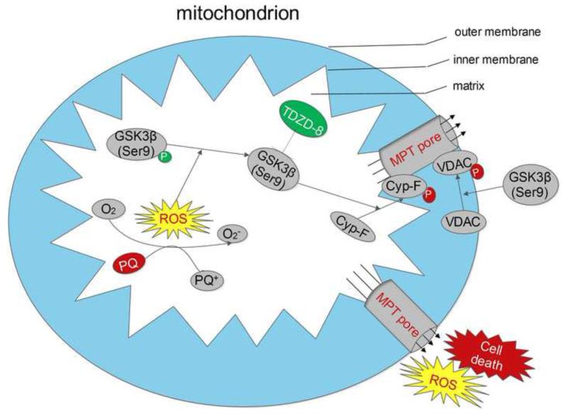
Mitochondrial dysfunction plays a pivotal role in necroapoptotic cell death and in the development of acute kidney injury (AKI). Evidence suggests that glycogen synthase kinase (GSK) 3β resides at the nexus of multiple signaling pathways implicated in the regulation of mitochondrial permeability transition (MPT). In cultured renal tubular epithelial cells, a discrete pool of GSK3β was detected in mitochondria. Coimmunoprecipitation assay confirmed that GSK3β physically interacts with cyclophilin F and voltage-dependent anion channel (VDAC), key MPT regulators that possess multiple GSK3β phosphorylation consensus motifs, suggesting that GSK3β has a direct control of MPT. Upon a strong burst of reactive oxygen species elicited by the pro-oxidant herbicide paraquat, the activity of the redox-sensitive GSK3β was drastically enhanced. This was accompanied by augmented phosphorylation of cyclophilin F and VDAC, associated with MPT and cell death. Inhibition of GSK3β by either the selective inhibitor 4-Benzyl-2-methyl-1,2,4-thiadiazolidine-3,5-dione (TDZD-8) or forced expression of a kinase-dead mutant obliterated paraquat-induced phosphorylation of cyclophilin F and VDAC, prevented MPT, and improved cellular viability. Conversely, ectopic expression of a constitutively active GSK3β amplified the effect of paraquat on cyclophilin F and VDAC phosphorylation and sensitized cells to paraquat-induced MPT and death. In vivo, paraquat injection elicited marked oxidant stress in the kidney and resulted in acute kidney dysfunction and massive tubular apoptosis and necrosis. Consistent with in vitro findings, the activity of GSK3β was augmented in the kidney after paraquat injury, associated with increased phosphorylation of cyclophilin F and VDAC and sensitized MPT. TDZD-8 blocked GSK3β activity in the kidney, intercepted cyclophilin F and VDAC phosphorylation, prevented MPT, attenuated tubular cell death, and ameliorated paraquat-induced AKI. Our data suggest that the redox-sensitive GSK3β regulates renal tubular injury in AKI by controlling the activity of MPT regulators.
Keywords: Acute kidney injury; Cyclophilin F; Free radicals; Glycogen synthase kinase 3β; Mitochondrial permeability transition; Paraquat; Voltage-dependent anion channel.
© 2013 Elsevier Inc. All rights reserved.
Figures
Similar articles
-
Inhibition of glycogen synthase kinase-3β prevents NSAID-induced acute kidney injury.Kidney Int. 2012 Apr;81(7):662-73. doi: 10.1038/ki.2011.443. Epub 2012 Jan 18. Kidney Int. 2012. PMID: 22258319 Free PMC article.
-
Pharmacological targeting of GSK3β confers protection against podocytopathy and proteinuria by desensitizing mitochondrial permeability transition.Br J Pharmacol. 2015 Feb;172(3):895-909. doi: 10.1111/bph.12952. Epub 2014 Dec 15. Br J Pharmacol. 2015. PMID: 25262943 Free PMC article.
-
Translocation of glycogen synthase kinase-3β (GSK-3β), a trigger of permeability transition, is kinase activity-dependent and mediated by interaction with voltage-dependent anion channel 2 (VDAC2).J Biol Chem. 2014 Oct 17;289(42):29285-96. doi: 10.1074/jbc.M114.563924. Epub 2014 Sep 3. J Biol Chem. 2014. PMID: 25187518 Free PMC article.
-
Role of the mitochondrial membrane permeability transition in cell death.Apoptosis. 2007 May;12(5):835-40. doi: 10.1007/s10495-006-0525-7. Apoptosis. 2007. PMID: 17136322 Review.
-
Regulation of necrotic cell death: p53, PARP1 and cyclophilin D-overlapping pathways of regulated necrosis?Cell Mol Life Sci. 2016 Jun;73(11-12):2309-24. doi: 10.1007/s00018-016-2202-5. Epub 2016 Apr 5. Cell Mol Life Sci. 2016. PMID: 27048819 Free PMC article. Review.
Cited by
-
Physiopathology of the Permeability Transition Pore: Molecular Mechanisms in Human Pathology.Biomolecules. 2020 Jul 4;10(7):998. doi: 10.3390/biom10070998. Biomolecules. 2020. PMID: 32635556 Free PMC article. Review.
-
Remote ischemic preconditioning for kidney protection: GSK3β-centric insights into the mechanism of action.Am J Kidney Dis. 2015 Nov;66(5):846-56. doi: 10.1053/j.ajkd.2015.06.026. Epub 2015 Aug 10. Am J Kidney Dis. 2015. PMID: 26271146 Free PMC article. Review.
-
Mitochondria ROS and mitophagy in acute kidney injury.Autophagy. 2023 Feb;19(2):401-414. doi: 10.1080/15548627.2022.2084862. Epub 2022 Jun 9. Autophagy. 2023. PMID: 35678504 Free PMC article.
-
Lithium in the Kidney: Friend and Foe?J Am Soc Nephrol. 2016 Jun;27(6):1587-95. doi: 10.1681/ASN.2015080907. Epub 2015 Nov 17. J Am Soc Nephrol. 2016. PMID: 26577775 Free PMC article. Review.
-
Gallic acid exerts anti-inflammatory, anti-oxidative stress, and nephroprotective effects against paraquat-induced renal injury in male rats.Naunyn Schmiedebergs Arch Pharmacol. 2021 Jan;394(1):1-9. doi: 10.1007/s00210-020-01931-0. Epub 2020 Jul 30. Naunyn Schmiedebergs Arch Pharmacol. 2021. PMID: 32734364
References
-
- Toth R, Breuer T, Cserep Z, Lex D, Fazekas L, Sapi E, Szatmari A, Gal J, Szekely A. Acute Kidney Injury Is Associated With Higher Morbidity and Resource Utilization in Pediatric Patients Undergoing Heart Surgery. Annals of Thoracic Surgery. 2012;93:1984–1991. - PubMed
-
- Valette X, Parienti J-J, Plaud B, Lehoux P, Samba D, Hanouz J-L. Incidence, morbidity, and mortality of contrast-induced acute kidney injury in a surgical intensive care unit: a prospective cohort study. Journal of critical care. 2012;27:322.e321–325. - PubMed
-
- Chertow GM, Burdick E, Honour M, Bonventre JV, Bates DW. Acute kidney injury, mortality, length of stay, and costs in hospitalized patients. Journal of the American Society of Nephrology. 2005;16:3365–3370. - PubMed
-
- Mergner WJ, Trump BF, Valigors Jm, Garbus J, Dees JH. STRUCTURAL AND FUNCTIONAL CHANGES IN HUMAN KIDNEY AND LIVER-MITOCHONDRIA IN ACUTE CELL INJURY AFTER SHOCK AND TRAUMA. Am. J. Pathol. 1972;66:A36.
Publication types
MeSH terms
Substances
Grants and funding
LinkOut - more resources
Full Text Sources
Other Literature Sources
