. 2013 Oct 4;288(40):28503-13.
doi: 10.1074/jbc.M113.471805. Epub 2013 Aug 21.

Effect of charged residues in the N-domain of Sup35 protein on prion [PSI+] stability and propagation

Affiliations

Effect of charged residues in the N-domain of Sup35 protein on prion [PSI+] stability and propagation

Stanislav A Bondarev et al. J Biol Chem. .

Abstract

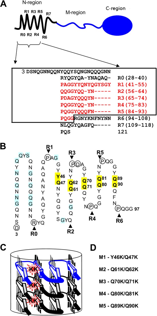
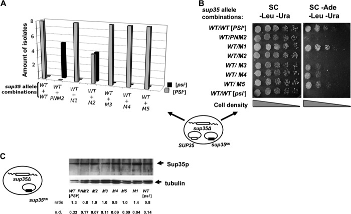
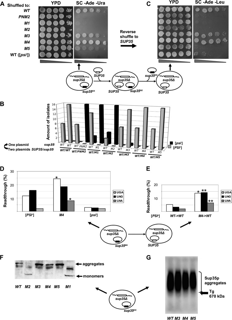
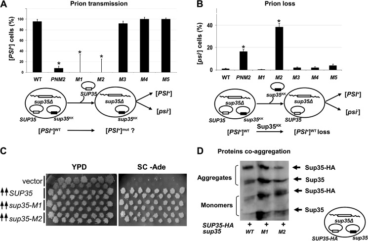
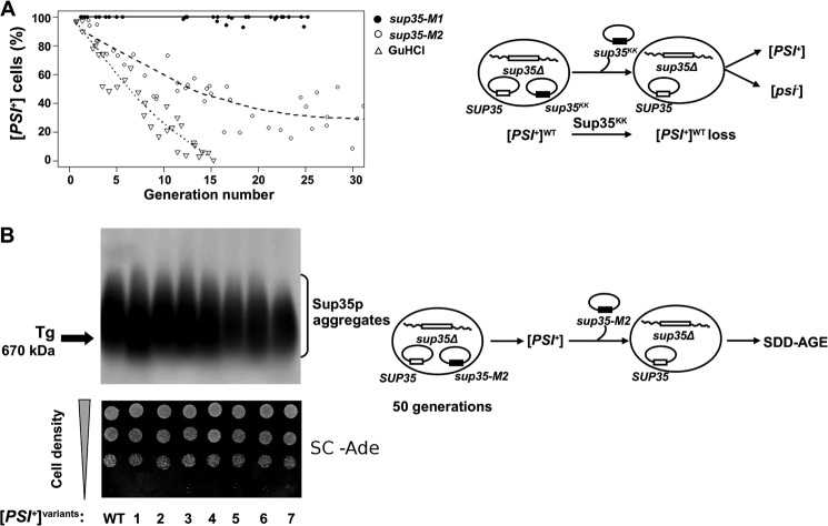
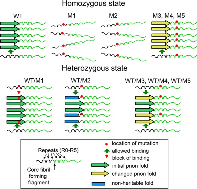
Recent studies have shown that Sup35p prion fibrils probably have a parallel in-register β-structure. However, the part(s) of the N-domain critical for fibril formation and maintenance of the [PSI(+)] phenotype remains unclear. Here we designed a set of five SUP35 mutant alleles (sup35(KK)) with lysine substitutions in each of five N-domain repeats, and investigated their effect on infectivity and ability of corresponding proteins to aggregate and coaggregate with wild type Sup35p in the [PSI(+)] strain. Alleles sup35-M1 (Y46K/Q47K) and sup35-M2 (Q61K/Q62K) led to prion loss, whereas sup35-M3 (Q70K/Q71K), sup35-M4 (Q80K/Q81K), and sup35-M5 (Q89K/Q90K) were able to maintain the [PSI(+)] prion. This suggests that the critical part of the parallel in-register β-structure for the studied [PSI(+)] prion variant lies in the first 63-69 residues. Our study also reveals an unexpected interplay between the wild type Sup35p and proteins expressed from the sup35(KK) alleles during prionization. Both Sup35-M1p and Sup35-M2p coaggregated with Sup35p, but only sup35-M2 led to prion loss in a dominant manner. We suggest that in the fibrils, Sup35p can bind to Sup35-M1p in the same conformation, whereas Sup35-M2p only allowed the Sup35p conformation that leads to the non-heritable fold. Mutations sup35-M4 and sup35-M5 influence the structure of the prion forming region to a lesser extent, and can lead to the formation of new prion variants.

Keywords: Amyloid; Prions; Protein Misfolding; Protein Structure; Saccharomyces cerevisiae; Superpleated β-Structure; Translation Release Factors; Translation Termination; [PSI+].


Figures

FIGURE 1.
FIGURE 1.
Localization of mutated Sup35 residues regarding the structural model of the N-terminal domain. A, top, schematic representation of Sup35p domain organization: the N-terminal region (1–123 amino acids) is shown with an array of eight repeats (R0–R7), discovered with the T-REKS program, the M-region (124–245 amino acids), C-terminal region (246–685 amino acids) has a globular-fold of the GTP-binding domain essential for robust translation termination. Bottom, new assignment of the Sup35p tandem repeats (T-REKS), which includes a previous one (8) colored in red. B, sequence of the N-terminal domain (the rectangle within the panel A) arranged in a serpentine with linear regions and bends, corresponding to putative β-strands and loop regions, respectively. The circled residues tend to localize to the loop regions (proline and charged residues). Positions where substitutions with charged residues lead to the PNM phenotype in previous reports (19, 36, 48) are highlighted in blue. Residues highlighted in yellow are mutated to lysines in this study. C, a model for the super-pleated β-structure that demonstrates how the introduction of charged residues in the β-strand region destabilizes the fibril by the electrostatic repulsion of same-sign charges along the fibril axis. D, sup35 mutations studied in this work (numbering of the mutations corresponds to the repeat number shown above).
FIGURE 2.
FIGURE 2.
sup35-M2 and PNM2 eliminate [PSI+] in the presence of wild type SUP35. A, transformants of yeast strain 10-7A-D832 (sup35Δ [PSI+]) contain two plasmids: pYCH-U2 (URA3) with a wild type copy of SUP35, and pRSU1 (LEU2) with wild type or mutant alleles of SUP35 (schematic representation of plasmid combination shown at bottom). Eight transformants were tested for each combination (except for PNM2 where 5 transformants were tested). In the case of sup35-M2 the [PSI+]-dependent suppression of ade1–14 was eliminated in 4 of 8 transformants. B, five serial dilutions of the indicated transformants were spotted onto SC medium selective for both plasmids with or without adenine, the tested sup35 allele combinations are shown on the left. A typical Ade transformant of sup35-M2 is shown. Yeast strain 7A-D832 (sup35Δ [psi]) bearing two plasmids with a wild type copy of SUP35 was used as a negative control. C, 10-7A-D832 (sup35Δ) derivatives bearing pRSU1-SUP35 or pRSU1-sup35KK plasmids were analyzed by Western blotting with an anti-eRF3 antibody. The captions below the lanes indicate the sup35KK allele. The “ratio” represents the relative abundance of Sup35p in each mutant compared with the amount of tubulin.
FIGURE 3.
FIGURE 3.
sup35KK alleles have the opposite effect on [PSI+] phenotype in the absence of wild type SUP35. A, five serial dilutions of 10-7A-D832 (sup35Δ) derivatives containing the pRSU1 plasmid with wild type or mutant alleles of SUP35. B, amount of [PSI+] and [psi] isolates after the loss of one of the plasmids. C, isolates shown on panel A were retransformed with pRSU2 (SUP35 (WT)) with subsequent loss of the pRSU1 plasmid, one representative clone from 16 tested in each case is shown. In both panels A and C one representative transformant was spotted onto YPD and SC-Ade. Derivatives of 7A-D832 (sup35Δ [psi]) containing the corresponding plasmids with a wild type copy of SUP35 were used as a negative control in panels A–C. Schematic representation of the plasmid shuffling assay is shown under panels A and C. Arrows indicate where the corresponding phenotype is demonstrated. D and E, read through efficiency in [PSI+] 10-7A-D832 derivatives bearing sup35-M4 mutation after direct (C) and reverse (D) shuffle measured by in vivo β-galactosidase activity assay. The efficiency of suppression was calculated as a ratio of β-galactosidase activity in cells harboring lacZ with premature termination codon to lacZ (WT) control. The results are from at least three separate experiments. Significant differences from wild type [PSI+] are shown by one or two asterisks (p < 0.05 and p < 0.01, respectively). F, SDS-PAGE with additional boiling of [PSI+] aggregates in 10-7A-D832 (sup35Δ) derivatives containing the pRSU1 plasmid with wild type SUP35 or sup35KK. The captions below the lanes indicate the sup35 allele. A schematic representation of the plasmid combination is shown near the panel. G, SDD-AGE analysis of Sup35p aggregates in transformants shown in panel A. The arrow marks the position of thyroglobulin (Tg, 670 kDa) in gel revealed by Coomassie staining.
FIGURE 4.
FIGURE 4.
Influence of sup35KK alleles on [PSI+] transmission, loss, induction, and protein coaggregation. A, [PSI+] transmission from the wild type to the indicated sup35 allele. A fraction of cells that retained prions after loss of the wild type allele is shown on the graph. B, [PSI+] loss induced by transient expression of the sup35KK alleles. The fraction of cells that have lost prions after loss of the sup35KK allele is shown. Significant differences from control variant (WT) in A and B are shown by asterisks (p < 0.05). C, transient overexpression of sup35-M1 or sup35-M2 leads to [PSI+] induction. The appearance of prions was detected by growth on the adenine-omitted medium. Transformants of the 7A-D832 strain (SUP35 WT [psi]) were incubated on -Ura/Gal medium and replica plated onto glucose -Ade medium. Plates were photographed after 5 days of incubation. D, cell lysates of 10-7A-D832 derivatives containing wild type Sup35p-HA together with mutant Sup35p were separated using SDS-PAGE with additional boiling. Blotted proteins were probed with anti-eRF3 antibody, which revealed Sup35p-HA and Sup35p migrating in monomeric and aggregate fractions. Schematic representations of experiments and the plasmid combinations are shown below each panel.
FIGURE 5.
FIGURE 5.
Kinetics of [PSI+] loss in the presence of sup35-M1 and sup35-M2 alleles. A, transformants of 10-7A-D832 bearing two plasmids (with SUP35WT and sup35KK) were grown in liquid medium selective for both plasmids. At regular time points, culture aliquots were taken and plated on 5-FOA solid medium (see ”Experimental Procedures“). 5-FOA resistant colonies were tested for the presence of [PSI+] by replica plating onto MD 1/5 Ade medium. The percentages of [PSI+] cells have been plotted as a function of generation number passed from the beginning of the experiment, which was estimated from measurements of A600. [PSI+] elimination in the presence of 4 mm GdnHCl was used as control. B, SDD-AGE analysis of [PSI+] aggregates in independent Ade+ isolates that have lost sup35-M2 after more than 50 generations of growth as in panel A (top panel). WT, wild type strain 10-7A-D832. The arrow marks the position of thyroglobulin (Tg, 670 kDa) in the gel as revealed by Coomassie staining. Ade+ phenotype of the same isolates (lower panel). Ten serial dilutions of one representative clone were spotted onto SC medium without adenine. Schematic representation of the plasmid shuffling assay is shown to the right of each panel.
FIGURE 6.
FIGURE 6.
The model describing interactions of mutant Sup35p with prion filaments. Mutations sup35-M1 and sup35-M2 are located within the core region of the parallel and in-register superpleated β-structure. In the homozygous state, both Sup35-M1p and Sup35-M2p do not form fibrils. In the heterozygous state, Sup35-M1p and Sup35-M2p can coaggregate with Sup35p. Sup35-M1p does not affect the prion, whereas Sup35-M2p leads to conformational changes, formation of non-heritable fibril-folds, and hence loss of [PSI+] in the majority of cells. We suggest that in the case of Sup35-M1p, homoaggregates are not permitted. Thus in the growing fibril Sup35p and Sup35 are perfectly interspersed, one following the other along the axis of the prion filament. We speculate that regions mutated in Sup35-M3p, Sup35-M4p, and Sup35-M5p lie outside of the superpleated β-structure of Sup35p in the conformation of the prion strain utilized in this study. Hence these alleles cannot eliminate [PSI+] under the conditions tested.

Similar articles

Cited by

References

    1. Prusiner S. B. (1998) Prions. Proc. Natl. Acad. Sci. U.S.A. 95, 13363–13383 - PMC - PubMed
    1. Wickner R. B. (1994) [URE3] as an altered URE2 protein. Evidence for a prion analog in Saccharomyces cerevisiae. Science 264, 566–569 - PubMed
    1. Wickner R. B., Edskes H. K., Shewmaker F. P., Kryndushkin D., Nemecek J., McGlinchey R., Bateman D. (2010) The relationship of prions and translation. Wiley Interdiscip. Rev. RNA 1, 81–89 - PMC - PubMed
    1. Crow E. T., Li L. (2011) Newly identified prions in budding yeast, and their possible functions. Semin. Cell Dev. Biol. 22, 452–459 - PMC - PubMed
    1. Tuite M. F., Lindquist S. L. (1996) Maintenance and inheritance of yeast prions. Trends Genet. 12, 467–471 - PubMed

MeSH terms

LinkOut - more resources