. 2013 Oct;19(10):1432-48.
doi: 10.1261/rna.038893.113. Epub 2013 Aug 20.

Comparison of EJC-enhanced and EJC-independent NMD in human cells reveals two partially redundant degradation pathways

Comparison of EJC-enhanced and EJC-independent NMD in human cells reveals two partially redundant degradation pathways

Stefanie Metze et al. RNA. 2013 Oct.

Abstract

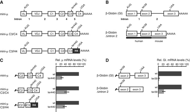
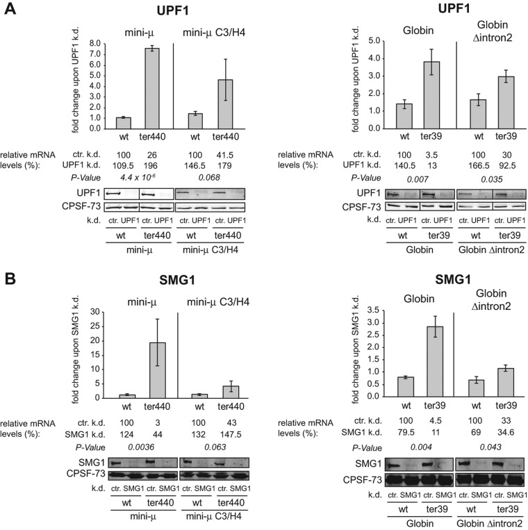
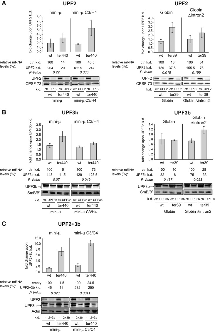
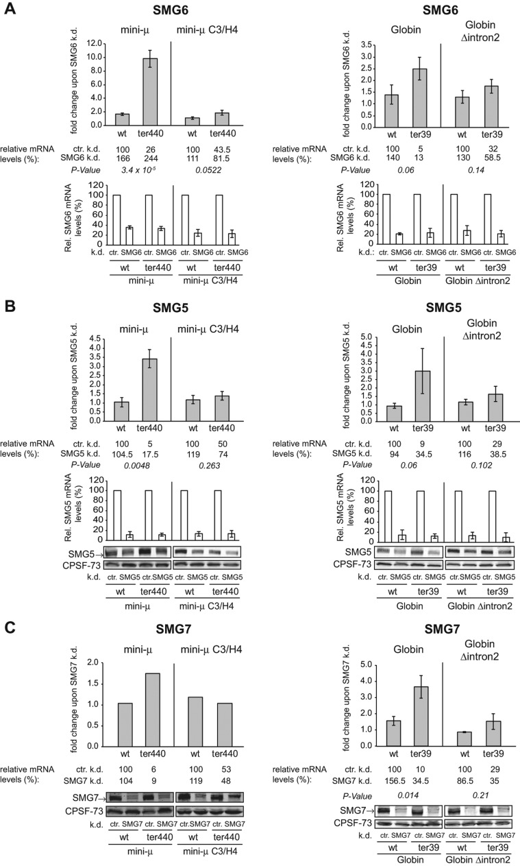
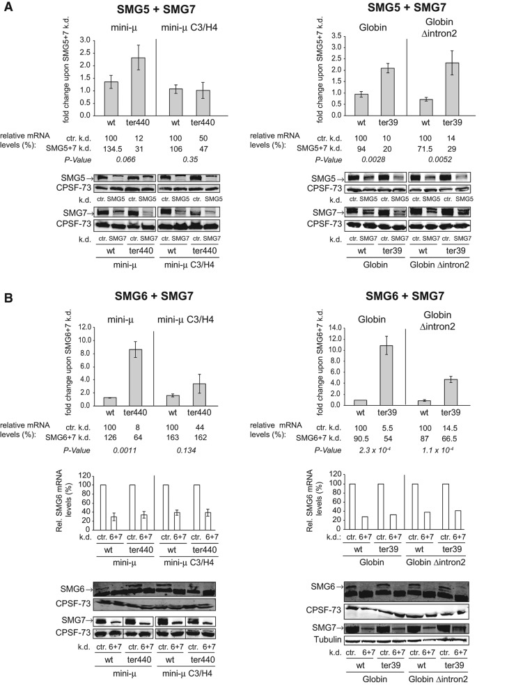
Nonsense-mediated mRNA decay (NMD) is a eukaryotic post-transcriptional gene regulation mechanism that eliminates mRNAs with the termination codon (TC) located in an unfavorable environment for efficient translation termination. The best-studied NMD-targeted mRNAs contain premature termination codons (PTCs); however, NMD regulates even many physiological mRNAs. An exon-junction complex (EJC) located downstream from a TC acts as an NMD-enhancing signal, but is not generally required for NMD. Here, we compared these "EJC-enhanced" and "EJC-independent" modes of NMD with regard to their requirement for seven known NMD factors in human cells using two well-characterized NMD reporter genes (immunoglobulin μ and β-Globin) with or without an intron downstream from the PTC. We show that both NMD modes depend on UPF1 and SMG1, but detected transcript-specific differences with respect to the requirement for UPF2 and UPF3b, consistent with previously reported UPF2- and UPF3-independent branches of NMD. In addition and contrary to expectation, a higher sensitivity of EJC-independent NMD to reduced UPF2 and UPF3b concentrations was observed. Our data further revealed a redundancy of the endo- and exonucleolytic mRNA degradation pathways in both modes of NMD. Moreover, the relative contributions of both decay pathways differed between the reporters, with PTC-containing immunoglobulin μ transcripts being preferentially subjected to SMG6-mediated endonucleolytic cleavage, whereas β-Globin transcripts were predominantly degraded by the SMG5/SMG7-dependent pathway. Overall, the surprising heterogeneity observed with only two NMD reporter pairs suggests the existence of several mechanistically distinct branches of NMD in human cells.

Keywords: endo- and exonucleolytic mRNA degradation; UPF1; UPF2; UPF3b; SMG1; SMG5; SMG6; SMG7; exon-junction complex; mRNA surveillance; mRNA turnover; nonsense-mediated mRNA decay; post-transcriptional gene regulation.


Figures

FIGURE 1.
FIGURE 1.
Relative abundance of mini-μ and β-Globin NMD reporter transcripts with and without an intron downstream from the PTC. (A) Schematic representation of the mini-μ reporter constructs, each of which exists in the wild-type (wt) and the ter440 version, the latter contains a single point mutation in exon C3 generating a PTC at amino acid 440 (GAA → TAA). In the constructs mini-μ C3/C4 and C3/H4, intron 5 was deleted. Additionally, exon C4 was replaced with a sequence of identical length from the histone H4 gene in construct mini-μ C3/H4. (B) Schematic representation of the β-Globin (Gl) reporter constructs that either code for the wild-type ORF (wt) or harbor a single point mutation in exon 2 generating a PTC at amino acid 39 (CAG → TAG; ter39). The β-Globin construct is a hybrid between the human and mouse β-Globin (Zhang et al. 1998). In β-Globin Δintron2, intron 2 was deleted. Exons and introns are illustrated by boxes and lines, respectively, and the positions of the start (AUG) and termination (UGA or UAA) codons, as well as of the PTCs (ter440 or ter39), are indicated. (C) Relative mini-μ mRNA levels expressed in transiently transfected HeLa cells were measured 48 h post-transfection by reverse transcription followed by quantitative real-time PCR (RT-qPCR) and normalized to relative mRNA levels of cotransfected pGEM5 IRE β-Globin wt. Mean values and ±SEM of three or four independent experiments are shown. (D) Relative β-Globin mRNA levels expressed in transiently transfected HeLa cells were measured 48 h post-transfection by RT-qPCR and normalized to relative mRNA levels of pβ mini-μ wt IRE. Mean values and ±SEM of three independent experiments are shown.
FIGURE 2.
FIGURE 2.
UPF1 and SMG1 are required for EJC-independent NMD. (A,B) Fold changes of relative mRNA levels of mini-μ and β-Globin constructs upon knockdown of UPF1 (A) or SMG1 (B) compared with a control knockdown (ctr.) in HeLa tTR-KRAB cells transiently transfected with the indicated reporter constructs are shown. Relative mRNA levels were determined 72 h post-transfection by RT-qPCR and normalized to the relative mRNA levels of cotransfected pGEM5 IRE β-Globin wt (for mini-μ) or pβ mini-μ wt IRE (for β-Globin). The relative mRNA levels are displayed below the diagrams, and the wt samples in the control knockdown were set as 100%. The efficiency of knockdowns was monitored by Western blotting. (k.d.) knockdown, (ctr.) control knockdown. (A) UPF1 or control knockdowns were induced by addition of DOX to HeLa tTR-KRAB-shUPF1 or HeLa tTR-KRAB-sh(scrambled) cells, except for mini-μ wt and mini-μ ter440, where the UPF1 and control knockdowns were induced by cotransfection of the respective pSUPERpuro plasmids. Mean values, fold changes, and ±SEM of three or four independent experiments are shown. P-values refer to the fold changes of PTC+ transcripts compared with the corresponding wt transcripts. (B) SMG1 or ctr. knockdowns were carried out in HeLa tTR-KRAB-shSMG1 or HeLa tTR-KRAB-sh(scrambled) cells as in A. Mean values, fold changes, and ±SEM of three independent experiments are shown. In all figures depicting data from knockdown experiments, the relative mRNA levels given below the histogram represent the mean of the corresponding relative mRNA levels in each independent experiment, whereas the “fold change” indicated in the histograms represents the mean of the corresponding fold changes in each independent experiment (for details, see Materials and Methods). Therefore, the depicted fold change in the histogram can deviate slightly from the ratio of the corresponding relative mRNA levels depicted below.
FIGURE 3.
FIGURE 3.
Transcript-specific roles for UPF2 and UPF3b and differential requirements in EJC-enhanced and EJC-independent NMD. (A) Fold changes of relative mini-μ and β-Globin mRNA levels upon knockdown of UPF2 in HeLa cells transiently transfected with reporter constructs are shown. Relative mini-μ and β-Globin mRNA levels were determined 72 h post-transfection by RT-qPCR and normalized to relative mRNA levels of cotransfected pNORM-7SL-AmdS. UPF2 or ctr. knockdowns were induced by addition of DOX to HeLa tTR-KRAB-shUPF2 or HeLa tTR-KRAB-sh(scrambled) cells. Mean values, fold changes, and ±SEM of three independent experiments are shown. The relative mRNA levels are displayed below the diagrams, and the wt samples in the control knockdown were set as 100%. The efficiency of the knockdowns was monitored by Western blotting. P-values were determined as in Figure 2A. (k.d.) knockdown; (ctr.) control knockdown. (B) As A, but with UPF3b knockdowns instead. (C) Fold changes of relative mini-μ mRNA levels upon knockdown of UPF2 and UPF3b in HeLa cells transiently transfected with reporter constructs are shown. Relative mini-μ mRNA levels were determined 96 h post-transfection by RT-qPCR and normalized to relative mRNA levels of cotransfected pmCMVrGPx1-TGC. Knockdowns with the mini-μ constructs were done by cotransfection of pSUPERpuro-shUPF2 and -shUPF3b plasmids or pSUPERpuro-empty as a control (empty/−) in HeLa cells. Data were analyzed as in A.
FIGURE 4.
FIGURE 4.
Single knockdowns of SMG6, SMG5, or SMG7 hardly affect EJC-independent NMD. (A–C) Fold changes of relative mRNA levels of mini-μ or β-Globin constructs upon knockdown of SMG6 (A), SMG5 (B), or SMG7 (C) compared with a control knockdown (ctr.) in HeLa tTR-KRAB cells transiently transfected with the indicated reporter constructs are shown. Relative mini-μ or β-Globin mRNA levels were determined 72 h post-transfection by RT-qPCR. SMG6, SMG5, SMG7, or ctr. knockdowns were induced by addition of DOX to HeLa tTR-KRAB-shSMG6, -shSMG5, -shSMG7, or -sh(scrambled) cells, respectively. Mean values, fold changes, and ±SEM of two to four independent experiments are shown. The relative mRNA levels are displayed below the diagrams, and the wt samples in the control knockdown were set as 100%. The efficiency of the knockdowns was monitored by RT-qPCR (SMG6 [A], SMG5 [B]) and/or Western blotting (SMG5 [B], SMG7 [C]). P-values were determined as in Figure 2A. ±SEM and P-values were calculated when n ≥ 3. (k.d.) knockdown; (ctr.) control knockdown. (A) Relative mini-μ or β-Globin mRNA levels were normalized to relative mRNA levels of cotransfected pGEM5 IRE β-Globin wt (for mini-μ) or pβ mini-μ wt IRE (for β-Globin). (B) Relative mini-μ or β-Globin mRNA levels were normalized to relative mRNA levels of cotransfected pNORM-7SL-AmdS. (C) Relative mini-μ or β-Globin mRNA levels were normalized to relative mRNA levels of cotransfected pNORM-7SL-AmdS (for mini-μ) or pβ mini-μ wt IRE and pNORM-7SL-AmdS (for β-Globin).
FIGURE 5.
FIGURE 5.
Codepletion of SMG6/SMG7 strongly inhibits EJC-enhanced and EJC-independent NMD, but a depletion of SMG5/SMG7 has much weaker effects. (A) Fold changes of relative mini-μ or β-Globin mRNA constructs upon a combined knockdown of SMG5 and SMG7 compared with a control knockdown (ctr.) in HeLa cells transiently transfected with the indicated reporter constructs are shown. Relative mini-μ or β-Globin mRNA levels were determined 96 h post-transfection by RT-qPCR and normalized to relative mRNA levels of cotransfected pNORM-7SL-AmdS. Knockdowns were performed by cotransfection into HeLa cells of pSUPERpuro-shSMG5 and -shSMG7 or pSUPERpuro-sh(scrambled) as a control. Mean values, fold changes, and ±SEM of three or four independent experiments are shown. The relative mRNA levels are displayed below the diagrams, and the wt samples in the control knockdown were set as 100%. The efficiency of the knockdowns was monitored by Western blotting. P-values were determined as in Figure 2A. (k.d.) knockdown; (ctr.) control knockdown. (B) As A, but with a combined knockdown of SMG6 and SMG7. For Western blotting, a different SMG7 antibody was used here from that in A and Figure 4C. Additionally, relative mRNA levels of SMG6 are shown.

Similar articles

Cited by

References

    1. Amrani N, Ganesan R, Kervestin S, Mangus DA, Ghosh S, Jacobson A 2004. A faux 3′-UTR promotes aberrant termination and triggers nonsense-mediated mRNA decay. Nature 432: 112–118 - PubMed
    1. Anders KR, Grimson A, Anderson P 2003. SMG-5, required for C. elegans nonsense-mediated mRNA decay, associates with SMG-2 and protein phosphatase 2A. EMBO J 22: 641–650 - PMC - PubMed
    1. Andersen CB, Ballut L, Johansen JS, Chamieh H, Nielsen KH, Oliveira CL, Pedersen JS, Seraphin B, Le Hir H, Andersen GR 2006. Structure of the exon junction core complex with a trapped DEAD-box ATPase bound to RNA. Science 313: 1968–1972 - PubMed
    1. Antoniou M, Geraghty F, Hurst J, Grosveld F 1998. Efficient 3′-end formation of human β-globin mRNA in vivo requires sequences within the last intron but occurs independently of the splicing reaction. Nucleic Acids Res 26: 721–729 - PMC - PubMed
    1. Behm-Ansmant I, Gatfield D, Rehwinkel J, Hilgers V, Izaurralde E 2007. A conserved role for cytoplasmic poly(A)-binding protein 1 (PABPC1) in nonsense-mediated mRNA decay. EMBO J 26: 1591–1601 - PMC - PubMed

Publication types

MeSH terms

LinkOut - more resources