. 2013;9(8):e1003531.
doi: 10.1371/journal.ppat.1003531. Epub 2013 Aug 1.

IL-1β suppresses innate IL-25 and IL-33 production and maintains helminth chronicity

Affiliations

IL-1β suppresses innate IL-25 and IL-33 production and maintains helminth chronicity

Mario M Zaiss et al. PLoS Pathog. 2013.

Abstract

Approximately 2 billion people currently suffer from intestinal helminth infections, which are typically chronic in nature and result in growth retardation, vitamin A deficiency, anemia and poor cognitive function. Such chronicity results from co-evolution between helminths and their mammalian hosts; however, the molecular mechanisms by which these organisms avert immune rejection are not clear. We have found that the natural murine helminth, Heligmosomoides polygyrus bakeri (Hp) elicits the secretion of IL-1β in vivo and in vitro and that this cytokine is critical for shaping a mucosal environment suited to helminth chronicity. Indeed in mice deficient for IL-1β (IL-1β(-/-)), or treated with the soluble IL-1βR antagonist, Anakinra, helminth infection results in enhanced type 2 immunity and accelerated parasite expulsion. IL-1β acts to decrease production of IL-25 and IL-33 at early time points following infection and parasite rejection was determined to require IL-25. Taken together, these data indicate that Hp promotes the release of host-derived IL-1β that suppresses the release of innate cytokines, resulting in suboptimal type 2 immunity and allowing pathogen chronicity.


Conflict of interest statement

The authors have declared that no competing interests exist.

Figures
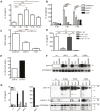
Figure 1
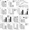
Figure 1. Hp infection elicits IL-1β secretion.
(A–E) Mice were administered 200 L3 Hp by oral gavage. IL-1β protein levels were measured by ELISA in (A) the peritoneal wash and (B) intestinal tissue culture supernatants at the indicated timepoints. (C) Hp worm counts were performed for defined segments of the small intestine from WT (C57BL/6) at 13 dpi. (D) IL-1β was measured by ELISA for duodenum tissue culture supernatants of day 6 infected bone marrow chimera mice (donor strain/recipient strain). Results are representative of at least 3 independent experiments (n = 5 per group) and expressed as mean ± SEM. (E) Intestinal lamina propria CD11b+ and CD11b cells were isolated at 6 dpi and analyzed by Western blot for IL-1β expression. Band intensity for IL-1β and β-actin was determined using Adobe Photoshop CS3. The ratio between IL-1β and the control protein β-actin band intensity was then determined. IL-1β is expressed as percent of β-actin intensity. (F) BMMs from WT (C57BL/6), Trif−/− or MyD88−/− mice were stimulated in vitro with HPL5 (100 µg/mL), HES (5 µg/mL) or P.HES (5 µg/mL) for 4 hours and cell extracts were analyzed for pro-IL-1β expression by Western blot. (G) BMMs from WT (C57BL/6), Asc−/− or Nlrp3−/− mice were stimulated in vitro with P.HES, HPL5 or LPS plus MSU and culture supernatants (SN) were analyzed for active IL-1β by ELISA. Cell extracts (XT) and SN were also analyzed for pro- or mature- IL-1β by Western blot. Pro-Casp1 was used as a control protein. XT blots for IL-1β and Casp1 demonstrate the pro-forms, and the observed lower bands in the IL-1β and Casp1 blots are cleavage products. Error bars represent means of triplicate cultures ± SEM and the experiment was repeated 3 times.
Figure 2
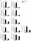
Figure 2. IL-1β promotes chronicity of Hp infection in mice.
Mice were administered 200 L3 Hp by oral gavage (A–M). The number of adult worms were determined for the entire and defined segments, of the small intestine of WT (C57BL/6) and IL-1β−/− mice at (A) 13 dpi and (B) 40 dpi. (C) Fecal egg counts were determined for WT (C57BL/6) and IL-1β−/− mice throughout the course of the experiment (data represents pooled feces of 2–3 individual cages per strain, n = 2–5 mice per cage). Numbers of type 2 granulomas were determined for the entire and defined segments of the small intestine of WT (C57BL/6) and IL-1β−/− mice at (D) 13 dpi and (E) 40 dpi. IL-1α (F) and IL-1ra (G) protein levels were measured by ELISA in duodenum tissue culture supernatants at the indicated timepoints after Hp infection. (H–I) WT (C57BL/6) mice were administered 200 L3 Hp by oral gavage and additionally received 100 µl of PBS (WT) or PBS plus 50 mg/kg Anakinra (WT+Anakinra) via i.p. injection every day from 0–10 dpi. The number of (H) adult Hp worms and (I) type 2 granulomas were determined for the entire and defined segments of the small intestine at 10 dpi. (J) Fecal egg counts were determined at 10 dpi (data represents pooled feces from 1 cage per strain, n = 5 mice per cage). (K–L) WT (C57BL/6) mice were administered 200 L3 Hp larvae by oral gavage and additionally received 100 µl of PBS (WT) or PBS plus 50 mg/kg Anakinra (WT+Anakinra (10 d)) via i.p. injection every day from 0–10 dpi. Mice were sacrificed at day 36 dpi and the number of (K) adult worms and (L) type 2 granulomas determined for the entire and defined segments of the small intestine. (M) Number of adult worms were determined for the entire and defined segments of the small intestine of WT (C57BL/6) and IL-1R1−/− mice at 13 dpi. All data are representative of 3 independent experiments (n = 5–10 per group) and expressed as mean ± SEM.
Figure 3
Figure 3. IL-1β negatively regulates the development of CD4+ Th2 cells following Hp infection.
Mice were administered 200 L3 Hp by oral gavage. At 13 dpi single cell suspensions were made from the (A, C, E) spleen and (B, D, F) MLN, and cells restimulated with HES as described in the Materials and Methods. The percentage of CD4+ T cells secreting (A, B) IL-4, (C, D) IL-13 and (E, F) IFNγ is shown for WT (C57BL/6) or IL-1β−/− mice. Serum was taken at 13 dpi and levels of (G) IL-4, (H) total IgE, (I) HES specific IgG2b, (J) total IgG and (K) HES specific IgG1 determined. All results are representative of 3 independent experiments (n = 5–7 per group) and expressed as mean ± SEM.
Figure 4
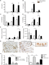
Figure 4. IL-1β attenuates Hp-induced IL-25 and IL-33 cytokine production.
WT (C57BL/6), WT (Balb/c), IL-1β−/− (C57BL/6), IL-25−/−(Balb/c), T1ST2−/− (Balb/c) and IL-17Br×T1ST2−/− (Balb/c) mice were administered 200 L3 Hp by oral gavage. (A, C) IL-25 and (B, D) IL-33 cytokine levels were measured by ELISA in (A–B) peritoneal wash and (C–D) intestinal duodenum tissue culture supernatants from at the indicated timepoints following Hp infection. mRNA expression for (E) IL-25 and (F) IL-33 in isolated intestinal epithelial cells were determined at 0, 3 and 6 dpi. (G) IL-1R1 expression was determined for the duodenum in naive and Hp infected WT (C57BL/6) at 6 dpi by immunohistochemistry. Scale bars represent 200 µm, and 50 µm for the inlet. (H) The mouse epithelial cell line (MSIE) was stimulated in vitro with HES (5 µg/ml) with or without additional rIL-1β and measured for IL-33 mRNA levels by RT-PCR. (I) Adult worm numbers determined for the entire small intestine at 45 dpi. All data are representative of 3 independent experiments (n = 5 per group), and expressed as mean ± SEM.
Figure 5
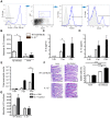
Figure 5. IL-1β attenuates Hp-induced increases in type 2 innate lymphoid cells (ILC2).
WT (C57BL/6), WT (Balb/c), IL-1β−/− and IL-17Br×T1ST2−/− mice were administered 200 L3 by oral gavage. (A) ILC2 cells were identified as lineage-negative (Gr-1, CD3, CD19), Sca-1, c-kit, T1/ST2 and ICOS positive live cells present in the spleen of mice at 6 dpi. (B) Absolute numbers of splenic ILC2 cells and levels of (C) IL-5 and (D) IL-13 protein present in the peritoneal wash at 3 and 6 dpi as determined by ELISA. (E) Absolute numbers of eosinophils present in the peritoneal wash at the indicated timepoints following Hp infection were determined by differential cell counting of cytospins. (F) Intestinal sections were stained with PAS and the percentage of intestinal epithelial cells represented by PAS+ goblet cells determined. Scale bars represent 200 µm. (G) WT (Balb/c), IL-25−/− and IL-17BR×T1ST2−/− mice were treated daily with 100 µl of PBS or PBS plus 50 mg/kg Anakinra for 6 consecutive days starting at day 0 of Hp infection. Absolute numbers of ILC2 cells were determined in the spleen at 6 dpi. All data are representative of at least 2 independent experiments (n = 5–10 per group) and expressed as mean ± SEM.
Figure 6
Figure 6. ILC2s alone are not sufficient enough to reject Hp worms and function mainly to support Th2 responses.
WT (Balb/c), RAG−/−, IL-25−/−, T1ST2−/− and IL-17Br×T1ST2−/− mice were administered 200 L3 Hp by oral gavage. (A) ILC2 numbers in WT (Balb/c), IL-25−/−, T1ST2−/− and IL-17Br×T1ST2−/− mice were determined in the spleen at 6 dpi. Hp infected RAG−/− mice additionally received 100 µl of PBS (RAG−/−) or PBS plus 50 mg/kg Anakinra (RAG−/−+Anakinra) via i.p. injection every day until 6 dpi to analyze, (B) IL-25 and (C) IL-33 cytokine levels in intestinal duodenum tissue cultures. (D) Absolute splenic ILC2 numbers. (E) Hp infected RAG−/− mice additionally received 100 µl of PBS or PBS plus 50 mg/kg Anakinra via i.p. injection from 0–10 dpi and total worm numbers were analyzed at 13 dpi. (F) IL-5 and (G) IL-13 protein present in the peritoneal wash at 6 dpi as determined by ELISA. (H) Absolute numbers of eosinophils present in the peritoneal wash at 6 dpi were determined by differential cell counting of cytospins. All data are representative 3 independent experiments (n = 5–10 per group) and expressed as mean ± SEM.
Figure 7
Figure 7. Hp induced IL-1β production promotes helminth chronicity.
(A) Parasites induce IL-1β secretion by lamina propria (LP) cells. (B) IL-1β inhibits parasite induced IL-25 and IL-33 secretion by intestinal tissue cells and intestinal epithelial cells (IECs). (C) IL-25 secretion induce innate lymphoid cells type 2 (ILC2s). (D) ILC2s in combination with IL-25 promote type 2 cytokine production. (E) Type 2 immunity, including goblet cell hyperplasia and smooth muscle contraction result in expulsion of the adult Hp worms.

Similar articles

Cited by

References

    1. de Silva NR, Brooker S, Hotez PJ, Montresor A, Engels D, et al. (2003) Soil-transmitted helminth infections: updating the global picture. Trends Parasitol 19: 547–551. - PubMed
    1. WHO (2010) Working to overcome the global impact of neglected tropical diseases, First WHO report on Neglected Tropical Diseases. - PubMed
    1. Anthony RM, Rutitzky LI, Urban JF Jr, Stadecker MJ, Gause WC (2007) Protective immune mechanisms in helminth infection. Nat Rev Immunol 7: 975–987. - PMC - PubMed
    1. Harris N, Gause WC (2010) To B or not to B: B cells and the Th2-type immune response to helminths. Trends Immunol 32: 80–88. - PMC - PubMed
    1. Monroy FG, Enriquez FJ (1992) Heligmosomoides polygyrus: a model for chronic gastrointestinal helminthiasis. Parasitol Today 8: 49–54. - PubMed

Publication types

MeSH terms

Grants and funding

The research leading to these results has received funding from the European community seventh framework program [FP7/2009–2014] under EC-GA no[241642] and part of this work was funded by grants from the Swiss National Science Foundation and the Institute of Arthritis Research. NLH is supported by the Swiss Vaccine Research Institute. KMM is supported by EMBO long-term fellowship and the Australian NHMRC post-doctoral fellowship. The funders had no role in study design, data collection and analysis, decision to publish, or preparation of the manuscript.