Comparative Study
. 2013 Jul 1:2013:518034.
doi: 10.1155/2013/518034. Print 2013.

Comparative analysis and systematic mapping of the labial sensilla in the Nepomorpha (Heteroptera: Insecta)

Affiliations
Comparative Study

Comparative analysis and systematic mapping of the labial sensilla in the Nepomorpha (Heteroptera: Insecta)

Jolanta Brożek. ScientificWorldJournal. .

Abstract

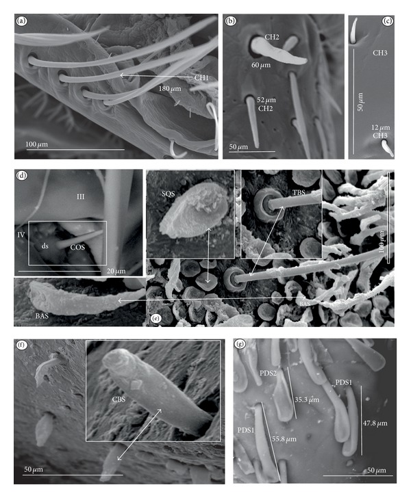
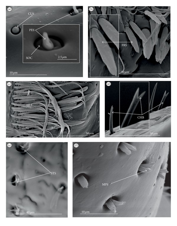
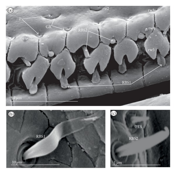
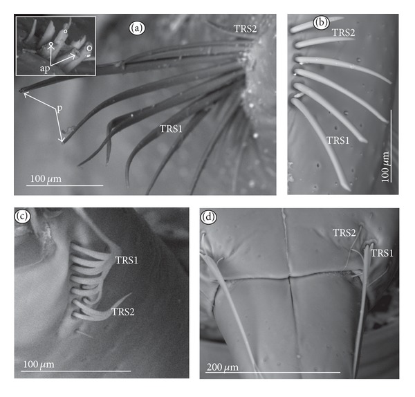
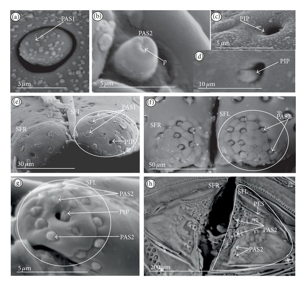
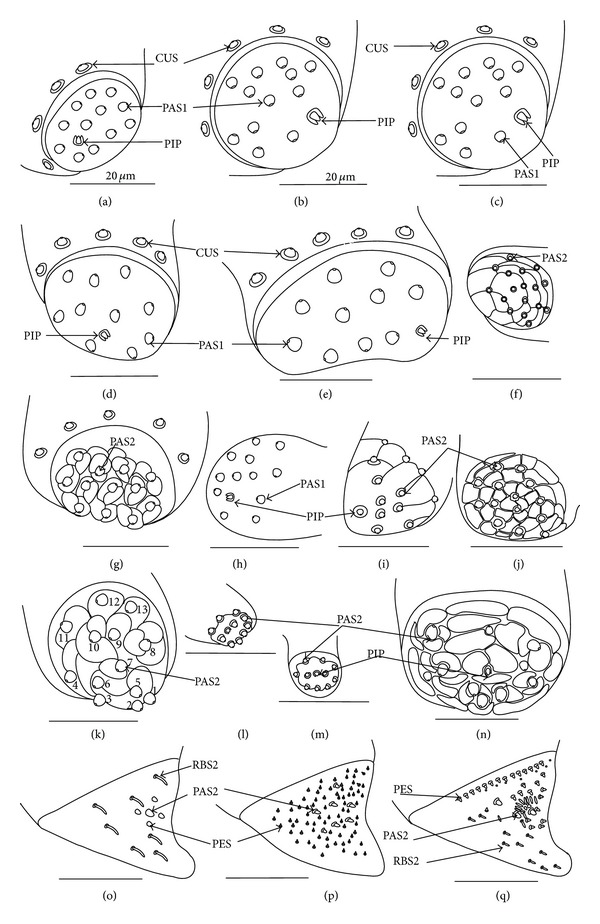
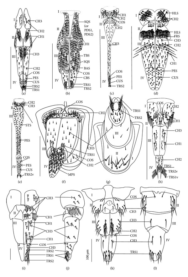
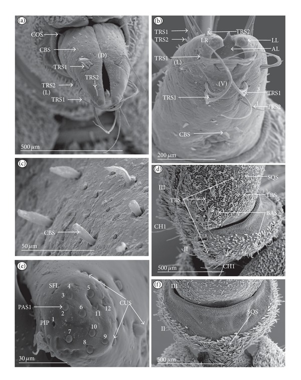
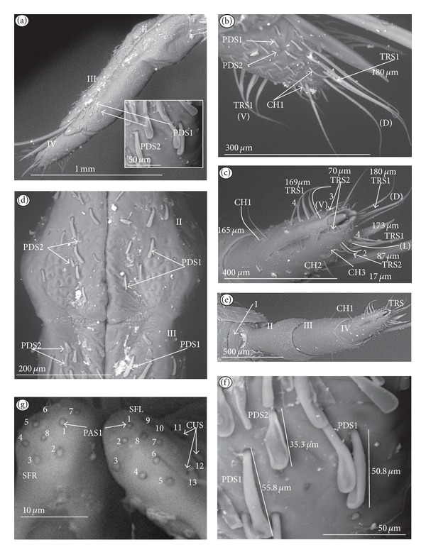
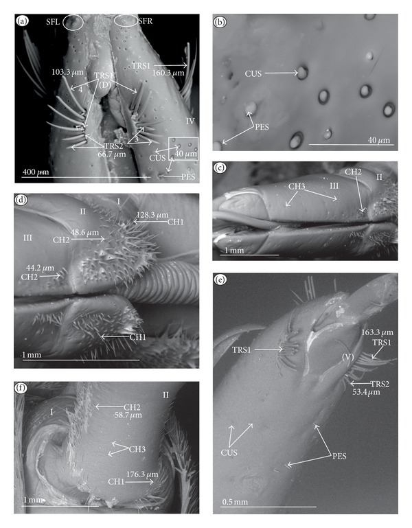
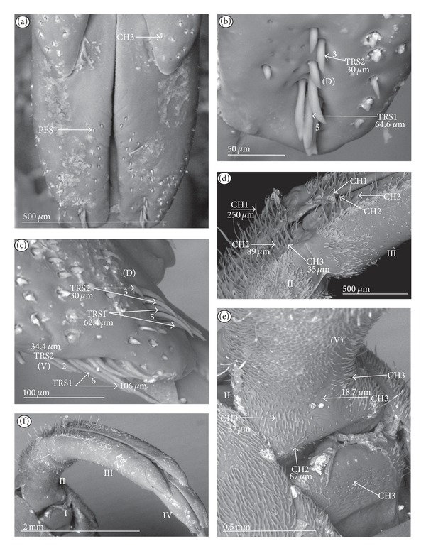
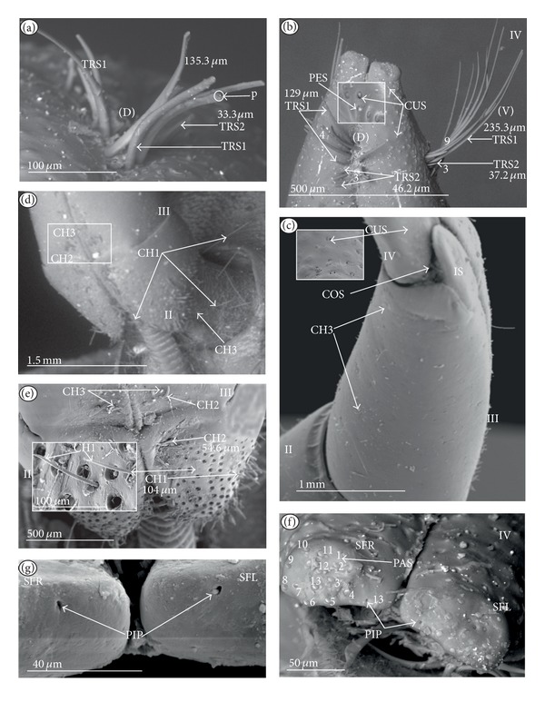
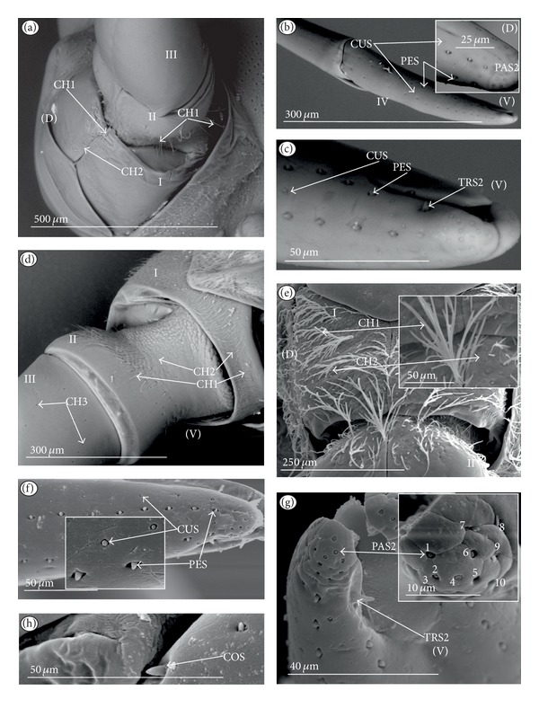
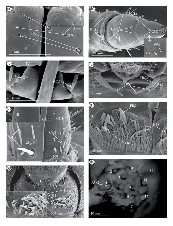
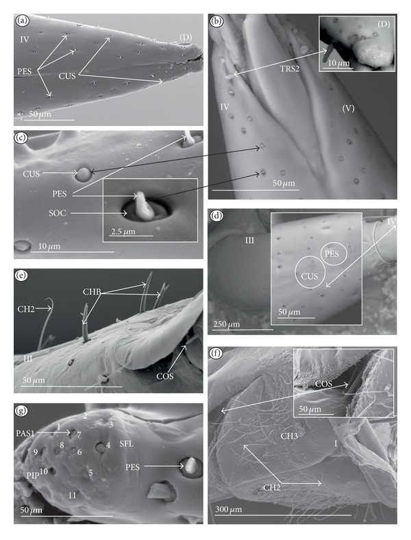
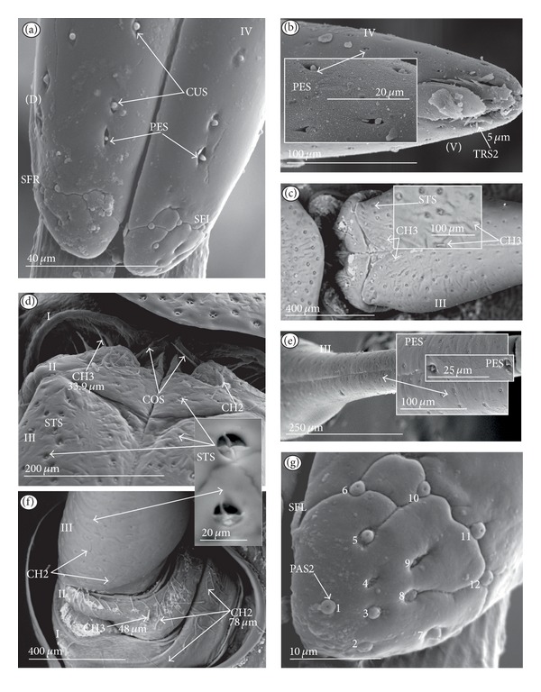
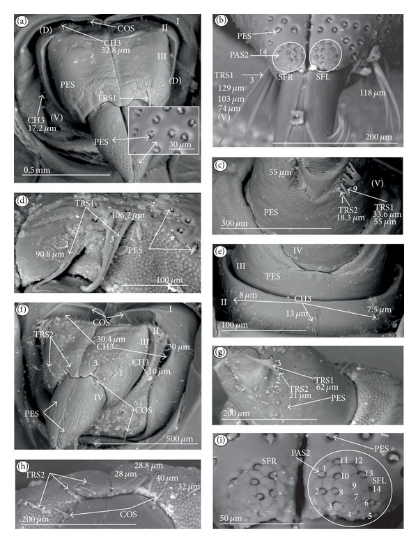
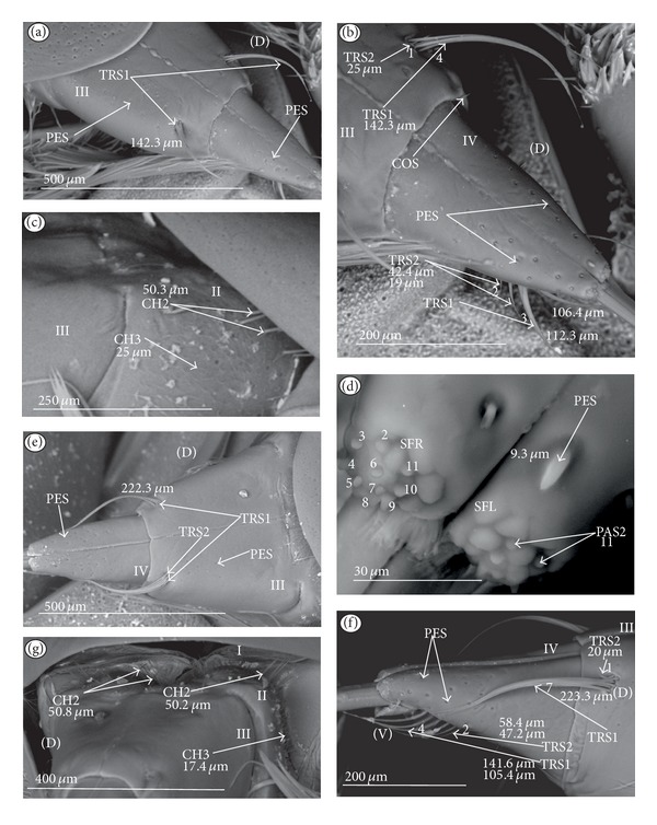
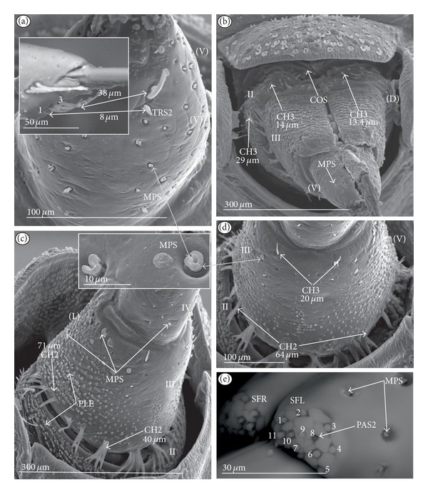
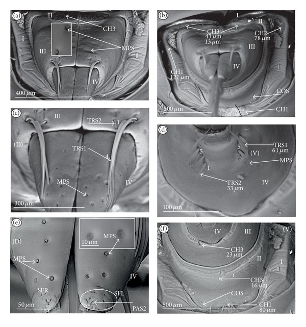
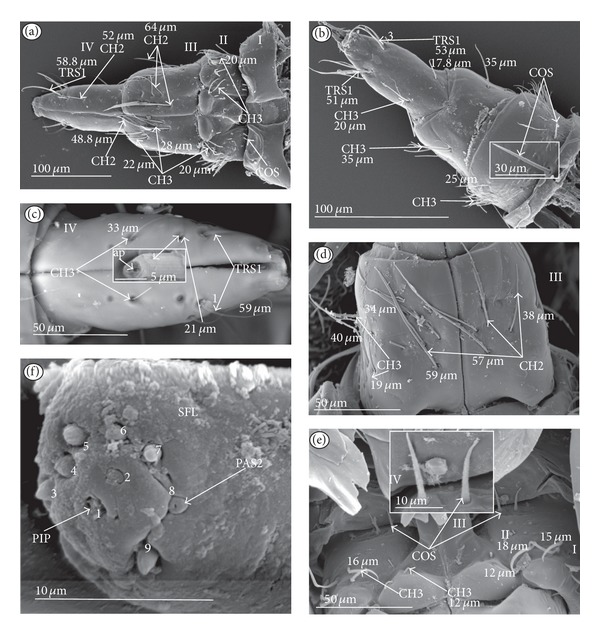
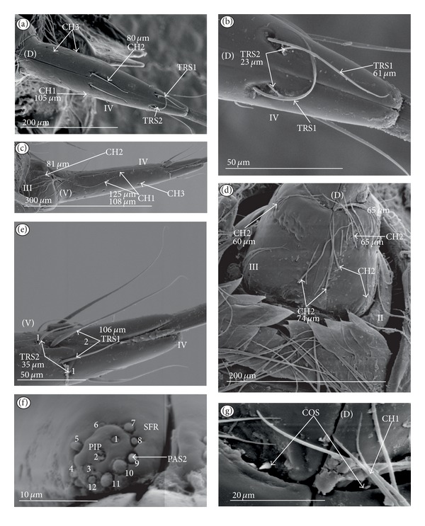
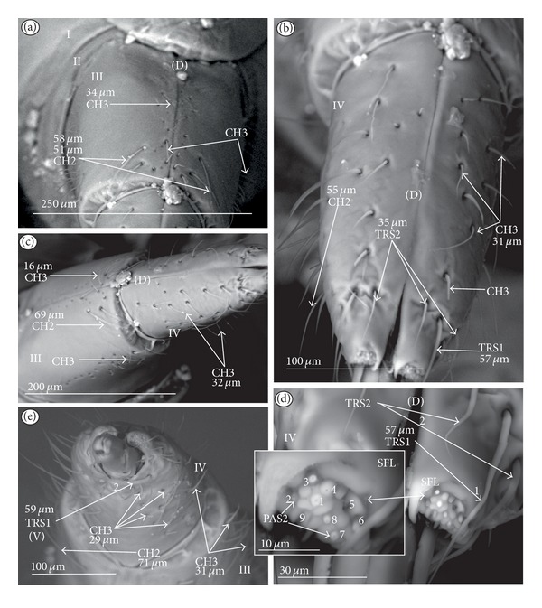
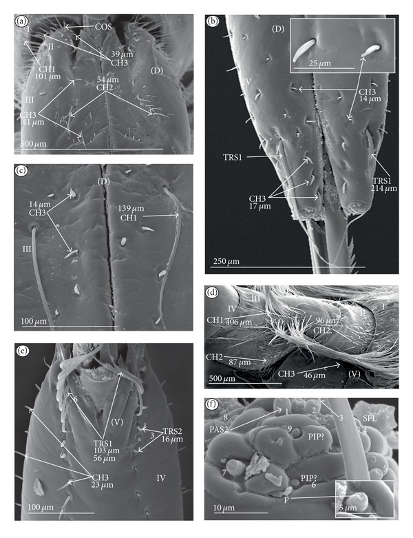
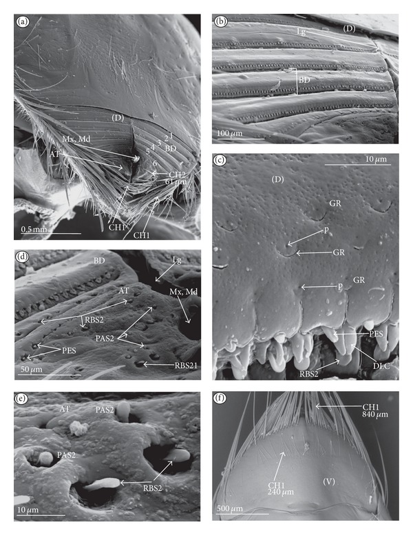
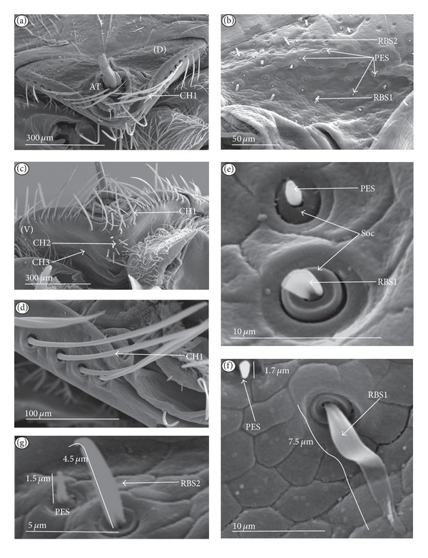
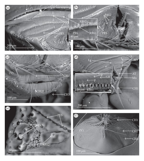
The present study provides new data concerning the morphology and distribution of the labial sensilla of 55 species of 12 nepomorphan families (Heteroptera: Nepomorpha) using the scanning electron microscope. On the labial tip, three morphologically distinct types of chemosensilla have been identified: two types of papillae sensilla and one type of peg-in-pit sensilla. Twenty-one morphologically distinct types of the mechanosensilla as well as two types of the trichoid sensilla (contact-chemoreceptive sensillum) have been identified on all labial segments in representatives of subfamilies. In Nepomorpha, morphological ground plan of the labial sensory structures is represented by an apical sensory field with 10-13 pairs of papillae sensilla and the peg-in-pit ones placed more laterally; numerous trichoid sensilla are placed on the IV segment; the chaetica sensilla are present and placed in groups or rows distributed along the labium near the labial groove on the dorsal side, and also several chaetica sensilla are unevenly scattered on the surface of that segment; the cupola and peg sensilla are numerous and evenly scattered on the fourth labial segment; the prioprerecptive sensilla, one pair is positioned on the dorsal side and on the fourth segment of the labium. The new apomorphical characters have been established for the labial sensilla in the Nepomorpha.


Figures

Figure 1
Figure 1
Types and sizes of the mechanosensilla of the Nepomorpha. (a) CH1 chaetic sensillum, long. (b) CH2 chaetic sensillum, medium length. (c) CH3 chaetic sensillum, short. (d) COS conical sensillum (prioprereceptive). (e) SQS squamiform sensillum, TBS trichobothrium sensillum, and BAS basiconic sensillum. (f) CBS clubbed-like sensillum. (g) PDS1 paddle-like sensillum, long, PDS2 paddle-like sensillum, short.
Figure 2
Figure 2
Types and sizes of the labial mechanosensilla of the Nepomorpha. (a) CUS cupola sensillum, PES peg sensillum. (b) FRS finger-like sensillum. (c) HLS freniale-like sensillum. (d) CHB chaetic sensillum with a bisected tip. (e) STS star-like sensillum. (f) MPS multilobed sensillum.
Figure 3
Figure 3
Types and sizes of the labial sensilla of the Nepomorpha (Corixoidea). (a) RBS ribbon-like sensillum. (b) RBS1 ribbon-like sensillum, long. (c) RBS2 ribbon-like sensillum, short, PES peg sensillum. g: groove, p: pore, rw1, rw2: the first and second row of semicircular grooves, rw3: the third row with the pores, rw4, rw5: the fourth and fifth rows with the ribbon-like sensillum.
Figure 4
Figure 4
Types and sizes of the trichoid sensilla of the Nepomorpha. (a) TRS1 trichoid sensillum, long, TRS2 trichoid sensillum, short; these sensilla are placed dorsally near the apex of the labium at the fourth segment; in the broken shank of the sensillum, a hole (ap) is visible, and it displays a dendrite running to the tip of the sensillum. (b) TRS1, TRS2 trichoid sensilla; these are individually placed dorsally near the apex of the labium in one row. (c) TRS1, TRS2 trichoid sensilla; these are placed ventrally, near the apex of the labium. (d) TRS1, TRS2 trichoid sensilla; they are placed dorsally on the third labial segment.
Figure 5
Figure 5
Types and arrangements of the labial apical sensilla of the Nepomorpha. (a) PAS1 papilla-like sensillum, flattened. (b) PAS2 papilla-like sensillum, rounded. (c) PIP peg-in-pit sensillum, only the pit is visible. (d) Peg-in-pit sensillum, the peg is visible. (e) PAS1 sensilla are circularly placed on the smooth, rounded labial tip with a slightly lateral position of the PIP. These are arranged symmetrically on the right (SFR) and left (SFL) sensory field. (f) PAS2 sensilla are circularly placed on the folded labial tip. The PIP are invisible. They are arranged symmetrically on the right (SFR) and left (SFL) sensory fields. (g) PAS2 sensilla are circularly placed on the labial tip with the central position of the PIP sensillum; the left (SFL) sensory field is shown. (h) PAS2 sensilla are irregularly placed on the labial tip. These sensilla are arranged symmetrically on the right (SFR) and left (SFL) sensory fields. PES form a short row; RBS ribbon-like sensilla (mechanosensilla) are not numerous.
Figure 6
Figure 6
Types of distribution of the labial apical sensilla in the Nepomorpha: Type A Nepidae, Belostomatidae, and Nerthrinae. (a) Nepa cinerea. (b) Laccotrephes japonensis. (c) Ranatra chinensis. (d) Belostomatinae; Appasus major. (e) Lethocerinae; Lethocerus deyrollei. (f) Type B, Ochteridae; Ochterus marginatus. (g) Type C, Gelastocorinae; Gelastocoris oculatus. (h) Type A, Nerthrinae; Nerthra nepaeformis. (i) Type B, Aphelocheiridae; Aphelocheirus aestivalis. (j) Type C, Cheirochelinae; Coptocatus oblongulus. (k) Limnocorinae; Limnocoris lutzi. (l) Helotrephidae; Helotrephes semiglobosus. (m) Type D, Pleidae; Paraplea frontalis. (n) Type C, Notonectinae; Notonecta glauca. (o) Type E, Cymatiinae; Cymatia coleoptrata. (p) Diaprepocoridae; Diaprepocoris zealandiae. (q) Micronectidae; Micronecta quadristrigata. PAS1 papilla sensillum, flattened; PAS2 papilla sensillum, rounded; PIP peg-in-pit sensillum; CUS cupola-like sensillum located outside the labial tip; RBS ribbon-like sensillum; PES peg sensillum.
Figure 7
Figure 7
Types of distribution of the mechanosensilla on the labial segments in the Nepomorpha (except for the Corixoidea). Sensilla numerous, grouped, and unevenly arranged on the labium ((a) Belostomatidae). Sensilla densely and evenly arranged on the labium ((b) Nepinae, Ranatrinae: I segment reduced dorsally); ((c) Ochteridae); ((d) Gelastocorinae). Sensilla less numerous and numerous evenly arranged ((e) Aphelocheiridae); ((f) dorsal side (g) ventral side of Cheirochelinae, Laccocorinae, Limnocorinae, Cryphocricinae, and Naucorinae). Sensilla are not very numerous and scattered unevenly ((h) Notonectinae); ((i) dorsal side, (j) lateral side of Pleidae); ((k) dorsal side, (l) ventral side of Helotrephidae).
Figure 8
Figure 8
Types of distribution of the mechanosensilla on the dorsal surface of the labium in the Corixoidea. Sensilla placed in the transverse bands or scattered unevenly on the dorsal labial surface ((a) and (b)) and sensilla scattered unevenly on the labial surface (c). (a) Diaprepocoridae; Diaprepocoris zealandiae, two and half of bands, three rows (r1, r2, r3) of semicircular grooves with a pore, two rows of ribbon-like sensilla. (b) Corixidae; six bands, three rows (r1, r2, r3) of semicircular grooves with a pore, two rows of ribbon-like sensilla. (c) Cymatiinae; ribbon-like sensilla (RBS1, RBS2) and peg sensilla (PES).
Figure 9
Figure 9
Types and sizes of sensilla of the Nepinae (Laccotrephes japonensis). (a) TRS1, TRS2 are placed dorsally (D) near of apex on the last segment of the labium, and TRS1, TRS2 are distributed laterally (L), CBS are distributed all over the surface of the last segment of the labium, and COS are visible in the proximal part of the IV segment. (b) TRS 1, TRS2 are placed ventrally (V) near the apex of the labium. LL: lateral left lobe, LR: lateral right lobe, Al, apical plate. (c) CBS are numerous and evenly distributed. (d) TBS, BAS, SQS, and CH1 are located dorsally and ventrally on III and II labial segment. (e) PAS1 (no. 2–12) and PIP (no. 1). (f) SQS are densely distributed over the ventral side.
Figure 10
Figure 10
Types, sizes and distribution of the sensilla of the Ranatrinae (Ranatra chinensis). (a) PDS1, PDS2, CH1, and CH2 are irregularly distributed on the segment II, III, and IV, dorsal view. (b) TRS1 are placed dorsally (D) near the apex of the labium (fourth segment). (c) TRS1, TRS2 distributed laterally (L), and ventrally (V); CH1 and CH2 are distributed all over the surface of the last segment of the labium. (d) PDS1, PDS2 evenly distributed, dorsal view of the II and III segment. (e) Ventral view of the I, II, and III segments, sensilla are not very numerous; they seem more numerous on the IV labial segment. (f) Shapes and sizes of the PDS1 and PDS2. (g) PASI (no 1–10) distributed apically, PIP is invisible.
Figure 11
Figure 11
Types, sizes, and distribution of the sensilla of the Belostomatinae (Belostoma flumineum). (a) TRS l, TRS2 are placed dorsally (D) near the apex of the labium. (b) TRS1, TRS2 are distributed ventrally (V); CUS, CH2 are distributed sparsely and unevenly all over the surface of the last segment of labium. (c) CUS and PES are placed dorsally; they are numerous and unevenly distributed over segment IV. (d) CH1, CH2, and CH3 sensilla are sparsely and unevenly distributed, ventral view on the I and II segments are one pair of the COS is situated ventrally; on the border between the I and II segments. (e) Lateral view of the I and II segment; sensilla are densely distributed ventrally, on segment I can be seen one group of the CH1 situated dorsally. (f) PAS1 (no. 1–10), PIP (no. 11) are distributed apicaily on the sensory fields: right (SFR) and left (SFL).
Figure 12
Figure 12
Types, sizes, and distribution of the sensilla of the Belostomatinae (Hydrocyrius colombiae). (a) TRS1 (four), TRS2 (three) form one row and are placed dorsally (D) near the apex of the labium; sensory fields (SFR, SFL); CUS and PES, placed dorsally, are numerous and unevenly distributed on segment IV. (b) CUS and PES (magnified). (c) CH3 are sparsely and unevenly distributed, dorsal view of segment III, several CH2 grouped near the labial groove. (d) CH2 are numerous, placed on the dorsal side, on segment II. (e) TRS1, TRS2 are distributed ventrally (V); CUS, PES, and CH3 are sparsely and unevenly distributed ventrally on the IV segment. (f) CH1 are visible at the base of segment II on the ventral side, CH2 are densely arranged along the surface of the second segment, and CH3 are slightly visible and unevenly distributed over segment II.
Figure 13
Figure 13
Types, sizes, and distribution of the sensilla of the Belostomatinae (Limnogeton fieberi). (a) PES are specifically situated only in the small area near the apex. (b) TRS2 (three), TRS1 (five) form a single row and are placed dorsally (D) near the apex of the labium. (c) TRS2 (two), TRS1 (six) form a single row and are placed ventrally (V) near the apex of the labium. (d) CH1, CH2, and CH3 densely cover the dorsal surface of the labium on segments II and III. (e) CH3 are numerous and is cover all surface of the ventral side of segment II, one pair of CH2 and at the base of the second segment, CH3 are densely distributed over segment I (ventrally). (f) Total view on the labial segments showing type (A) distribution of the labial sensilla.
Figure 14
Figure 14
Types, sizes, and distribution of the sensilla of the Belostomatinae (Lethocerus deyrollei). (a) TRS2 and TRS1 are placed dorsally (D) near the apex of the labium; the pore (ap) is visible at the end of TRS1. (b) TRS2 (three), TRS1 (four) form a single row dorsally (D), TRS2 (three) and TRS1 (nine) form a tuft ventrally (V), and CUS and PES are numerous on the IV segment. (c) CH3 are spread sparsely on III segment, and COS is situated on segment IV and hidden under IS (intercalary sclerites) of the III segment. (d) CH1, CH2, and CH3 densely cover the dorsal and ventral surfaces of the labium on segments II and III. (e) CH3 and CH2 are less numerous and placed dorsally in a small group on segment III. CH1 are numerous on the dorsal surface of the II segment. (f) PAS1 (no. 1–13) and PIP (no. 14) are distributed apically over sensory fields (SFR, SFL). (g) PIP situated in the lateral positions on SRF and SFR are clearly visible.
Figure 15
Figure 15
Types and sizes of the sensilla of the Ochteridae ((a)–(d) Ochterus piliferus, (e)–(g) O. marginatus). ((a) and (d)) CH1, CH2 are numerous and distributed dorsally (D) and ventrally (V) on the I and II segments of the labium. ((b) and (c)) PES are numerous and cover the III and IV segments of the labium in even rows, and between two rows of pegs there is a row of CUS. PAS2 are distributed on the labial tip. One pair of TRS2 is situated near the tip of the labium. (e) CH1, CH2 are densely distributed dorsally (D) on I and II segments of the labium. (f) PES are numerous and cover the III and IV segment of the labium in even rows, and between two rows of pegs there is a row of CUS. (g) PAS2 (no. 1–10) are on the labial tip; PIP is not visible. (h) COS (prioprereceptive sensilla) are situated on segment IV, dorsally, near the base of segment III.
Figure 16
Figure 16
Types and sizes of the sensilla of the Gelastocoridae: Gelastocorinae (Gelastocoris oculatus). (a) On the dorsal surface PES are numerous and cover the IV segment of the labium in even rows, and between two rows of pegs there is a row of CUS. (b) CUS and PES are numerous and cover the ventral surface of the IV segment, a few CH1 are situated laterally (L) on the III segment, one pair of CH2 is placed ventrally (V), and a few CH2 are more situated more laterally on the III segment. (c) COS (prioprereceptive sensilla) are situated dorsally on the IV segment near the base of the III segment. (d) TRS2 (three) are situated ventrally near the tip of the labium. (e) CH1, CH2 are less numerous and situated dorsally (D) on the III segment of the labium, and CH3 are numerous and slightly visible. (f) Several FRS are present on the dorsal surface of the II segment, and HLS are very numerous and distributed in bands along the width of segment II. (g) CH2 and HLS are numerous and densely cover the dorsal surface on the labial segment I; COS are situated at the basal edge of the II segment (h) PAS2 (no. 1–13) are on the left SFL, and PIP in not visible.
Figure 17
Figure 17
Types and sizes of the sensilla of the Gelastocoridae: Nerthrinae (Nerthra nepaeformis). (a) On the dorsal surface PES and CUS are numerous and evenly cover the IV segment of the labium. (b) TRS2 (one) is situated ventrally near the tip of the labium. (c) The shapes of CUS and PES, magnified. The socket is clearly visible. (d) PES and CUS are numerous and cover the ventral surface of the II and III segments. (e) CHB are unevenly distributed on the dorsal surface of the III segment, and COS (prioprereceptive sensilla) are situated on the dorsal surface of the IV segment near the base of the III segment. (f) CH2 are numerous and situated dorsally (D) on segment I of the labium, and COS are placed at the base of the edge of segment II. (g) PAS1 (no. 1–11) are distributed over SFL.
Figure 18
Figure 18
Types and sizes of the sensilla of the Aphelocheiridae (Aphelocheirus aestivalis). (a) PES and CUS are numerous and distributed unevenly in rows on the IV segment (dorsal view). (b) TRS2 (one) is situated ventrally near the tip of the labium; PES and CUS are distributed similarly as on the dorsal side. (c) STA and CH3 are in the proximal part of the III segment (dorsal view). (d) STS, CH2, CH3, and COS are situated on the II segment (dorsal view). (e) PES are unevenly distributed on the dorsal surface of the distal part of segment III. (f) CH2 are not numerous and are situated ventrally (V) on segments I and II of the labium. (g) PAS2 (no. 1–12) are distributed over SFL.
Figure 19
Figure 19
Types and sizes of the sensilla of the Naucoridae: Cheirochelinae ((a)–(e), Cheirochela feana, (f)–(i), Gestroiella limnocoroides). (a) PES are numerous and distributed evenly on the IV segment (dorsal view), PES are present only in the distal area of the III segment, CH3 are less numerous and distributed over the II segment (dorsally and ventrally), and COS are situated on segment II (dorsal view). (b) PAS2 (no. 1–14) are distributed over SFL and SFR; TRS1 (three) are visible only ventrally (V) (the first specimen). (c) TRS1 (nine) and TRS2 (one) are distributed ventrally (V) near the apex of segment IV (the second specimen), and PES are numerous. (d) TRS1 are distributed in two tufts (2 + 3) on the distal edge of the III segment, and several PES are visible near the TRS1. (e) CH3 are sparsely distributed over segment II; several PES are visible on the III segment (ventral view). (f) PES are numerous and distributed evenly over the IV segment, TRS2 (four) are distributed separately (3 + 1) on the distal edge of the III segment, several PES are visible near TRS1, CH3 are less numerous and distributed over the II segment, and COS are situated on segments IV and II (ventral view). (g) TRS1 (three) and TRS2 (two) are distributed ventrally (V) near the apex of segment IV. (h) TRS2 and COS magnified. (i) PAS2 (no. 1–14) are distributed over SFL and SFR.
Figure 20
Figure 20
Types and sizes of the sensilla of the Naucoridae: Laccocorinae ((a)–(d), Laccocoris hoogstraali, (e)–(g), Heleocoris humeralis). (a) PES are numerous and distributed evenly on the IV segment (dorsal view). (b) TRS1 (three) and TRS2 (two) are distributed on the ventral (V) and dorsal (D) sides, and TRS1 (four) and TRS (two) are situated d in one tuft on the distal edge of the III segment. (c) CH3 densely cover the dorsal surface on the II segment; several CH2 are present. (d) PAS2 (no. 1–11) are distributed over SFL and SFR. (e) TRS1 and TRS2 are placed in one tuft on the distal edge of the III segment; PES are distributed evenly on the dorsal surface of the IV segment but are less numerous on the III segment. (f) PES are numerous and distributed evenly on the IV segment (lateral view); TRS2 (two) and TRS1 (four) are distributed ventrally (V) and dorsally (D); TRS1 (seven) and TRS2 (one) are distributed on the distal edge of the III segment. (g) CH3 densely cover the dorsal surface (D) of the II segment; only several CH2 are present.
Figure 21
Figure 21
Types and sizes of the sensilla of the Naucoridae: Limnocorinae (Limnocoris lutzi). (a) MPS are numerous and distributed in rows over the IV segment (dorsal view). (b) PAS (no. 1–12) are located on the labial tip (SFL). (c) TRS1 (four) and TRS2 (one) are situated on the ventral side (V) in one row near the apex of the IV segment; MPS are also numerous ventrally and distributed in rows. (d) CH2 are less numerous and sparsely distributed over the ventral surface of the III and II segments. (e) TRS2 (three) are distributed separately on the distal edge of the III segment, MPS are less numerous and sparsely situated on the dorsal surface of the III segment, COS are situated on the IV segment, and on the surface of the III segment there are numerous, characteristic plates (generally unidentified structures, PLE-type). (f) Two (or four) COS-p are visible on the external distal edge of the II segment, and one is situated internally closer to the labial groove (Figure 21(f)). Two the COS-d are situated in the middle of the II segment on the proximal edge.
Figure 22
Figure 22
Types and sizes of the sensilla of the Naucoridae: Cryphocricinae (Cryphocricos hungerfordi). (a) MPS are numerous and spread evenly on the IV segment (ventral view); TRS2 (four) are distributed over the ventral side (V) in one row near the apex of the IV segment. (b) MPS are numerous and distributed evenly over the IV segment (dorsal view), CH3 are less numerous and situated on the dorsal surface of the II segment, and COS are situated on the II segment (dorsal view). (c) MPS are less numerous, and only a few are situated on the dorsal surface of the III segment; on this segment there are present numerous, unidentified plates (PLE); CH2 are situated around the edge of the II segment on the ventral side. (d) One pair of CH3 is situated ventrally near the distal edge of the III segment. (e) PAS2 (no. 1–11) are situated on the labial tip (SFL).
Figure 23
Figure 23
Types and sizes of the sensilla of the Naucoridae: Cryphocricinae (Ambrysus occidentalis). (a) MPS are less numerous and spread evenly over the III segment (dorsal view); several CH3 are present on the II segment (dorsal view). (b) CH2 and CH1 cover the dorsal and ventral surfaces of the I segment; COS are situated ventrally on the II segment. (c) TRS1 (four) and TRS2 (one) are situated on the dorsal side (D), in one tuft located distally on the III segment. (d) TRS1 (five) and TRS2 (one) are distributed over the ventral side (V) in one row near the apex of the labium. (e) MPS are less numerous and distributed evenly over the IV segment (dorsal view); PAS2 are distributed on the labial tip (SFR, SFL). (f) One pair of CH3 is situated ventrally near the distal edge of the III and II segments, CH1 are situated oil the edge of the I segment on the ventral side, and COS are located on the II segment on the ventral.
Figure 24
Figure 24
Types and sizes of the sensilla of the Naucoridae: Naucorinae ((a) and (b) Naucoris maculatus, (c)–(e) Namtokocoris siamensis, (f) and (g) Neomacrocoris handlirschi). (a) CH2 are numerous and form a collar around the distal edge of the II segment on the ventral side; individual CH2 can be seen on the III segment (lateral view); TRS1 and TRS2 are visible ventrally and dorsally. (b) TRS1 (four) are distributed over the ventral side (V) in one row near the apex of the labium; MPS are less numerous and spread evenly over the IV segment (dorsal view); TRS1 (two) are distributed over the dorsal side (D), in one tuft placed distally on the III segment; COS are situated dorsally on the IV segment. (c) CH2 and CH1 are densely spread over the ventral side of the III and II segments; COS are distributed dorsally over the IV segment. (d) TRS1 (four) are distributed over the dorsal side (D), in one tuft located distally on the III segment; MPS are numerous and spread evenly over the IV segment (dorsal view). (e) TRS1 (four) and TRS2 (two) are distributed over the ventral side (V) in one row near the apex of the labium; PAS2 (no. 1–10) and PIP (no. 11) are distributed on the labial tip. (f) TRS1 (twelve) arc distributed over the dorsal side (D), in one tuft placed distally on the III segment; MPS are less numerous and distributed evenly over the IV segment (dorsal view). (g) Several CH2 are visible on the ventral side of the II segment; less numerous CH3 are present on the II and III segments.
Figure 25
Figure 25
Types and sizes of the sensilla of the Pleidae (Paraplea frontalis). (a) CH2 and CH3 are less numerous and spread unevenly over all segment; TRS1 (one) is placed dorsally near the apex; one pair of COS is situated on the II segment. (b) TRS1 (three) are located ventrally near the apex; one pair of CH3 is situated near the proximal edge of the IV segment; one pair of CH3 is situated ventrally on the III segment; several CH3 are visible on the ventral side of the II segment, and some COS are located near the edge of the I segment; no sensila can be seen on segment I (ventral view). (c) TRS1 (one), TRS2 (one), and CH3 (two) are situated on the dorsal surface of the IV segment. (d) CH2 (several) are situated on the dorsal side and CH3 (several) are situated on the lateral side of the III segment. (e) Several CH3 are present on the III segment dorsally as well as one pair of COS. (f) PAS2 (no. 2–9) are placed in a circle on the labial tip, and PIP (no. 1) is situated centrally.
Figure 26
Figure 26
Types and sizes of the sensilla of the Helotrephidae (Hydrotrephes visayasinensis). (a) Several CH3, one CH2, and one CH1 are spread dorsally (D) oil the IV segment. (b) TRS1 (one) and TRS2 (one) are situated dorsally (D) near the apex. (c) Several CH3 and four CH1 are situated ventrally on the IV segment; several CH2 are situated on the III segment. (d) CH2 are placed near the labial groove and also near the distal edge of the III segment. (e) TRS1 (two) and TRS2 (two) are situated in two tufts, ventrally, near the apex of the labium. (f) Sensory field (SFR) with PAS2 (no. 1, 3–13) and PIP sensilla (no. 2). (g) COS are present on the dorsal surface of the IV segment, and several CH1 are present on the first segment.
Figure 27
Figure 27
Types and sizes of the sensilla of the Notonectidae: Anisopinae (Buenoa uhleri). (a) Several CH3 and CH2 are present on the III segment (dorsal view). (b) Several CH3 and CH2 are present on the IV segment (dorsal view). (c) Several CH3 are scattered all over the surface of the IV segment (lateral view), two CH2 are situated near the distal edge of the III segment, and CH3 are situated in rows dorsally and laterally. (d) TRS1 (one) and TRS2 (two) are distributed over the dorsal side (D) near the apex of the labium; PAS2 (no. 1–9) are situated on the sensory field (left-side view; SFL). (e) Several CH3 are visible on the ventral side of the IV and III segments; TRS1 (two) are situated ventrally near the apex.
Figure 28
Figure 28
Types and sizes of the sensilla of the Notonectidae: Notonectinae (Notonecta glauca). (a) The arrangement of several CH2, CH3, and CH1 on the dorsal surface of the II segment; COS is situated near the edge of the I segment. (b) TRS1 (one pair) placed dorsally, near the apex; CH3 are numerous and distributed all over the surface of the IV segment (dorsal view). (c) CH1 and CH3 are situated in the distal part of the III segment. (d) CH1 are numerous and densely cover the ventral side of the II and I segments; CH2 are numerous and situated on the lateral side of the III segment; CH2 and CH3 are numerous and densely spread over the II and I segments (ventral view). (e) TRS1 (six) and TRS2 (three) are placed near the apex; CH3 are situated in two rows (ventral view, IV segment). (f) PAS2 (no. 1–7) and probably PIP (no. 6 or 9) are located on the sensory field (left-side view; SFL).
Figure 29
Figure 29
Types and sizes of the sensilla of the Corixidae: Corixinae (Corixa dentipes). (a) BD (no. 1–6) are spread evenly on the dorsal surface of the labium; several CH1 and CH2 are arranged on the lateral and ventral edges of the membranous labial lip (AT); CH1 located on the lateral edge of the labium below the apical tip. (b) BD magnified. (c) Three rows of porous sensilla (PLS) and RBS2 and RBS1 sensilla situated in two rows. (d) The apical tip (AT) with PES, PAS2, and RBS2 sensilla unevenly spread except for PES (they form a row). (e) PAS2 and RBS2 magnified. (f) Ventral view, CH1 are numerous and arranged on the lateral and frontal edges of the labium; shorter CH1 arranged in one row on the ventral side of the base of the labium.
Figure 30
Figure 30
Types and size of the sensilla of the Corixidae: Cymatiinae (Cymatia coleoptrata). (a) Several of the CH1 are placed on the lateral edge of the labium and below the apex; several PES and RBS2 are visible on AT. (b) RBS1, RBS2, and PES are spread unevenly on the dorsal surface of the labium. (c) CH1, CH2, and CH3 are less numerous and situated ventrally. (d) CH1 magnified (lateral view). (e) PES and RBS1 magnified, Soc: socket. (f) RBS1, magnified. (g) The sizes of PES and RBS2.
Figure 31
Figure 31
Types and sizes of the sensilla of the Diaprepocoridae and Micronectidae ((a)–(c) Diaprepocoris zealandiae, (d)–(f) Micronecta quadristrigata). (a) BD (no. 1−2.5) are spread evenly on the dorsal surface. (b) AT in magnification; PES or PAS2 are numerous and unevenly cover the tip. (c) Several of the CH1 are arranged on the lateral edge of the membranous labial tip (AT); the CH1 are placed on the lateral edge of the labium; the CH2 and CH3 are placed below the apex, on the ventral side. (d) BD (no. 1–4) are spread evenly on the dorsal surface; the CH1 are placed on the lateral edge of the labium. (e) BD in magnification, PES, RBS2, and PAS on the apex (AT). (f) The CH1 are numerous and situated below the apex (ventral view), the CH2 are situated in a short row below the CH1, and the CH3 are not numerous and placed below the CH2.

Similar articles

Cited by

References

    1. Cobben RH. Evolutionary Trends in Heteroptera—Part 2: Mouthpart-Structures and Feeding Strategies. Meded, Landbou-whogeschool Wageningen; 1978.
    1. Popov YA. Historical development of the hemipterous infraorder Nepomorpha. Trudy Paleontological Institute Academy of Science, Nauk, USSR. 1971;129:1–228. (Rus).
    1. Backus EA. Anatomical and sensory mechanisms of leafhopper and planthopper feeding behavior. In: Nault LR, Rodriguez JG, editors. The Leafhoppers and Planthoppers. New York, NY, USA: John Wiley & Sons; 1985. pp. 163–194.
    1. Chapman RF. Mechanoreception. Chemoreception. In: Chapman RF, editor. The Insects, Structure and Function. 4th edition. Cambridge, UK: Cambridge University Press; 1998. pp. 610–653.
    1. Miles PW. The stylet movements of a plant-sucking bug, Oncopeltus fasciatus Dall. Heteroptera: Lygaeidae. Proceedings of the Royal Entomological Society of London. 1958;33:15–20.

Publication types

MeSH terms

LinkOut - more resources