. 2013 Sep 27;288(39):27849-60.
doi: 10.1074/jbc.M112.445098. Epub 2013 Aug 9.

Noncanonical control of vasopressin receptor type 2 signaling by retromer and arrestin

Affiliations

Noncanonical control of vasopressin receptor type 2 signaling by retromer and arrestin

Timothy N Feinstein et al. J Biol Chem. .

Abstract

The vasopressin type 2 receptor (V2R) is a critical G protein-coupled receptor (GPCR) for vertebrate physiology, including the balance of water and sodium ions. It is unclear how its two native hormones, vasopressin (VP) and oxytocin (OT), both stimulate the same cAMP/PKA pathway yet produce divergent antinatriuretic and antidiuretic effects that are either strong (VP) or weak (OT). Here, we present a new mechanism that differentiates the action of VP and OT on V2R signaling. We found that vasopressin, as opposed to OT, continued to generate cAMP and promote PKA activation for prolonged periods after ligand washout and receptor internalization in endosomes. Contrary to the classical model of arrestin-mediated GPCR desensitization, arrestins bind the VP-V2R complex yet extend rather than shorten the generation of cAMP. Signaling is instead turned off by the endosomal retromer complex. We propose that this mechanism explains how VP sustains water and Na(+) transport in renal collecting duct cells. Together with recent work on the parathyroid hormone receptor, these data support the existence of a novel "noncanonical" regulatory pathway for GPCR activation and response termination, via the sequential action of β-arrestin and the retromer complex.

Keywords: Arrestin; G Protein-coupled Receptors (GPCR); Kidney Metabolism; Membrane Trafficking; Signaling; V2R.


Figures
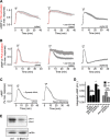
FIGURE 1.
FIGURE 1.
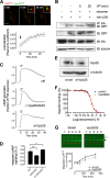
Internalization and arrestin binding regulate the time course of cAMP signaling mediated by V2R. A and B, averaged time course of cAMP generation (black) and PKA activity (red) measured by FRET-based biosensors in HEK293 cells expressing the human V2R and a cytoplasmic sensor for cAMP (Epac-CFP/YFP) or a sensor for PKA activity (AKAR3). Cells were perfused with buffer or with ligand (horizontal bars representing 30 s of a saturating concentration of either VP (100 nm) (A) or OT (10 μm). Data represent the mean ± S.E., of N ≥ 3 experiments and n ≥ 28 cells. The expression level of β-arrestins was titrated by either overexpression of a dominant-active mutant (βarr1(IV-AA), middle panel) or depletion of both β-arrestins by siRNA (right panel). C, averaged time course of cAMP generation in HEK293 cells expressing V2R and a dominant-negative mutant of dynamin 1 that prevents receptor internalization (K44A, right panel) or in which cAMP detection was limited to the plasma membrane by expression of a plasma membrane-limited mutant of the biosensor epac1CFP/YFP (right panel). Note that FRET was detected using TIRF microscopy. D, total cAMP production of data shown in A–C as measured by the integrated area under the curve. All manipulations are significant at least to p ≤ 0.05 by one-way analysis of variance and pairwise comparison with controls challenged with VP (black) or OT (gray); in all cases N ≥ 3. A.U., arbitrary unit. *, p < 0.05; **, p < 0.01; ***, p < 0.001. E, native β-arrestins were depleted from HEK293 cells as described under “Experimental Procedures,” and the reduction of native protein was shown by Western blotting. ctrl, control.
FIGURE 2.
FIGURE 2.
Arrestin control of V2R signaling. A, Western blot showing expression of both recombinant β-arrestin1(IV-AA) (left panel) and β-arrestin2YFP (right panel). B, FRET between V2R-CFP and βarr2-YFP, as measured by TIRF microscopy (green, blue) or by epifluorescence (black). Horizontal bar indicates that cells were challenged for 30 s with either 100 nm VP or 10 μm OT and then washed with buffer. Data represent the mean ± S.E. of N ≥ 3 experiments and n ≥ 28 cells. C and D, time course (C) and basal activity (D) of [35S]GTPγS binding to GαS was measured in plasma membrane extracts of HEK293 cells expressing V2R with or without purified β-arrestin2 (red line). ANOVA, analysis of variance. Data represent mean ± S.E. of n = 4 independent experiments. ctrl, control. **, p < 0.01.
FIGURE 3.
FIGURE 3.
Retromer binds V2R and desensitizes VP-mediated cAMP generation. A, co-localization of V2RCFP (red) and Vps29YFP (green) in HEK293 cells challenged with 100 nm VP (arrow) and imaged using time-lapse confocal microscopy. Co-localization between V2R and retromer is shown (insets) and was quantified using Pearson's coefficient. Bars represent mean ± S.E. of N = 3 experiments and n = 14 cells. The bar represents 10 μm. B, binding of V2R and retromer was measured in HEK293 cells transfected with HA-V2R along with either Vps29YFP, Vps26, and Vps35 after a challenge with VP (100 nm) or carrier followed by immunoprecipitation (IP) and Western blot analysis. IB, immunoblot. Image is representative of n = 4 independent experiments. C, time course of cAMP generation in HEK293 cells transfected with cDNAs encoding V2R and Epac-CFP/YFP along with either siRNA against the retromer subunit Vps35 (si-Vps35) or plasmids encoding the retromer subunits Vps26, Vps29, and Vps35 (retromer). The horizontal bar indicates a 30-s challenge with 100 nm VP followed by washout with buffer. Vertical bars represent mean ± S.E. of N ≥ 3 experiments and n ≥ 28 cells. ctrl, control. D, total cAMP signaling of cAMP curves shown in A and B, as measured by the integrated area under the curve. *, p < 0.05; **, p < 0.01. E, measurement of Vps35 depletion by Western blot. F, affinity of V2R cells in the presence of overexpressed retromer was measured by competition binding assay in HEK293 cells. G, degradation of VP-challenged GFPV2R in HEK293 cells (left panel) and HEK293 cells overexpressing Vps26/29 (right panel) was measured by Western blot.
FIGURE 4.
FIGURE 4.
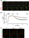
Significance of internalization for V2R signaling. A, co-localization of β-arr1tom (red, top row) or β-arr1(IV-AA)tom (red, bottom row) and V2RCFP (green) in HEK293 cells after a challenge with vasopressin (100 nm). The bar represents 10 μm. B, time course of cAMP generation in HEK293 transfected with β-arr1(IV-AA)tom (red) or a control plasmid (green). Gray bars represent the mean ± S.E., of N ≥ 3 experiments and n ≥ 28 cells. Time points indicated in B correspond to panel numbers in A. C, time-lapse confocal imaging of V2RCFP (red) and the retromer subunit Vps29YFP (green) at time points (min) shows no internalization of V2R after challenge with a saturating concentration of OT. Bar, 10 μm.
FIGURE 5.
FIGURE 5.
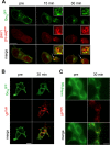
Internalization of a functional V2R-β-arrestin signaling complex. A, co-internalization of β-arrestin and GαS was demonstrated by challenging HEK293 cells expressing V2R along with GαS tagged with GFP (GαSGFP) and β-arrestin1(IV-AA) labeled at its N terminus with dTomato (dTomβarr1(IV-AA)). B and C, HEK293 cells expressing HA-V2R and GαSGFP (B) or stained with fluorescent forskolin (bodipyFSK) and then fixed (C) were challenged with VPTMR and visualized at the designated time points using confocal microscopy. Bar, 10 μm.
FIGURE 6.
FIGURE 6.
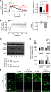
Differential actions of oxytocin and vasopressin on kidney cells. A, cAMP was measured in mpkCCDC14 cells using the epac1-CFP/YFP biosensor, as described above. Means ± S.E. of integrated cAMP generation (right panel), N ≥ 13. A.U., arbitrary unit. *, p < 0.05; **, p < 0.001. B, transepithelial current of polarized mpkCCDC14 cells was measured electrophysiologically by mounting permeable supports in Ussing chambers. Addition of VP or OT to the basolateral chamber (black bar) was followed by washout of the basolateral chamber with 5× volume of fresh medium (broken bar). The decrease in conductance at this time is an artifact attributable to washing. Vertical deviations (green arrows) represent ±2-mV voltage clamp pulses induced once each minute to verify the continued epithelial integrity. Amiloride was added at the end of each experiment to verify that >95% of the measured current was attributable to ENaC (right panel). Means ± S.E. of the slope of transepithelial current versus time after peak current at ∼6 min, N ≥ 6. *, p < 0.05. C, phosphorylation of AQP2 residues Ser-256 and Ser-269 after challenge with VP or OT for 10 min and measured without ligand washout (10 min) or after a 20-min ligand washout (10 min, w/o 20 min). D, corresponding band intensities from A were quantified by densitometry. Data represent the mean ± S.E., of n = 3 separate experiments. **, p < 0.01; ***, p < 0.001. E, translocation of AQP2 from the cytosol (center of cell) to the apical plasma membrane (apical PM) of a polarized monolayer of MDCK cells after challenge with VP or OT for 10 min and measured without ligand washout (10 min), or after a 20 min ligand washout (10 min, w/o 20 min). Bar, 10 μm.
FIGURE 7.
FIGURE 7.
Model of regulation of water transport in epithelial cells of the kidney by V2R. VP binding to the V2R induces β-arrestin1/2 binding (red) and internalization of a VP-V2R-arrestin complex, which continues to generate cAMP via adenylyl cyclase (AC, gray) from endosomes until bound by retromer (orange). cAMP-activated protein kinase A (PKA) stimulates phosphorylation of the water channel AQP2 (blue) on serine 269, stabilizing its localization to the apical plasma membrane. OT also activates PKA via generation of cAMP from the basolateral plasma membrane, but OT does not induce a sustained cAMP response upon β-arrestin1/2 mobilization or internalization of active receptor. This causes a transient phosphorylation of AQP2 and transient localization of AQP2 in the apical plasma membrane.

Similar articles

Cited by

References

    1. Lohse M. J., Benovic J. L., Codina J., Caron M. G., Lefkowitz R. J. (1990) β-Arrestin: a protein that regulates β-adrenergic receptor function. Science 248, 1547–1550 - PubMed
    1. Laporte S. A., Miller W. E., Kim K. M., Caron M. G. (2002) β-Arrestin/AP-2 interaction in G protein-coupled receptor internalization: identification of a β-arrestin binding site in β2-adaptin. J. Biol. Chem. 277, 9247–9254 - PubMed
    1. Nelson C. D., Perry S. J., Regier D. S., Prescott S. M., Topham M. K., Lefkowitz R. J. (2007) Targeting of diacylglycerol degradation to M1 muscarinic receptors by β-arrestins. Science 315, 663–666 - PubMed
    1. Perry S. J., Baillie G. S., Kohout T. A., McPhee I., Magiera M. M., Ang K. L., Miller W. E., McLean A. J., Conti M., Houslay M. D., Lefkowitz R. J. (2002) Targeting of cyclic AMP degradation to β2-adrenergic receptors by β-arrestins. Science 298, 834–836 - PubMed
    1. Luttrell L. M., Lefkowitz R. J. (2002) The role of β-arrestins in the termination and transduction of G-protein-coupled receptor signals. J. Cell Sci. 115, 455–465 - PubMed

Publication types

MeSH terms

LinkOut - more resources