. 2012 Mar;6(1-2):19-32.
doi: 10.1080/21553769.2012.761163. Epub 2013 Mar 21.

Twins, quadruplexes, and more: functional aspects of native and engineered RNA self-assembly in vivo

Affiliations
Free PMC article

Twins, quadruplexes, and more: functional aspects of native and engineered RNA self-assembly in vivo

Richard A Lease et al. Front Life Sci. 2012 Mar.
Free PMC article

Abstract

The primacy and power of RNA in governing many processes of life has begun to be more fully appreciated in both the discovery and inventive sciences. A variety of RNA interactions regulate gene expression, and structural self-assembly underlies many of these processes. The understanding sparked by these discoveries has inspired and informed the engineering of novel RNA structures, control elements, and genetic circuits in cells. Many of these engineered systems are built up fundamentally from RNA-RNA interactions, often combining modular, rational design with functional selection and screening. It is therefore useful to review the particular class of RNA-based regulatory mechanisms that rely on RNA self-assembly either through homomeric (self-self) or heteromeric (self-nonself) RNA-RNA interactions. Structures and sequence elements within individual RNAs create a basis for the pairing interactions, and in some instances can even lead to the formation of RNA polymers. Example systems of dimers, multimers, and polymers are reviewed in this article in the context of natural systems, wherein the function and impact of self-assemblies are understood. Following this, a brief overview is presented of specific engineered RNA self-assembly systems implemented in vivo, with lessons learned from both discovery and engineering approaches to RNA-RNA self-assembly.

Keywords: RNA engineering; RNA structure; dimeric and oligomeric RNA; noncoding RNA; self-assembly.


Figures

Figure 1.
Figure 1.
Heteromeric RNA–RNA assembly involving non-coding RNA. (A) Regulatory RNA:mRNA interaction affects translation by forming base-pairs with the Shine-Dalgarno ribosome-binding sequence (green box), thus occluding ribosome binding. (B) Base-pairing may also target both the mRNA and sRNA for degradation (stoichiometric turnover). (C) An mRNA that contains an intrinsic translational operator sequence in its 5’ –untranslated region (5′-UTR) blocks the translation of mRNA. (D) In the presence of its cognate trans-antisense sRNA (+sRNA), the translational operator is paired to the sRNA, leading to a structural rearrangement of the 5′-UTR. The ribosome binding site becomes accessible to the ribosome and the mRNA is translated. (E) In the absence of a proterminator regulatory RNA, cotranscriptional folding blocks the formation of an intrinsic terminator and leads to transcription read-through (antitermination). Alternatively, interaction of a trans-proterminator regulatory RNA (below) with the nascent transcript leads to a structural rearrangement that favors the transcription terminator. Premature transcription termination results, and no protein is made. In these diagrams, RNA 5′-ends are denoted by a ball and 3′-ends are denoted by broad arrowheads. The mRNAs are in blue, except terminator sequences, which are in green and aqua, and the sRNAs are in red. Blue-shaded ovals symbolize translating ribosomes, with a ‘tail’ symbolizing protein synthesis.
Figure 2.
Figure 2.
The basis of dimerization and multimerization by structured RNAs. The 5′-end of each RNA is labeled as a ball; the 3′-end is depicted as a broad arrowhead. (A–C) A stem–loop multimerization model is depicted in blue. (D) A loop–loop interaction involving Watson-Crick base-pairing between two complementary RNA loops is depicted in green. (E–H) A loop–receptor multimerization model, which usually involves non-Watson–Crick base-pairing, is depicted in red (Hansma et al. 2003). (A) Stem–loop structures are by definition largely self-complementary (here P–P′ and Q–Q′). (B) Dimerization occurs by replacement of intramolecular pairing (P–P′) with intermolecular base-pairing to an exact RNA copy (P–p′ and p–P′, where the second RNA complementary region is in lowercase). (C) Polymeric self-assembly of monomeric RNA is enabled by conversion of additional intramolecular pairing regions (Q–Q′) to intermolecular pairings (Q–q′ and q–Q′) with more copies of the same RNA (blue dashed lines). (D) For loop–loop interactions, the loop sequences must be complementary (see, e.g. Figure 3). (E–H) A scenario is depicted for loop–receptor interactions, wherein L–R indicates complementary loop-receptor interactions. In (E), the loop and receptor are on separate heterologous molecules. In (F), the RNA contains an internal loop-receptor interaction pair (red dashed arrow). In (G), a dimer forms by pairing (red dashed arrows) of loop and receptor between two copies of the same RNA. In (H), the RNA polymerizes via loop-receptor interactions.
Figure 3.
Figure 3.
RNA dimerization. (A) HIV-1 RNA dimerization. This RNA dimerization process involves the dimerization initiation site (DIS) of two RNAs (Ennifar & Dumas 2006). The formation of a DIS loop–loop complex promotes dimerization, which is considered to be converted into a stable extended duplex in the presence of viral nucleocapsid protein Ncp7. The 3-D image was generated in Jmol software using PDB file 1XP7. (B) Self-assembly of bcd mRNA. The six-nucleotide complementary sequences in loops of domains III of bcd mRNA are shown. The RNA can form intermolecular loop–loop interactions leading to the formation of dimers or eventually (C) to multimers (Wagner et al. 2004). This process may be controlled by bicoid mRNA concentration and depends on the mRNA gradient in the embryo.
Figure 4.
Figure 4.
RNA multimerization. (A). Structure of G-quadruplexes and hypothetical functions for their roles in RNA UTRs. Presence of a stable RNA G-quadruplex may prevent translation by disrupting the scanning process toward the start codon or may cause RNA dimerization or tetramerization. Both parallel and antiparallel G-quadruplexes can form in trans. (B) Phage Φ29 pRNA self-assembly. pRNA is one component of the Φ29 DNA-packaging motor. Through the interaction between loops in adjacent RNAs, pRNA forms a multimeric ring-shaped structure. The pentameric or hexameric oligomerization state of pRNA in the prohead is still under debate. (G-quadruplex graphic image was contributed by R. Sinden.)
Figure 5.
Figure 5.
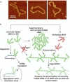
Polymerization of sRNA into long filaments. (A) Atomic force microscopy visualization of nanostructures formed by DsrA transcripts, an abundant 87nt long Escherichia coli ncRNA (O. Pietremont, IGR, Villejuif, personal communication). (B) Schematic drawing illustrating how RNA self-assembly could target supernumerary or misfolded small noncoding RNAs for degradation. As DsrA polymers are degraded much faster than monomers, the formation of DsrA polymers cannot be a storage form of the sRNA, but plausibly either regulates the sRNA concentration in vivo or helps to eliminate misfolded RNAs.
Figure 6.
Figure 6.
Hybrid sensor–actuator component design of RNA transduces RNA binding by ligands or proteins into regulatory outcomes. (A) A small RNA sensor–actuator binds either a small ligand or MS2 RNA-binding protein to activate regulation. (B) An engineered ligand-binding aptamer domain stabilizes pre-miRNA structures and blocks Drosha processing that produces the miRNA. Removal of the ligand restores the activity by creating an unstructured precursor recognized by Drosha that can be processed into a RISC complex for RNA interference. RNA 5′-ends are denoted by a ball and 3′-ends are denoted by broad arrowheads. The sensor components are in blue and actuators are in red.

Similar articles

References

    1. Adamson DN, Lim HN. Essential requirements for robust signaling in hfq dependent small RNA networks. PLoS ComputBiol. 2011;7:e1002138–e1002138. - PMC - PubMed
    1. Afonin KA, Bindewald E, Yaghoubian AJ, Voss N, Jacovetty E, Shapiro BA, Jaeger L. In vitro assembly of cubic RNA-based scaffolds designed in silico. Nat Nanotechnol. 2010;5:676–682. - PMC - PubMed
    1. Aguilera A, Garcia-Muse T. R loops: from transcription byproducts to threats to genome stability. Mol Cell. 2012;46:115–124. - PubMed
    1. Bayer TS, Smolke CD. Programmable ligand-controlled riboregulators of eukaryotic gene expression. Nat Biotechnol. 2005;23:337–343. - PubMed
    1. Beisel CL, Bayer TS, Hoff KG, Smolke CD. Model-guided design of ligand-regulated RNAi for programmable control of gene expression. Mol Syst Biol. 2008;4:224. - PMC - PubMed

LinkOut - more resources