. 2013 Jul;25(7):2679-98.
doi: 10.1105/tpc.113.112342. Epub 2013 Jul 31.

Multisite light-induced phosphorylation of the transcription factor PIF3 is necessary for both its rapid degradation and concomitant negative feedback modulation of photoreceptor phyB levels in Arabidopsis

Affiliations

Multisite light-induced phosphorylation of the transcription factor PIF3 is necessary for both its rapid degradation and concomitant negative feedback modulation of photoreceptor phyB levels in Arabidopsis

Weimin Ni et al. Plant Cell. 2013 Jul.

Abstract

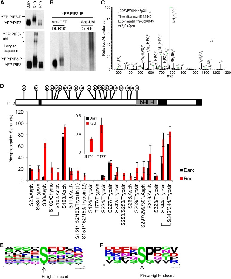
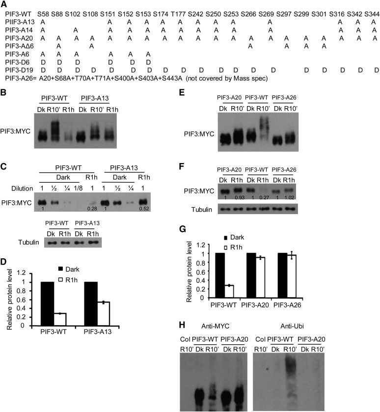
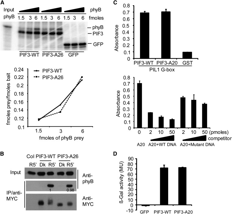
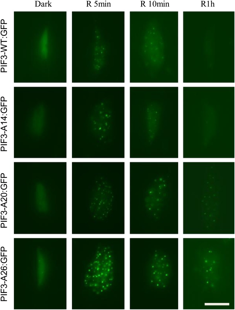
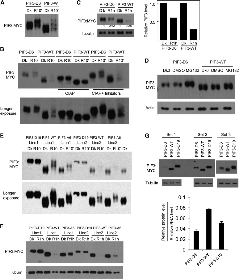
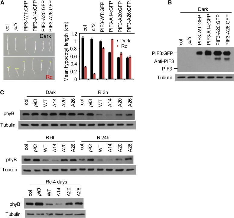
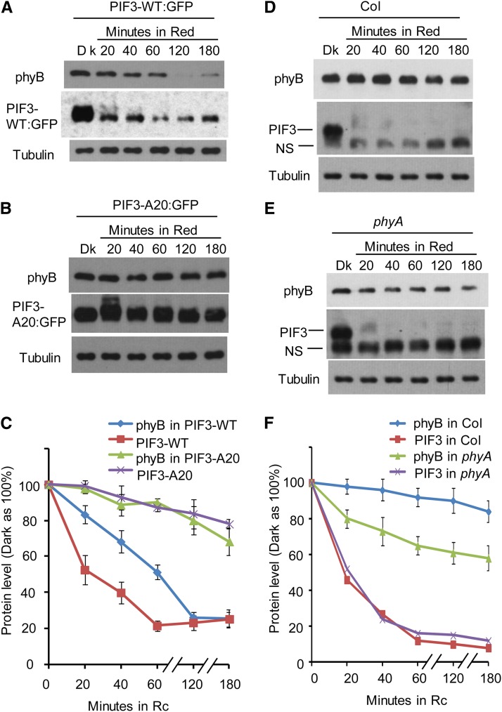
Plants constantly monitor informational light signals using sensory photoreceptors, which include the phytochrome (phy) family (phyA to phyE), and adjust their growth and development accordingly. Following light-induced nuclear translocation, photoactivated phy molecules bind to and induce rapid phosphorylation and degradation of phy-interacting basic Helix Loop Helix (bHLH) transcription factors (PIFs), such as PIF3, thereby regulating the expression of target genes. However, the mechanisms underlying the signal-relay process are still not fully understood. Here, using mass spectrometry, we identify multiple, in vivo, light-induced Ser/Thr phosphorylation sites in PIF3. Using transgenic expression of site-directed mutants of PIF3, we provide evidence that a set of these phosphorylation events acts collectively to trigger rapid degradation of the PIF3 protein in response to initial exposure of dark-grown seedlings to light. In addition, we show that phyB-induced PIF3 phosphorylation is also required for the known negative feedback modulation of phyB levels in prolonged light, potentially through codegradation of phyB and PIF3. This mutually regulatory intermolecular transaction thus provides a mechanism with the dual capacity to promote early, graded, or threshold regulation of the primary, PIF3-controlled transcriptional network in response to initial light exposure, and later, to attenuate global sensitivity to the light signal through reductions in photoreceptor levels upon prolonged exposure.


Figures

Figure 1.
Figure 1.
R Light Induces Rapid Enhancement of Multisite Phosphorylation in PIF3 in Vivo. (A) Top panel: YFP:PIF3-N507 protein from dark (Dk)-grown transgenic Arabidopsis seedlings undergoes rapid light-induced mobility shift (phosphorylation) and degradation similar to native PIF3. Dark-grown YFP:PIF3-N507-expressing transgenic Arabidopsis seedlings were either kept in the dark or given a saturating R light pulse, then moved into darkness for a total time of 10 min or 1 h before protein extraction and immunoblot with GFP antibody. Bottom panel: YFP:PIF3-N507 protein was also detected in the very high molecular weight region (likely polyubiquitinated product) after a longer autoradiograph exposure time. (B) Immunoblot against ubiquitin shows that light induces rapid YFP:PIF3-N507 phosphorylation (mobility shift; left panel [anti-GFP]) and polyubiquitination (high molecular weight region; right panel [anti-Ubi]) in the transgenic seedlings. YFP:PIF3-N507 protein was affinity purified with polyclonal GFP antibody and then blotted with either monoclonal GFP antibody (anti-GFP) or with ubiquitin antibody (anti-Ubi). Transgenic seedlings were grown and treated with R light (10 min) as in (A). IP, immunoprecipitation. (C) Collision-induced dissociation mass spectrum showing phosphorylation of Ser-102, a light-induced phosphorylation site in PIF3. YFP:PIF3-N507 protein from transgenic plants was affinity purified as in (B) before being subjected to in-gel digestion with AspN. (D) The phosphopeptide signal increases rapidly in response to R light exposure of seedlings. Top panel: Distribution of all phosphorylation sites identified in PIF3. The total mass spectrometry peptide coverage for PIF3 is ∼85% (uncovered regions are marked with back boxes). Bottom panel: Comparison of phosphopeptide signal (%) for PIF3 from dark control and R light–exposed (red 10 min) seedlings. The signal of the phosphopeptide containing a given phosphorylation site is expressed as the ion intensity of the phosphopeptide divided by the total ion intensity signal for this peptide (unmodified + phosphorylated) × 100 (see Supplemental Figure 1C online). The values do not represent absolute phosphorylation stoichiometries, but the relative percent values for R versus dark samples do correspond to the change in phosphorylation stoichiometry between these dark and R light–treated samples (red 10 min). Peptides were derived from in-gel digestion with trypsin, chymotrypsin (chymo), or AspN. Data are represented as the mean of biological triplicates ± se. Inset: Two light-induced sites with low phosphopeptide signal values. (E) and (F) No strict consensus motif was identified for either light-induced (E) or non-light-induced (F) PIF3 phosphorylation sites using Weblogo. The middle Ser or Thr is the phosphorylation site.
Figure 2.
Figure 2.
R Light–Induced PIF3 Phosphorylation in Vivo Is Required for Rapid Degradation. (A) Amino acid substitutions in various mutant PIF3 constructs used for protein expression in vivo. The series of point mutants introduced in the 20 residues at the 15 light-induced phosphorylation sites identified here, shown below the wild-type (WT) sequence. A, Ser/Thr-to-Ala mutation; D, Ser/Thr-to-Asp mutation. Construct designations denote the nature and number of concomitant residue substitutions engineered in each mutant polypeptide. For in vivo functional analysis in Arabidopsis seedlings, all the constructs were transformed into the pif3 mutant and expressed transgenically under the control of the cauliflower mosaic virus 35S promoter. (B) to (D) Targeted Ser/Thr-to-Ala mutations in a subset of the light-induced phosphorylation sites results in reduced phosphorylation and degradation of PIF3 induced by light in transgenic Arabidopsis. (B) The PIF3-A13 protein shows a partial reduction in light-induced phosphorylation and degradation. Seedlings of transgenic lines expressing PIF3-WT:MYC or PIF3-A13:MYC fusion proteins were grown for 2.5 d in darkness and then either maintained in the dark (Dk) or given a 1-min saturating R light pulse and returned to darkness for 9 min (R10’) or 59 min (R1h) before extraction into denaturing buffer and immunoblot analysis using antibody against the MYC epitope. (C) Visual quantitative assessment of the degree of light-induced PIF3-A13 degradation. Immunoblot analysis of the same protein extracts as in (B). The PIF3-WT:MYC and PIF3-A13:MYC proteins at R1h were compared with a dilution series of their corresponding dark samples. Tubulin was used as loading control. Numbers directly on blot image denote the relative PIF3 protein levels normalized to tubulin. (D) Quantification of the comparative degrees of PIF3-WT and PIF3-A13 degradation at R1h from immunoblot scans. Dark protein levels were set at 1 for each construct. Data are represented as the mean of biological triplicates ± se. (E) to (H) Targeted Ser/Thr-to-Ala mutations in all identified light-induced sites strongly reduce the phosphorylation, ubiquitination, and degradation of PIF3 induced by light in transgenic plants. (E) and (F) Immunoblot analysis of R light–induced phosphorylation (R10’) (E) and degradation (R1h) (F) of PIF-A20 and PIF-A26 variants using anti-MYC antibody. Seedlings of transgenic lines expressing PIF3-WT:MYC, PIF3-A20:MYC, or PIF3-A26:MYC fusion proteins were grown and treated with R light as in (B). Immunoblot analysis was similar to (B) and (C). Numbers directly on blot image in (F) denote the relative PIF3 protein levels normalized to tubulin. (G) Quantification of the comparative degrees of light-induced PIF3-WT, PIF3-A20, and PIF3-A26 degradation at R1h as in (D). Data represent the mean of biological triplicates ± SE. (H) R light induces a significant accumulation of polyubiquitinated PIF3-WT protein but not PIF3-A20 in transgenic lines. Proteins extracted from Col, PIF3-WT:MYC, and PIF3-A20:MYC transgenic lines were affinity purified with goat anti-MYC antibody and subjected to immunoblot analysis on two duplicate blots, one using monoclonal anti-MYC antibodies and the other antiubiquitin antibodies, for detection of the respective proteins. Seedlings were grown and treated with R light (R10’) as in (B).
Figure 3.
Figure 3.
Multisite Mutations at the Light-Induced PIF3 Phosphorylation Sites Do Not Affect Its Binding Affinity for the phyB Protein, Recognition of a Target Gene Promoter in Vitro, or Transcriptional Activation Activity in Yeast. (A) PIF3-WT and PIF3-A26 have similar binding affinity for Pfr phyB in vitro. Top panel: Autoradiographs showing interactions of PIF3 with different fold dilutions of phyB prey. All proteins were produced in a TNT in vitro expression system labeled with [35S]Met. PIF3-WT, PIF3-A26, and GFP were first immunoprecipitated with an antibody against the MYC epitope present in each of these constructs and then used as baits to coimmunoprecipitate Pfr phyB. Bottom panel: Quantitative analysis of the data obtained in the top panel. The amount of each bait and prey used was calculated from a standard curve using a known amount of [35S]Met. The femtomoles of prey/femtomoles of bait are plotted against increasing amount of the phyB prey used. (B) PIF3-WT and PIF3-A26 have similar conformer-specific, apparent binding affinity for Pfr phyB upon coextraction from R light–irradiated seedlings. Proteins were extracted from dark-grown transgenic seedlings that had been irradiated (R5′) or not (Dk) with R light for 5 min. Transgenically expressed MYC epitope–tagged PIF3 was immunoprecipitated (IP) with goat polyclonal MYC antibody and subjected to immunoblot analysis using monoclonal MYC (PIF3 bait) or phyB (prey) antibody. Proteins from nontransgenic Col seedlings were used as the negative control. Numbers directly on the blot image denote the relative phyB protein levels normalized to the PIF3:MYC bait. (C) PIF3-WT and PIF3-A20 show similar apparent binding affinity for a G-box DNA sequence motif from the PIL1 gene promoter in vitro using a DPI-ELISA assay. Top panel: In vitro binding assay with a PIL1 G-box DNA probe using recombinant GST:PIF3:WT or GST:PIF3:A20 proteins, and GST protein as a negative control. Bottom panel: Competition binding assay of GST:PIF3-A20 with different concentrations of wild-type or mutant G-box probes. Data represent the means of independent duplicates ± se. WT DNA, wild-type competitor probes; mutant DNA, competitor probes mutated in the G-box motif at positions known to eliminate sequence-specific binding by the PIFs. (D) PIF3-WT and PIF3-A20 have similar transcriptional activation activity in yeast. PIF3-WT and PIF3-A20 were fused to the LexA DNA binding domain and tested for autonomous transcriptional activation activity in yeast using a standard liquid ONPG assay. A GFP fusion protein was used as negative control. MU, miller units. Data represent the means of independent duplicates ± se.
Figure 4.
Figure 4.
Light-Induced PIF3 Phosphorylation Is Not Required for Its Subnuclear Localization in Photobodies (Speckles). Epifluorescent imaging of GFP fluorescence in hypocotyl cell nuclei of transgenic seedlings expressing the various PIF3:GFP fusions of the PIF3 variants indicated. Seedlings were grown for 3 d in darkness and then either maintained in the dark or given a 1-min saturating R light pulse and returned to darkness for 4, 9, or 59 min before imaging. Bar = 10 µm.
Figure 5.
Figure 5.
Phosphomimic Mutations of a Subset of Strongly Light-Induced Phosphorylation Sites in PIF3 Promote Its Degradation in Vivo in the Absence of Light. (A) PIF3-D6 exhibits a mobility shift in the dark (Dk) in transgenic Arabidopsis compared with the PIF3-WT control. Seedlings of transgenic lines expressing PIF3-D6:MYC or PIF3-WT:MYC fusion proteins were grown for 2.5 d in darkness, then either kept in the dark or given a saturating R light pulse and returned to darkness for 10 min before extraction into denaturing buffer and immunoblot analysis using MYC antibody. (B) The same protein extracts as in (A) were treated with calf intestine alkaline phosphatase (CIAP) or phosphatase plus phosphatase inhibitors (CIAP + inhibitors) before immunoblot analysis. (C) PIF3-D6 exhibits less light-induced degradation than the PIF3-WT control. Seedling growth, light treatment, and immunoblot analysis were as in (A), except that proteins were extracted at 1 h after light treatment and compared with those from dark samples. Tubulin was used as a control. Numbers directly on the blot image denote the relative PIF3 protein levels normalized to tubulin. Right panel shows quantification of relative PIF3 protein levels (normalized to the dark level set as 1). (D) PIF3-D6 proteins are degraded through the 26S proteasome–mediated pathway in the absence of light. The 2.5-d-old dark-grown (Dk0) PIF3-D6:MYC– or PIF3-WT:MYC–expressing seedlings were treated with proteasome inhibitor MG132 or DMSO for a further 4 h in the dark before extraction and immunoblot analysis using antibodies against the MYC-epitope tag or against actin as a control. (E) and (F) Light-induced mobility shift and degradation analysis of the PIF3-D19 and PIF3-A6 proteins from transgenic plants. (E) Phosphomimic mutations of all light-induced sites identified in PIF3 (PIF3-D19) generate a mobility shift of the protein in the dark, similar to that induced by light at 10 min in the PIF3-WT protein, and no additional light-induced phosphorylation is observed. By contrast, the PIF3-A6 mutant protein exhibits reduced light-induced phosphorylation compared with PIF3-WT. (F) The PIF3-D19 protein shows no apparent light-induced degradation, whereas the PIF3-A6 variant exhibits reduced light-induced degradation. Light treatment and immunoblot analysis were as in (A) and (C). (G) PIF3-D6 and PIF3-D19 protein levels are lower than that of PIF3-WT after normalization to their corresponding RNA levels. Top panels: Triplicates of biological protein samples from 3-d-old dark-grown seedlings of the PIF3-D6, PIF3-WT, and PIF3-D19 were assayed by immunoblot using MYC antibody. Tubulin was used as a loading control. Bottom panel: Relative PIF3 protein levels after normalization to the corresponding RNA levels. Data represent the mean of biological triplicates ± se.
Figure 6.
Figure 6.
Light-Induced PIF3 Phosphorylation and Degradation Are Necessary for Negative Feedback Modulation of phyB Levels. (A) The PIF3-A20 and PIF3-A26 transgenic lines are not as tall as the PIF3-WT and PIF3-A14 lines in prolonged continuous R light. Left panel: Four-day-old seedlings transgenically expressing PIF3:GFP fusions of the various PIF3 variants indicated in the pif3 mutant are shown in comparison with the Col wild type and the pif3 mutant. Seedlings were grown either in the dark (top image) or continuous R light (Rc) (bottom image) for 4 d. Bars = 1 cm. Right panel: Mean hypocotyl lengths of the genotypes shown on the left. Approximately 30 seedlings of each genotype were used for each analysis. Error bars represent se. (B) PIF3 protein levels in the various lines shown in (A). Proteins were extracted from dark-grown seedlings and subjected to immunoblot analysis using affinity-purified anti-PIF3 antibody. A longer exposure would be required to observe endogenous PIF3 (see Supplemental Figure 6C online). Tubulin was used as control. (C) PIF3-A20 and PIF3-A26 transgenic lines retain more phyB protein in the R light than PIF3-WT and PIF3-A14 lines. phyB protein level in the same PIF3 transgenic lines as shown in (A) were analyzed by immunoblot using a monoclonal anti-phyB antibody or antitubulin antibody as a control. Top two panels: Three-day-old dark-grown seedlings were either kept in the dark or irradiated with continuous R light for 3, 6, or 24 h before protein extraction. Bottom panel: Seeds were grown in continuous R light for 4 d before protein extraction.
Figure 7.
Figure 7.
Light-Induced phyB Degradation Kinetics Correlate Robustly with the Differential Rates of Degradation Displayed by the Wild-Type and Phosphorylation Refractory A20 PIF3 Mutant Proteins. (A) The PIF3-WT overexpression transgenic line shows rapid light-induced degradation of both phyB and PIF3-WT. Three-day-old, dark-grown PIF3-WT:GFP–expressing seedlings were either kept in the dark (Dk) or exposed to continuous R light for the periods indicated. Extracted proteins were subjected to immunoblot analysis using either anti-phyB, anti-GFP, or antitubulin antibodies. (B) The PIF3-A20 overexpression line shows comparatively slower light-induced degradation of both phyB and PIF3-A20. Seedlings were grown, exposed to light, and analyzed as in (A). (C) Quantification of phyB and PIF3-GFP degradation kinetics in the PIF3-WT:GFP and PIF3-A20:GFP lines, from replicated immunoblot scans. Dark protein levels were set at 100% for each protein. Data are represented as the mean of biological triplicates ± se. Rc, continuous R light. (D) Light-induced phyB degradation in wild-type Col is relatively slow. Col seedlings were grown and treated with R light as in (A) before protein extraction and immunoblot analysis using antibodies against phyB, PIF3, or tubulin. (E) R light–induced phyB degradation in the phyA mutant is faster than that in Col. phyA seedling growth, R light treatment, and immunoblot analysis were the same as for the Col seedlings in (D). (F) Quantification of phyB and PIF3 degradation kinetics in Col and the pif3 mutant. Dark protein levels were set at 100% for each protein. Data are represented as the mean of biological triplicates ± se. [See online article for color version of this figure.]

Similar articles

Cited by

References

    1. Al-Sady B., Kikis E.A., Monte E., Quail P.H. (2008). Mechanistic duality of transcription factor function in phytochrome signaling. Proc. Natl. Acad. Sci. USA 105: 2232–2237 - PMC - PubMed
    1. Al-Sady B., Ni W., Kircher S., Schäfer E., Quail P.H. (2006). Photoactivated phytochrome induces rapid PIF3 phosphorylation prior to proteasome-mediated degradation. Mol. Cell 23: 439–446 - PubMed
    1. Bao M.Z., Shock T.R., Madhani H.D. (2010). Multisite phosphorylation of the Saccharomyces cerevisiae filamentous growth regulator Tec1 is required for its recognition by the E3 ubiquitin ligase adaptor Cdc4 and its subsequent destruction in vivo. Eukaryot. Cell 9: 31–36 - PMC - PubMed
    1. Bauer D., Viczián A., Kircher S., Nobis T., Nitschke R., Kunkel T., Panigrahi K.C., Adám E., Fejes E., Schäfer E., Nagy F. (2004). Constitutive photomorphogenesis 1 and multiple photoreceptors control degradation of phytochrome interacting factor 3, a transcription factor required for light signaling in Arabidopsis. Plant Cell 16: 1433–1445 - PMC - PubMed
    1. Brand L.H., Kirchler T., Hummel S., Chaban C., Wanke D. (2010). DPI-ELISA: A fast and versatile method to specify the binding of plant transcription factors to DNA in vitro. Plant Methods 6: 25. - PMC - PubMed

Publication types

MeSH terms

LinkOut - more resources