

The use of microfluidics in hemostasis: clinical diagnostics and biomimetic models of vascular injury
- PMID: 23872531
- PMCID: PMC9004362
- DOI: 10.1097/MOH.0b013e3283642186
The use of microfluidics in hemostasis: clinical diagnostics and biomimetic models of vascular injury
Abstract
Purpose of review: This article reviews the application of microfluidic technologies in hemostasis. The emphasis is on promising developments in devices for clinical applications and novel approaches to modeling complex hemodynamics.
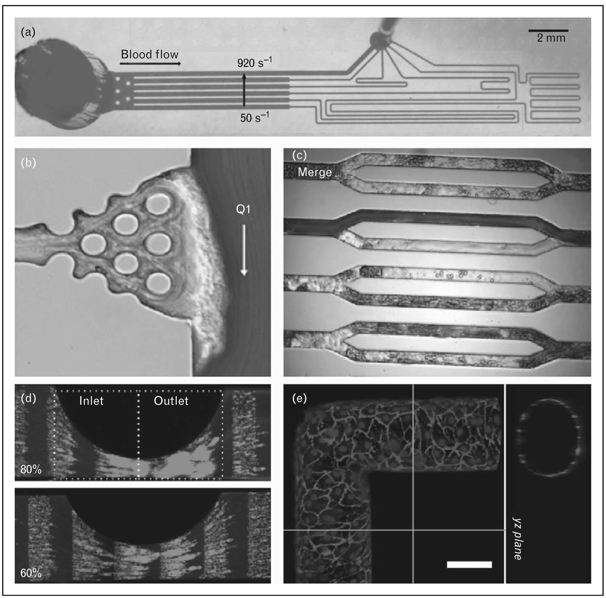
Recent findings: Microfluidics combined with micropatterning of prothrombotic substrates provides devices for measuring platelet function and coagulation with low blood volumes (∼100 μl) over a wide range of shear stresses. This technology has been applied to the diagnosis of bleeding and thrombotic disorders, as well as to dosing and monitoring of anticoagulation and antiplatelet agents. Microfluidic devices that mimic vascular geometries such as bifurcations, stenosis, and complex interconnected networks yield complex flow fields that have revealed new mechanisms of platelet adhesion and aggregation. Applying techniques from tissue engineering by endothelializing these networks is beginning to close the gap between in-vitro and in-vivo models of vascular injury.
Summary: Microfluidic technology enables researchers to create in-vitro models of vascular disease with unprecedented control of the biochemical and biophysical conditions. Two promising directions are flow-dependent clinical assays and biomimetic vascular networks. These approaches are particularly well suited for modeling the microvasculature. However, caution should be used when extrapolating results from microfluidic channels to the pathophysiology of thrombosis in large arteries and veins.
Conflict of interest statement
Conflicts of interest
There are no conflicts of interest.
Figures
Similar articles
-
Application of microfluidic devices in studies of thrombosis and hemostasis.Platelets. 2017 Jul;28(5):434-440. doi: 10.1080/09537104.2017.1319047. Epub 2017 Jun 5. Platelets. 2017. PMID: 28580870 Free PMC article. Review.
-
Recent advances in microfluidic platelet function assays: Moving microfluidics into clinical applications.Clin Hemorheol Microcirc. 2019;71(2):249-266. doi: 10.3233/CH-189416. Clin Hemorheol Microcirc. 2019. PMID: 30584134
-
Flow chamber and microfluidic approaches for measuring thrombus formation in genetic bleeding disorders.Platelets. 2017 Jul;28(5):463-471. doi: 10.1080/09537104.2017.1306042. Epub 2017 May 22. Platelets. 2017. PMID: 28532218 Free PMC article. Review.
-
Monitoring in vitro thrombus formation with novel microfluidic devices.Platelets. 2012;23(7):501-9. doi: 10.3109/09537104.2012.709653. Epub 2012 Aug 8. Platelets. 2012. PMID: 22873212 Review.
-
Microfluidics and coagulation biology.Annu Rev Biomed Eng. 2013;15:283-303. doi: 10.1146/annurev-bioeng-071812-152406. Epub 2013 May 3. Annu Rev Biomed Eng. 2013. PMID: 23642241 Free PMC article. Review.
Cited by
-
Microfluidic technology as an emerging clinical tool to evaluate thrombosis and hemostasis.Thromb Res. 2015 Jul;136(1):13-9. doi: 10.1016/j.thromres.2015.05.012. Epub 2015 May 21. Thromb Res. 2015. PMID: 26014643 Free PMC article. Review.
-
Microvascular platforms for the study of platelet-vessel wall interactions.Thromb Res. 2014 Apr;133(4):525-31. doi: 10.1016/j.thromres.2013.12.039. Epub 2014 Jan 7. Thromb Res. 2014. PMID: 24438943 Free PMC article. Review.
-
Automated analysis of mitochondrial dimensions in mesenchymal stem cells: Current methods and future perspectives.Heliyon. 2023 Jan 18;9(1):e12987. doi: 10.1016/j.heliyon.2023.e12987. eCollection 2023 Jan. Heliyon. 2023. PMID: 36711314 Free PMC article. Review.
-
Hemostasis-On-a-Chip: Impedance Spectroscopy Meets Microfluidics for Hemostasis Evaluation.Micromachines (Basel). 2019 Aug 14;10(8):534. doi: 10.3390/mi10080534. Micromachines (Basel). 2019. PMID: 31416133 Free PMC article.
-
The effect of platelet storage temperature on haemostatic, immune, and endothelial function: potential for personalised medicine.Blood Transfus. 2019 Jul;17(4):321-330. doi: 10.2450/2019.0095-19. Blood Transfus. 2019. PMID: 31385802 Free PMC article. Review.
References
-
- Sakariassen KS, Turitto VT, Baumgartner HR. Recollections of the development of flow devices for studying mechanisms of hemostasis and thrombosis in flowing whole blood. J Thromb Haemost 2004; 2:1681–1690. - PubMed
-
- Zwaginga JJ, Sakariassen KS, King MR, et al. Can blood flow assays help to identify clinically relevant differences in von Willebrand factor functionality in von Willebrand disease types 1–3? J Thromb Haemost 2007; 5:2547–2549. - PubMed
-
- Roest M, Reininger A, Zwaginga JJ, et al., the Biorheology Subcommittee of the SSC of the ISTH. Flow chamber-based assays to measure thrombus formation in vitro: requirements for standardization. J Thromb Haemost 2011; 9:2322–2324. - PubMed
-
- Westein E, De Witt S, Lamers M, et al. Monitoring in vitro thrombus formation with novel microfluidic devices. Platelets 2012; 23:501–509. - PubMed
Publication types
MeSH terms
Grants and funding
LinkOut - more resources
Full Text Sources
Other Literature Sources
Medical
Research Materials
Miscellaneous
