

The use of bioinspired alterations in the glycosaminoglycan content of collagen-GAG scaffolds to regulate cell activity
- PMID: 23871542
- PMCID: PMC4090944
- DOI: 10.1016/j.biomaterials.2013.06.056
The use of bioinspired alterations in the glycosaminoglycan content of collagen-GAG scaffolds to regulate cell activity
Abstract
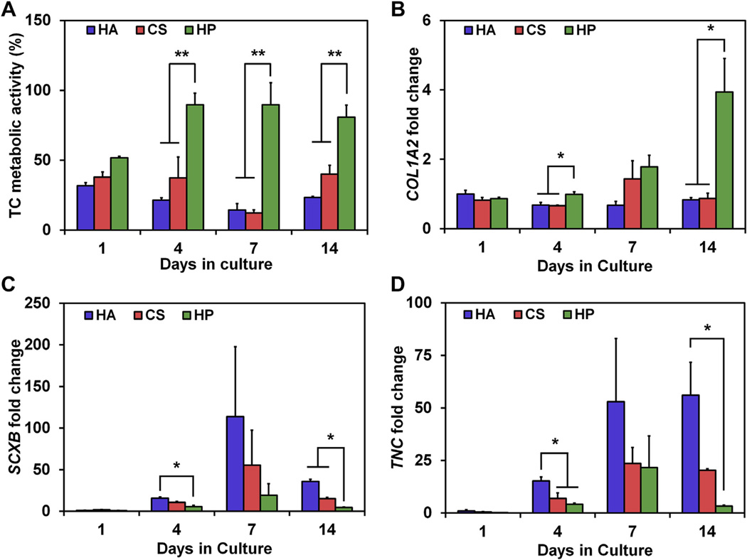
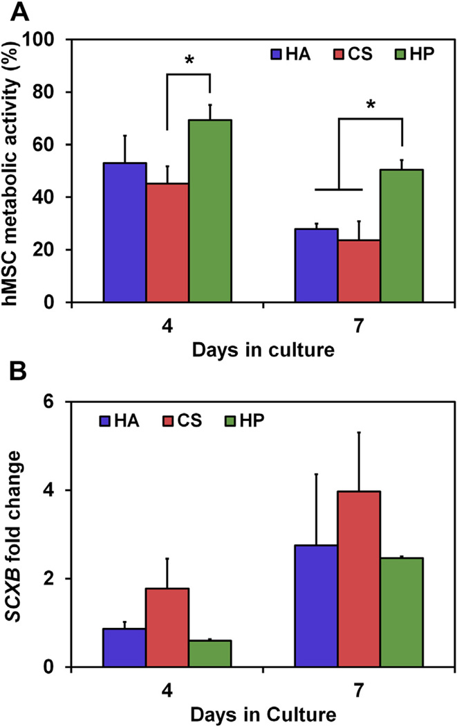
The design of biomaterials for regenerative medicine can require biomolecular cues such as growth factors to induce a desired cell activity. Signal molecules are often incorporated into the biomaterial in either freely-diffusible or covalently-bound forms. However, biomolecular environments in vivo are often complex and dynamic. Notably, glycosaminoglycans (GAGs), linear polysaccharides found in the extracellular matrix, are involved in transient sequestration of growth factors via charge interactions. Biomaterials mimicking this phenomenon may offer the potential to amplify local biomolecular signals, both endogenously produced and exogenously added. GAGs of increasing sulfation (hyaluronic acid, chondroitin sulfate, heparin) were incorporated into a collagen-GAG (CG) scaffold under development for tendon tissue engineering. Manipulating the degree of GAG sulfation significantly impacts sequestration of growth factors from the media. Increasing GAG sulfation improved equine tenocyte metabolic activity in normal serum (10% FBS), low serum (1% FBS), and IGF-1 supplemented media conditions. Notably, previously reported dose-dependent changes in tenocyte bioactivity to soluble IGF-1 within the CG scaffold were replicated by using a single dose of soluble IGF-1 in scaffolds containing increasingly sulfated GAGs. Collectively, these results suggest that CG scaffold GAG content can be systematically manipulated to regulate the sequestration and resultant enhanced bioactivity of growth factor signals on cell behavior within the matrix.
Keywords: Biomimetic material; Collagen; Growth factors; Mesenchymal stem cell; Scaffold; Tendon.
Copyright © 2013 Elsevier Ltd. All rights reserved.
Figures
Similar articles
-
Composite growth factor supplementation strategies to enhance tenocyte bioactivity in aligned collagen-GAG scaffolds.Tissue Eng Part A. 2013 May;19(9-10):1100-12. doi: 10.1089/ten.TEA.2012.0497. Epub 2013 Jan 4. Tissue Eng Part A. 2013. PMID: 23157454 Free PMC article.
-
The effect of glycosaminoglycan content on polyethylenimine-based gene delivery within three-dimensional collagen-GAG scaffolds.Biomater Sci. 2015 Apr;3(4):645-54. doi: 10.1039/C5BM00033E. Biomater Sci. 2015. PMID: 26097698 Free PMC article.
-
The development of collagen-GAG scaffold-membrane composites for tendon tissue engineering.Biomaterials. 2011 Dec;32(34):8990-8. doi: 10.1016/j.biomaterials.2011.08.035. Epub 2011 Aug 30. Biomaterials. 2011. PMID: 21880362 Free PMC article.
-
Glycosaminoglycan-Based Cryogels as Scaffolds for Cell Cultivation and Tissue Regeneration.Molecules. 2021 Sep 15;26(18):5597. doi: 10.3390/molecules26185597. Molecules. 2021. PMID: 34577067 Free PMC article. Review.
-
From molecules to matrix: construction and evaluation of molecularly defined bioscaffolds.Adv Exp Med Biol. 2006;585:279-95. doi: 10.1007/978-0-387-34133-0_19. Adv Exp Med Biol. 2006. PMID: 17120791 Review.
Cited by
-
The effect of intraurethral hyaluronic acid on healing and fibrosis in rats with experimentally induced urethral trauma.Int Urol Nephrol. 2022 Apr;54(4):757-761. doi: 10.1007/s11255-022-03128-1. Epub 2022 Jan 31. Int Urol Nephrol. 2022. PMID: 35099687
-
Chitooligomer-Immobilized Biointerfaces with Micropatterned Geometries for Unidirectional Alignment of Myoblast Cells.Biomolecules. 2016 Jan 15;6(1):12. doi: 10.3390/biom6010012. Biomolecules. 2016. PMID: 26784249 Free PMC article.
-
Biomaterial design strategies to address obstacles in craniomaxillofacial bone repair.RSC Adv. 2021;11(29):17809-17827. doi: 10.1039/d1ra02557k. Epub 2021 May 17. RSC Adv. 2021. PMID: 34540206 Free PMC article.
-
The Influence of Hyaluronic Acid and Glioblastoma Cell Coculture on the Formation of Endothelial Cell Networks in Gelatin Hydrogels.Adv Healthc Mater. 2017 Nov;6(22):10.1002/adhm.201700687. doi: 10.1002/adhm.201700687. Epub 2017 Sep 22. Adv Healthc Mater. 2017. PMID: 28941173 Free PMC article.
-
The Use of Platelet-Rich Plasma to Promote Cell Recruitment into Low-Molecular-Weight Fucoidan-Functionalized Poly(Ester-Urea-Urethane) Scaffolds for Soft-Tissue Engineering.Polymers (Basel). 2019 Jun 9;11(6):1016. doi: 10.3390/polym11061016. Polymers (Basel). 2019. PMID: 31181822 Free PMC article.
References
-
- Place ES, Evans ND, Stevens MM. Complexity in biomaterials for tissue engineering. Nat Mater. 2009;8:457–470. - PubMed
-
- Farrell E, O’Brien FJ, Doyle P, Fischer J, Yannas I, Harley BA, et al. A collagen–glycosaminoglycan scaffold supports adult rat mesenchymal stem cell differentiation along osteogenic and chondrogenic routes. Tissue Eng. 2006;12:459–468. - PubMed
-
- Gulotta LV, Rodeo SA. Growth factors for rotator cuff repair. Clin Sports Med. 2009;28:13–23. - PubMed
-
- Molloy T, Wang Y, Murrell G. The roles of growth factors in tendon and ligament healing. Sports Med. 2003;33:381–394. - PubMed
Publication types
MeSH terms
Substances
Grants and funding
LinkOut - more resources
Full Text Sources
Other Literature Sources
Miscellaneous
