

The expanded collagen VI family: new chains and new questions
- PMID: 23869615
- PMCID: PMC5248970
- DOI: 10.3109/03008207.2013.822865
The expanded collagen VI family: new chains and new questions
Abstract
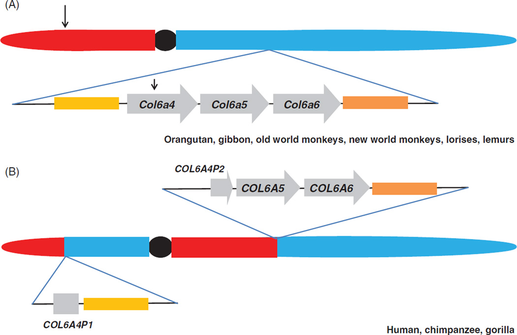
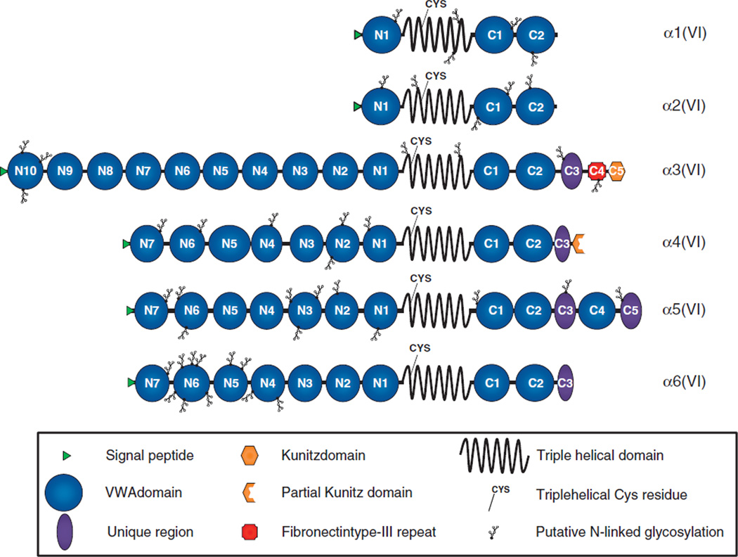
Collagen VI is a component of the extracellular matrix of almost all connective tissues, including cartilage, bone, tendon, muscles and cornea, where it forms abundant and structurally unique microfibrils organized into different suprastructural assemblies. The precise role of collagen VI is not clearly defined although it is most abundant in the interstitial matrix of tissues and often found in close association with basement membranes. Three genetically distinct collagen VI chains, α1(VI), α2(VI) and α3(VI), encoded by the COL6A1. COL6A2 and COL6A3 genes, were first described more than 20 years ago. Their molecular assembly and role in congenital muscular dystrophy has been broadly characterized. In 2008, three additional collagen VI genes arrayed in tandem at a single gene locus on chromosome 3q in humans, and chromosome 9 in mice, were described. Following the naming scheme for collagens the new genes were designated COL6A4. COL6A5 and COL6A6 encoding the α4(VI), α5(VI) and α6(VI) chains, respectively. This review will focus on the current state of knowledge of the three new chains.
Figures
Similar articles
-
Three novel collagen VI chains, alpha4(VI), alpha5(VI), and alpha6(VI).J Biol Chem. 2008 Jul 18;283(29):20170-80. doi: 10.1074/jbc.M710139200. Epub 2008 Apr 9. J Biol Chem. 2008. PMID: 18400749
-
COL6A3 protein deficiency in mice leads to muscle and tendon defects similar to human collagen VI congenital muscular dystrophy.J Biol Chem. 2013 May 17;288(20):14320-14331. doi: 10.1074/jbc.M112.433078. Epub 2013 Apr 5. J Biol Chem. 2013. PMID: 23564457 Free PMC article.
-
Expression of the collagen VI α5 and α6 chains in normal human skin and in skin of patients with collagen VI-related myopathies.J Invest Dermatol. 2011 Jan;131(1):99-107. doi: 10.1038/jid.2010.284. Epub 2010 Sep 30. J Invest Dermatol. 2011. PMID: 20882040
-
Collagen VI disorders: Insights on form and function in the extracellular matrix and beyond.Matrix Biol. 2018 Oct;71-72:348-367. doi: 10.1016/j.matbio.2017.12.008. Epub 2017 Dec 22. Matrix Biol. 2018. PMID: 29277723 Review.
-
Collagen VI related muscle disorders.J Med Genet. 2005 Sep;42(9):673-85. doi: 10.1136/jmg.2002.002311. J Med Genet. 2005. PMID: 16141002 Free PMC article. Review.
Cited by
-
Biopsy-Controlled Non-Invasive Quantification of Collagen Type VI in Kidney Transplant Recipients: A Post-Hoc Analysis of the MECANO Trial.J Clin Med. 2020 Oct 7;9(10):3216. doi: 10.3390/jcm9103216. J Clin Med. 2020. PMID: 33036366 Free PMC article.
-
Basement membrane collagens and disease mechanisms.Essays Biochem. 2019 Sep 13;63(3):297-312. doi: 10.1042/EBC20180071. Print 2019 Sep 13. Essays Biochem. 2019. PMID: 31387942 Free PMC article. Review.
-
Decreased collagen VI in the tunica media of pulmonary vessels during exposure to hypoxia: a novel step in pulmonary arterial remodeling.Pulm Circ. 2019 Jul-Sep;9(3):2045894019860747. doi: 10.1177/2045894019860747. Pulm Circ. 2019. PMID: 31187694 Free PMC article.
-
Collagen-VI supplementation by cell transplantation improves muscle regeneration in Ullrich congenital muscular dystrophy model mice.Stem Cell Res Ther. 2021 Aug 9;12(1):446. doi: 10.1186/s13287-021-02514-3. Stem Cell Res Ther. 2021. PMID: 34372931 Free PMC article.
-
Endotrophin, a Key Marker and Driver for Fibroinflammatory Disease.Endocr Rev. 2024 May 7;45(3):361-378. doi: 10.1210/endrev/bnad036. Endocr Rev. 2024. PMID: 38091968 Free PMC article. Review.
References
-
- Myllyharju J, Kivirikko KI. Collagens, modifying enzymes and their mutations in humans, flies and worms. Trends Genet. 2004;20:33–43. - PubMed
-
- Chu ML, Mann K, Deutzmann R, Pribula-Conway D, Hsu-Chen CC, Bernard MP, Timpl R. Characterization of three constituent chains of collagen type VI by peptide sequences and cDNA clones. Eur J Biochem. 1987;168:309–317. - PubMed
-
- Chu ML, Pan TC, Conway D, Kuo HJ, Glanville RW, Timpl R, Mann K, Deutzmann R. Sequence analysis of alpha 1(VI) and alpha 2(VI) chains of human type VI collagen reveals internal triplication of globular domains similar to the A domains of von Willebrand factor and two alpha 2(VI) chain variants that differ in the carboxy terminus. EMBO J. 1989;8:1939–1946. - PMC - PubMed
Publication types
MeSH terms
Substances
Grants and funding
LinkOut - more resources
Full Text Sources
Other Literature Sources
Miscellaneous