. 2013 Jul 16:12:76.
doi: 10.1186/1476-4598-12-76.

Lyn, a Src family kinase, regulates activation of epidermal growth factor receptors in lung adenocarcinoma cells

Affiliations

Lyn, a Src family kinase, regulates activation of epidermal growth factor receptors in lung adenocarcinoma cells

Parnetta Sutton et al. Mol Cancer. .

Abstract

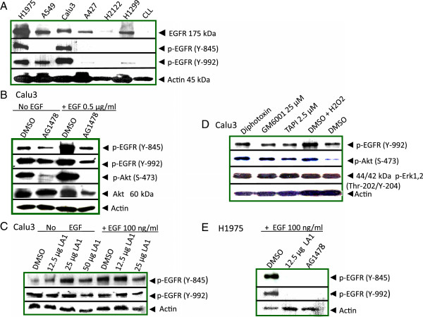
Background: Activation of receptors for growth factors on lung epithelial cells is essential for transformation into tumor cells, supporting their viability and proliferation. In most lung cancer patients, EGFR is constitutively activated without evidence of mutation. Defining mechanisms for constitutive activation of EGFR could elucidate additional targets for therapy of lung cancers.

Methods: The approach was to identify lung cancer cell lines with constitutively activated EGFR and use systematic selection of inhibitors to evaluate their effects on specific EGFR phosphorylations and downstream signaling pathways. Interactions between receptors, kinases, and scaffolding proteins were investigated by co-immunoprecipitation plus Western blotting.

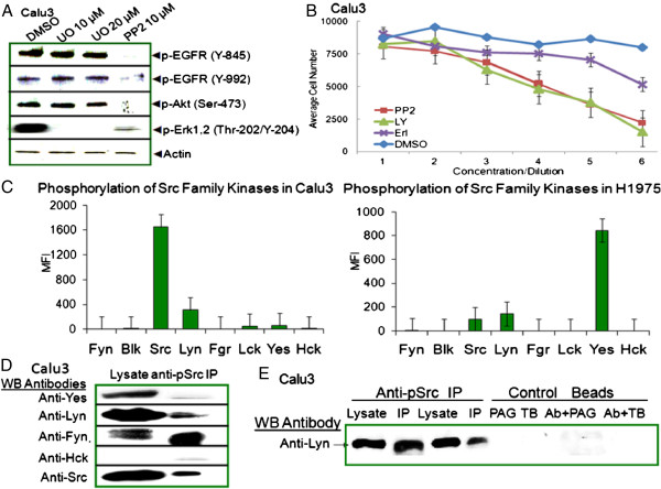
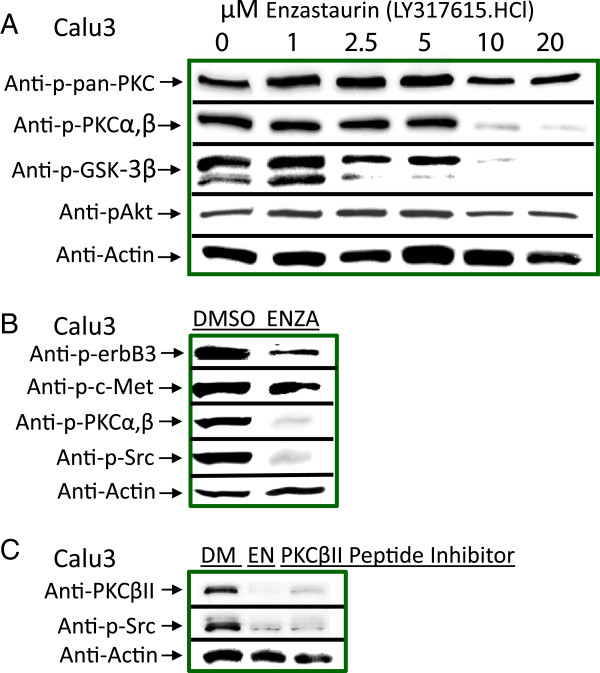
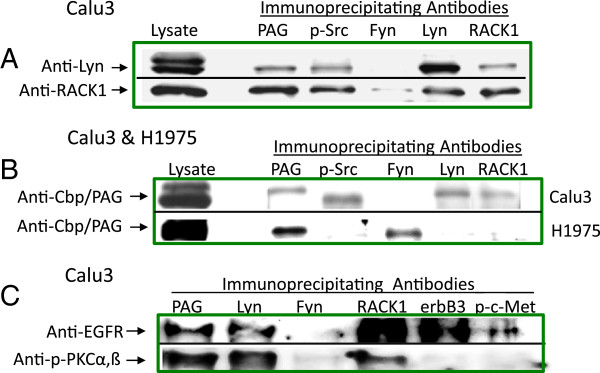
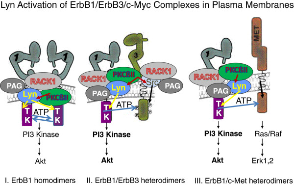
Results: The results revealed a dependence on Src family of tyrosine kinases for downstream signaling and cell growth. Lyn, a Src family kinase functional in normal and malignant B-lymphocytes, was a defining signal transducer required for EGFR signaling in Calu3 cell line. Src family kinase activation in turn, was dependent on PKCßII. Lyn and PKC exist in membrane complexes of RACK1 and in association with EGFR which pairs with other receptor partners. Silencing of Lyn expression with interfering siRNA decreased EGFR activation and cell viability.

Conclusions: The importance of Src family kinases and PKCßII in the initiation of the EGFR signaling pathway in lung tumor cells was demonstrated. We conclude that phosphorylation of EGFR is mediated through PKCßII regulation of Lyn activation, and occurs in association with RACK1 and Cbp/PAG proteins. We suggest that protein complexes in cell membranes, including lipid rafts, may serve as novel targets for combination therapies with EGFR and Src Family Kinase inhibitors in lung cancer.


Figures

Figure 1
Figure 1
Constitutive phosphorylation of EGFR in non-small cell lung cancer cell lines (NSCLC). (A) Constitutive phosphorylation of EGFR at Y-845 and Y-992. Western blots from lysates of unstimulated NSCLC cell lines and chronic lymphocytic leukemia (CLL) cells were probed with anti-phospho-EGFR (Y-845), anti-phospho-EGFR (Y-992), anti-EGFR or anti-actin antibodies as loading controls. (B) EGFR triggered autophosphorylation is not responsible for constitutive EGFR (Y845) or (Y-992) phosphorylation in Calu3. Calu3 cells were incubated with EGFR kinase inhibitor @ 1 μM AG1478, or an equal volume of DMSO solvent, for 1 hour with or without addition of 0.5 μg EGF in the final 10 minutes before lysates were prepared and Western blotted with anti-phospho-EGFR (Y845 and Y-992), anti-phospho-Akt (ser-473), anti-Akt, or anti-actin. (C) EGFR ligands are not responsible for constitutive phosphorylation in Calu3 cells. EGFR neutralizing antibodies, LA1 at 12.5, 25 or 50 μg were incubated for 18 hours with Calu3 cells with or without 100 ng EGF in the final 5 minutes before lysates were prepared and Western blotted. (D) Transactivation by membrane associated ligands was not responsible for constitutive phosphorylation of EGFR Y-992 or downstream phosphorylation of Akt or Erk1,2. Calu3 cells were serum cultured with Corynebacterium diphtheriae toxin @ 10 μg/ml, 25 μM GM6001, 2.5 μM TAPI, 100 μM H2O2, or an equivalent volume of DMSO for 1 hour before lysates were prepared and Western blotted. (E) EGFR neutralizing antibodies blocked phosphorylation in H1975 NSCLC cell line. LAI at 12.5 μg was added to H1975 cells for 18 hours. DMSO or 1 μM AG1478 was added for 1 hour. Lysates were prepared for SDS-PAGE and Western blotting with anti-phospho-EGFR(Y-992) and anti-phospho-EGFR (Y-845) or anti-actin. Caco-2 cells (ATCC #HTB-37, human colorectal adenocarcinoma) served as positive controls for the TACE (ADAM 17) inhibitors, GM6001 and TAPI [35] (data not presented).
Figure 2
Figure 2
Src Family Kinase (SFK) expression and activation. (A) Src-kinase inhibitor PP2, blocked phosphorylation of EGFR at Y-845 and Y-992 and downstream phosphorylations of Akt and Erk1,2. Calu3 cells were serum starved for 6 hours, then cultured with 10 μM PP2, 10 or 20 μM U0126, or an equivalent volume of DMSO for 1 hour with addition of recombinant EGF @ 1 μg/ml for the final 10 minutes; then lysates prepared for SDS-PAGE and Western blotting with indicated antibodies. (B) PP2 decreased viability of Calu3 cells in a concentration dependent manner. Calu3 cells were cultured in triplicate in 96-well plates, and serum starved for 8 hours before addition of the respective inhibitors with highest concentrations for PP2 @ 20 μM; LY29004 @ 40 μM; Erlotinib @ 20 μM; and DMSO @ 4 μl; then dilutions of one-half through seven serial titrations (7 → 1). After 70 hours, Calcein AM was added two hours before harvesting. (C) Distinct patterns of SFK activation were revealed in a quantitative Milliplex assay of unstimulated Calu3 and H1975 cell lysates. MFI (median fluorescence intensity) equals sample - background. (D) Expression of phosphorylated Lyn, Src, and an isoform of Fyn in Calu3 cell lysates were confirmed by Western blotting. Anti-phospho-Src (Y-416) immunoprecipitates were blotted and probed separately with anti-SFK member antibodies including Yes, Lyn, Fyn, Hck, and v-Src. Anti-Hck served as a specificity control in that no non-specific bands were observed in either the lysates or IP. (E) Lyn expression and phosphorylation were further confirmed by direct immunoprecipitation. Calu3 cell lysates were incubated with anti-phospho-Src (Y-417), anti-vimentin isotype control or no antibody. Duplicate immunoprecipitations were performed with recombinant protein A\G conjugated beads (PAG) (wells 2,5,7) or TrueBlot ® anti-light chain beads (TB, wells 4,6,8). Control immunoprecipitations demonstrated no extraneous bands near mw of SFKs, 58–66 kDa.
Figure 3
Figure 3
EGFR is physically associated with distinct Src family kinases, c-Met, and other ErbB family members. (A) Phosphorylated c-Met and EGFR from are pulled down together in anti-phospho-c-Met immunoprecipitations. Western blots of unstimulated Calu3 lysates, anti-phospho-c-Met (Y-1230,1234,1235) immunoprecipitates (IP), and “unbound” (*ub) proteins from IP supernatants were probed with anti-EGFR (Y-845) and anti-c-Met. While all phosphorylated c-Met was pulled down, a portion of phospho-EGFR remained, likely unbound to phosphorylated c-Met. (B) Unstimulated and serum-starved Calu3 cells were cultured with DMSO (DM) or 10 μM erlotinib (erl) for 3 hours, and 5 μM SU11274 (SU), 1 μM AG1478 (AG), or 10 μM PP2 for 1 hour before lysates and Western blots were prepared and probed with anti-phospho-c-Met (Y-1230,1234,1235), anti-phospho-Erk1,2 (Thr-202/Y-204), anti-phospho-ErbB3 (Y-1289), or anti-actin. (C) Immunoprecipitation of unstimulated Calu3 lysates with anti-ErbB3 also pulled down ErbB2, phosphorylated EGFR, and phosphorylated ErbB3. Anti-ErbB2 reciprocally immunoprecipitated ErbB3 from unstimulated Calu3 lysates. (D) Anti-Lyn pulled down EGFR but not phosphorylated c-Met (Left panels) while anti-Fyn pulled down neither EGFR nor c-Met (Right panels) from unstimulated Calu3 lysates. Western blots of mouse anti-Lyn IPs (top left panel) were probed with anti-EGFR and anti-phospho-Src (Y-416) while rabbit anti-Lyn IPs (lower left panels) were probed with anti-phospho-c-Met (Y-1230,1234,1235) and anti-phospho-Src (Y-416). (E) Yes was associated with EGFR but not ErbB3 in H1975. Lysates from untreated H1975 cells were immunoprecipitated with antibodies to ErbB3, phospho-Src (Y-416), EGFR, and vimentin. Western blots of the IPs were probed with anti-Yes and anti-phospho-Src (Y-416). Anti-vimentin immunoprecipitates served as specificity controls.
Figure 4
Figure 4
Lyn siRNA inhibits EGFR activation, downstream phosphorylation of c-Met but not phosphorylation of ErbB3. (A) Lyn specific siRNA inhibits Lyn and EGFR phosphorylation. Calu3 cells were cultured with 12.5 nM Lyn siRNA and NC siRNA for 72, 96, and 144 hours before lysates were prepared and 30 μg protein from each treatment loaded into SDS-PAGE wells for separation and Western blotting. Blots were probed with anti-phospho-Lyn, anti-Lyn, anti-phospho-EGFR (Y-1068), anti-phospho-ErbB3 (Y-1289), and actin. (B) EGF triggered reexpression and phosphorylation of Lyn was not evident after 144 hours of culture with 12.5 nM Lyn siRNA while NC siRNA had little effect on Lyn or EGFR phosphorylation levels. EGFR phosphorylations were substantially decreased by Lyn siRNA. Serum-starved Calu3 cells were treated with Lyn and NC siRNA, then cultured for 144 hours. EGF at 100 ng/ml was added for the final 10 minutes before lysates were prepared and 30 μg protein from each treatment loaded into SDS-PAGE wells for separation and Western blotting. Multiple blots were probed with antibodies against phospho-Lyn, non-phosphorylated Lyn control, phospho-EGFR (Y-845), phospho-EGFR (Y-1068), non-phosphorylated ErbB control, phospho-Src (Y-416) or β-actin. (C) Lyn siRNA @ 12.5 nM significantly decreased cell survival after 72 hours of culture. Lyn siRNA decreased viability to highly significant levels, p = <0.001 and 0.0433. Some nonspecific decrease in viability with NC siRNA was also evident but did not reach a similar level of significance. Calcein AM uptake was used to measure cell viability during the last 2 hours of culture. Mann Whitney unit analysis test was applied to relative fluorescent units (RFU) data from 10 replicate wells.
Figure 5
Figure 5
PKCß inhibition reduced constitutive activation of Src, decreased phosphorylation of c-Met and ErbB3, and had downstream effects on Akt and GSK-3ß in both Calu3 and H1975 cells. (A) PCKß inhibitor enzastaurin demonstrates that PKCß is required for Src activation. Concentrations of enzastaurin from 0–20 μM were added to unstimulated, serum-starved cells for 3.5 hours before lysates were prepared for Western blotting. Multiple blots were probed with antibodies against phospho-pan PKC, phospho-PKCα,ß, phospho-GSK-3ß, Akt and actin. (B) PKC inhibitor blocks constitutive activation. Unstimulated, serum-starved Calu3 and H1975 cells were cultured for 3.5 hours with 10 μM enzastaurin before lysates were prepared for Western blotting. Multiple blots were probed with antibodies against phospho-ErbB3 (Y-1289), phospho-c-Met (Y-1230,1234,1235), phospho-PKCα,ß, phospho-Src and actin. (C) Specificity of inhibition for PKCßII was confirmed by culture of unstimulated Calu3 cells with 80 μM PKCßII specific peptide inhibitor. Western blots were probed with anti-PKCβII antibodies, anti-phospho-Src (Y-416) and anti-actin.
Figure 6
Figure 6
Immunoprecipitations identify scaffolding and regulatory proteins pulled down with Lyn, PKCß, and EGFR. (A) Anti-phospho-Src (Y-416), Lyn, RACK1 and Cbp/PAG co-immunoprecipitate each other. Immunoprecipitates from unstimulated, serum starved Calu3 cell lysates were performed with antibodies to Cbp/PAG, phospho-Src (Y-416), Lyn, Fyn and RACK1. Western blots were probed with anti-Lyn, and anti-RACK. No Lyn and minimally detectable quantities of RACK1 were immunoprecipitated by anti-Fyn. (B) Cbp/PAG was associated with phosphorylated Src, Lyn and RACK1 in Calu3 while anti-Fyn pulled down Cbp\PAG only in H1975 cells. Western blots of immunoprecipitates from unstimulated, serum starved Calu3 and H1975 cell lysates were probed with anti-Cbp/PAG. (C) EGFR, PKCα or β, Cbp/PAG, and RACK1 are all pulled down with anti-Lyn. Immunoprecipitates with antibodies to Cbp/PAG, Lyn, Fyn, RACK1, ErbB3 and phosphorylated-c-Met from Calu3 lysates were probed in Western blots with antibodies to EGFR and phospho-PKCα,ß.
Figure 7
Figure 7
Hypothetical working model of ErbB1 homodimers and heterodimers in lipid rafts with RACK1, PKCßII, Lyn and/or Src, Cbp/PAG. EGFR/ErbB1 homodimers are drawn together, independent of ligand, within a single RACK1 complex while ligand-independent heterodimers of ErbB1 and ErbB3 or ErbB1 and c-Met may result from the merging of two or more RACK1complexes. Heterodimers lead to enhanced PI3 kinase activity as illustrated by bolding.

Similar articles

Cited by

References

    1. Hirsch FR, Varella-Garcia M, Bunn PA Jr, Di Maria MV, Veve R, Bremmes RM, Baron AE, Zeng C, Franklin WA. Epidermal growth factor receptor in non-small-cell lung carcinomas: correlation between gene copy number and protein expression and impact on prognosis. J Clin Oncol. 2003;21(20):3798–3807. doi: 10.1200/JCO.2003.11.069. - DOI - PubMed
    1. Holbro T, Civenni G, Hynes NE. The ErbB receptors and their role in cancer progression. Exp Cell Res. 2003;284(1):99–110. doi: 10.1016/S0014-4827(02)00099-X. - DOI - PubMed
    1. Kanematsu T, Yano S, Uehara H, Bando Y, Sone S. Phosphorylation, but not overexpression, of epidermal growth factor receptor is associated with poor prognosis of non-small cell lung cancer patients. Oncol Res. 2003;13(5):289–298. - PubMed
    1. Sebastian S, Settleman J, Reshkin SJ, Azzariti A, Bellizzi A, Paradiso A. The complexity of targeting EGFR signalling in cancer: from expression to turnover. Biochim Biophys Acta. 2006;1766(1):120–139. - PubMed
    1. Yarden Y, Sliwkowski MX. Untangling the ErbB signalling network. Nat Rev Med Cell Biol. 2001;2(2):127–137. doi: 10.1038/35052073. - DOI - PubMed

Publication types

MeSH terms