. 2013 Sep;12(9):1715-27.
doi: 10.1158/1535-7163.MCT-12-1174. Epub 2013 Jul 16.

AZD3514: a small molecule that modulates androgen receptor signaling and function in vitro and in vivo

Affiliations

AZD3514: a small molecule that modulates androgen receptor signaling and function in vitro and in vivo

Sarah A Loddick et al. Mol Cancer Ther. 2013 Sep.

Abstract

Continued androgen receptor (AR) expression and signaling is a key driver in castration-resistant prostate cancer (CRPC) after classical androgen ablation therapies have failed, and therefore remains a target for the treatment of progressive disease. Here, we describe the biological characterization of AZD3514, an orally bioavailable drug that inhibits androgen-dependent and -independent AR signaling. AZD3514 modulates AR signaling through two distinct mechanisms, an inhibition of ligand-driven nuclear translocation of AR and a downregulation of receptor levels, both of which were observed in vitro and in vivo. AZD3514 inhibited testosterone-driven seminal vesicle development in juvenile male rats and the growth of androgen-dependent Dunning R3327H prostate tumors in adult rats. Furthermore, this class of compound showed antitumor activity in the HID28 mouse model of CRPC in vivo. AZD3514 is currently in phase I clinical evaluation.


Figures

Figure 1
Figure 1
Chemical structure of AZD3514.
Figure 2
Figure 2
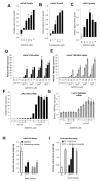
(A-C) LNCaP and LAPC4 growth in steriod free media supplemented with 1 nM DHT and AZD3514 or enzalutamide. Cell number determined after 7 days. Data shown is growth relative to vehicle control and is the mean of 3 (AZD3514) or 2 (enzalutamide) independent experiments. Error bars are SEM. (D-G) PSA and TMPRSS2 gene expression normalized to levels of 18s after AZD3514 treatment of cells +/− DHT. Data shown is gene expression relative to the vehicle control in the absence of ligand and is a mean of 2 independent experiments. Error bars are SEM. (H) AZD3514 or abiraterone acetate was administered by oral gavage once daily for 6 days to intact 42 day old male rats at the doses indicated. On day 7 animals were terminated and seminal vesicles weighed. Data shown are the mean values of 5 animals. Error bars are SEM. (I) Testosterone proprionate (TP) (0.4 mg/kg) was administered by subcutaneous injection along with either vehicle; AZD3514 or abiraterone acetate were administered by oral gavage once daily for 6 days to castrated 42d old male rats at the doses indicated. On day 7 animals were terminated and seminal vesicles weighed. Data shown are the mean values of 5 animals. Error bars are SEM.
Figure 3
Figure 3
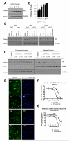
(A-B) LNCaP cells treated with AZD3514 for 24 hours. Graph shows AR levels normalized to GAPDH and relative to the vehicle control. Data is represented as Mean of 3 independent experiments. Error bars are SEM. (C) LNCaP cells grown in steroid free conditions were incubated with 10 μM AZD3514, enazalutamide or vehicle control for the indicated times. Cells were then treated −/+ 1 nM DHT for 30 minutes. Effects on total AR were assessed by Western blot and normalized to levels of GAPDH. * indicates reduced AR expression. (D) LNCaP cells grown in steroid free conditions were treated with 1 or 10 μM of AZD3514 or enzalutamide for 2 hours and then −/+ 1 nM of DHT for 30 minutes. Levels of cytoplasmic and nuclear AR were assessed by Western blotting. Levels of PARP and GAPDH were used to assess nuclear and cytoplasmic fractionation. (E-G) AR U2OS cells, Recombinant U2OS cells stably expressing human androgen receptor (AR) fused to the C-terminus of enhanced green fluorescent protein, were dosed with AZD3514 or enzalutamide for 30 minutes before treatment of cells with 0.3 nM DHT for 2 hours prior to fixation. Representive images at 20x magnification include (i) Vehicle (ii) DHT (iii) 10 μM AZD3514 + DHT (iv) 10 μM enzalutamide + DHT. (F-G) Inhibition of DHT induced AR foci formation and AR translocation as measured on Arrayscan VTI using a compartmental analysis bioapplication. Data are representative from one experiment. Error bars are SD.
Figure 4
Figure 4
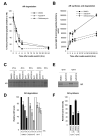
(A-B) LNCaP cells grown in steroid free conditions in SILAC media containing 5% dialyzed fetal calf serum, 13C615N4 arginine and 13C6 lysine (to label proteins as “heavy”) were treated with 10 μM AZD3514 or vehicle for 24 hours, then switched to grow in SILAC media containing unlabeled arginine (to label newly synthesized protein as “light”). Cells previously dosed with AZD3514 were treated for a further 24 hours with 10 μM AZD3514. Cells previously dosed with vehicle were treated for a further 24 hours with either 3 μM geldanamycin or vehicle. Samples were collected at indicated timepoints following the media switch and protein lysates prepared. AR protein levels were measured in the samples by immunoprecipitation with an AR specific antibody followed by liquid chromatography– mass spectrometry quantifying levels of N-terminal peptide of AR. (A) AR shown is % of heavy labeled AR peptide at each time point relative to the level of AR present at the time of the media switch. (B) AR shown is the increase in light labeled AR peptide at each time point following the media switch. Both graphs are representative data of 3 separate experiments −/+ SD. Half-life values obtained in 3 independent experiments are shown in supplementary table 3. (C-D) Radio-labeled pulse chase analysis of AR degradation in LNCaP cells. Cells were incubated with radiolabel, washed and label-free media added containing 10 μM AZD3514 or 1 nM DHT. Lysates were prepared, levels of radio-labeled AR measured and expressed relative to AR levels at time 0. Data are represented as Mean levels from 2 independent experiments. Error bars are SD. (E-F) Radio-labeled pulse chase analysis of AR synthesis in LNCaP cells. Cells were treated with 10 μM AZD3514 or vehicle control −/+ 1 nM DHT for 24 hr. Labeling Media containing treatments was added, and the cells incubated 1 hr. Lysates were prepared and levels of radio-labeled AR measured and expressed relative to the DMSO control -DHT. The graph shows the mean AR levels from 2 independent experiments. Error bars are SD.
Figure 5
Figure 5
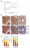
(A-D) AZD3514 was administered by oral gavage once daily to Copenhagen rats bearing established R3327H tumors at the doses indicated. (A) tumor volumes are plotted against time. (B) i, representative images showing AR IHC from animals administered with vehicle or AZD3514 at 50 or 100 mg/kg for 3 days and ii, the associated Genie™ annotation grading intensity of nuclear AR. Graphical representation of the percentage of tumor nuclei with 0+, 1+, 2+ or 3+ intensity of AR protein as quantified by Genie™ (C) and scored by a pathologist (D).
Figure 6
Figure 6
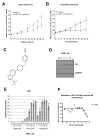
(A-B) ARD1 was administered at 100 mg/kg by oral gavage twice daily (bid) to either (A) intact or (B) castrated male nude mice bearing established HID28 tumors. Tumor volumes are plotted against time. Error bars are SEM. (C) Chemical structure of ARD1. (D) LNCaP cells grown in stripped serum conditions were treated for 24 hours with a dose response to ARD1. Effects on total AR levels were assessed by Western blot and normalized to levels of GAPDH. (E) LNCaP cells in steroid depleted media were treated with a dose response to AZD3514 and the indicated levels of DHT for 24 hours. Expression of PSA was determined by quantitative RT-PCR and normalized to levels of 18s. Data shown is expression relative to the vehicle control in the absence of ligand and is representative data. (F) Inhibition of DHT induced AR translocation in AR U2OS cells, recombinant U2OS cells stably expressing human androgen receptor (AR) fused to the C-terminus of enhanced green fluorescent protein. Cells were dosed with ARD1 30 minutes before treatment with 0.3 nM DHT for 2 hours prior to fixation. Error bars are SD.

Similar articles

Cited by

References

    1. Jemal A, Siegel R, Ward E, Hao Y, Xu J, Murray T, et al. Cancer statistics 2008. CA Cancer J Clin. 2008;58:71–96. - PubMed
    1. Huggins C, Hodges CV. Studies on prostatic cancer I the effect of castration, of estrogen and androgen injection on serum phosphatases in metastatic carcinoma of the prostate. CA Cancer J Clin. 1972;22:232–40. - PubMed
    1. Scher HI, Sawyers CL. Biology of progressive, castration-resistant prostate cancer: Directed therapies targeting the androgen-receptor signaling axis. J Clin Oncol. 2005;23:8253–61. - PubMed
    1. Attard G, Cooper CS, de Bono JS. Steroid hormone receptors in prostate cancer: A hard habit to break? Cancer Cell. 2009;16:458–62. - PubMed
    1. Knudsen KE, Penning TM. Partners in crime: Deregulation of AR activity and androgen synthesis in prostate cancer. Trends Endocrinol Metab. 2010;21:315–24. - PMC - PubMed

Publication types

MeSH terms