

Residue-level prediction of HIV-1 antibody epitopes based on neutralization of diverse viral strains
- PMID: 23843642
- PMCID: PMC3753990
- DOI: 10.1128/JVI.00984-13
Residue-level prediction of HIV-1 antibody epitopes based on neutralization of diverse viral strains
Abstract
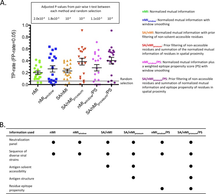
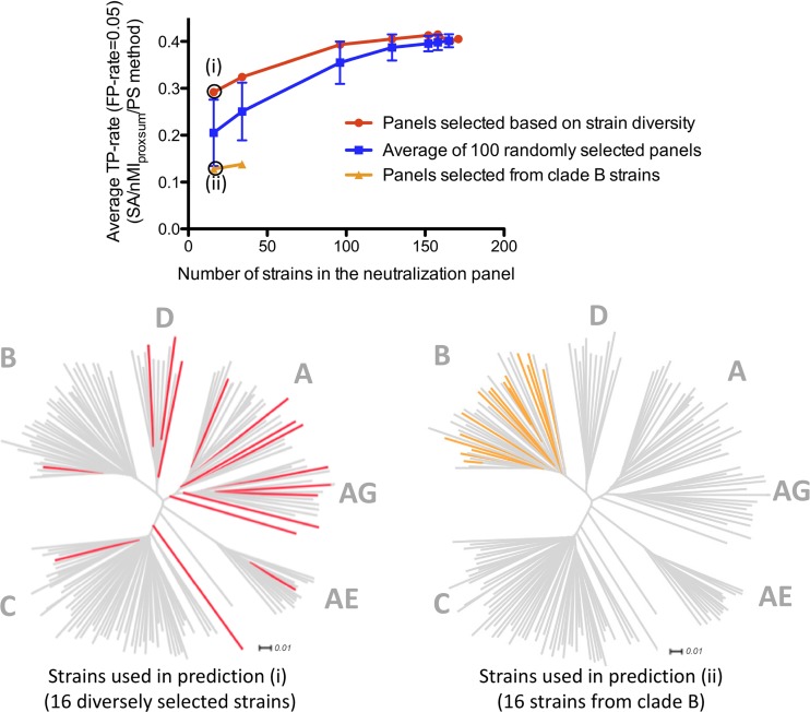
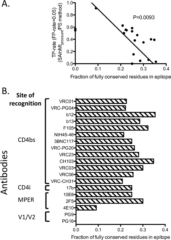
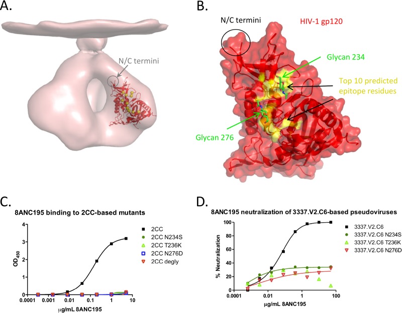
Delineation of antibody epitopes at the residue level is key to understanding antigen resistance mutations, designing epitope-specific probes for antibody isolation, and developing epitope-based vaccines. Ideally, epitope residues are determined in the context of the atomic-level structure of the antibody-antigen complex, though structure determination may in many cases be impractical. Here we describe an efficient computational method to predict antibody-specific HIV-1 envelope (Env) epitopes at the residue level, based on neutralization panels of diverse viral strains. The method primarily utilizes neutralization potency data over a set of diverse viral strains representing the antigen, and enhanced accuracy could be achieved by incorporating information from the unbound structure of the antigen. The method was evaluated on 19 HIV-1 Env antibodies with neutralization panels comprising 181 diverse viral strains and with available antibody-antigen complex structures. Prediction accuracy was shown to improve significantly over random selection, with an average of greater-than-8-fold enrichment of true positives at the 0.05 false-positive rate level. The method was used to prospectively predict epitope residues for two HIV-1 antibodies, 8ANC131 and 8ANC195, for which we experimentally validated the predictions. The method is inherently applicable to antigens that exhibit sequence diversity, and its accuracy was found to correlate inversely with sequence conservation of the epitope. Together the results show how knowledge inherent to a neutralization panel and unbound antigen structure can be utilized for residue-level prediction of antibody epitopes.
Figures


Similar articles
-
Computational analysis of anti-HIV-1 antibody neutralization panel data to identify potential functional epitope residues.Proc Natl Acad Sci U S A. 2013 Jun 25;110(26):10598-603. doi: 10.1073/pnas.1309215110. Epub 2013 Jun 10. Proc Natl Acad Sci U S A. 2013. PMID: 23754383 Free PMC article.
-
Predicting HIV-1 broadly neutralizing antibody epitope networks using neutralization titers and a novel computational method.BMC Bioinformatics. 2014 Mar 19;15:77. doi: 10.1186/1471-2105-15-77. BMC Bioinformatics. 2014. PMID: 24646213 Free PMC article.
-
Murine Antibody Responses to Cleaved Soluble HIV-1 Envelope Trimers Are Highly Restricted in Specificity.J Virol. 2015 Oct;89(20):10383-98. doi: 10.1128/JVI.01653-15. Epub 2015 Aug 5. J Virol. 2015. PMID: 26246566 Free PMC article.
-
A Structural Update of Neutralizing Epitopes on the HIV Envelope, a Moving Target.Viruses. 2021 Sep 5;13(9):1774. doi: 10.3390/v13091774. Viruses. 2021. PMID: 34578355 Free PMC article. Review.
-
Structural Features of Broadly Neutralizing Antibodies and Rational Design of Vaccine.Adv Exp Med Biol. 2018;1075:73-95. doi: 10.1007/978-981-13-0484-2_4. Adv Exp Med Biol. 2018. PMID: 30030790 Review.
Cited by
-
Comparison of Antibody-Dependent Cell-Mediated Cytotoxicity and Virus Neutralization by HIV-1 Env-Specific Monoclonal Antibodies.J Virol. 2016 Jun 10;90(13):6127-6139. doi: 10.1128/JVI.00347-16. Print 2016 Jul 1. J Virol. 2016. PMID: 27122574 Free PMC article.
-
Determinants of HIV-1 broadly neutralizing antibody induction.Nat Med. 2016 Nov;22(11):1260-1267. doi: 10.1038/nm.4187. Epub 2016 Sep 26. Nat Med. 2016. PMID: 27668936
-
Role of framework mutations and antibody flexibility in the evolution of broadly neutralizing antibodies.Elife. 2018 Feb 14;7:e33038. doi: 10.7554/eLife.33038. Elife. 2018. PMID: 29442996 Free PMC article.
-
Progress and challenges in predicting protein interfaces.Brief Bioinform. 2016 Jan;17(1):117-31. doi: 10.1093/bib/bbv027. Epub 2015 May 13. Brief Bioinform. 2016. PMID: 25971595 Free PMC article. Review.
-
Vaccine-Elicited Tier 2 HIV-1 Neutralizing Antibodies Bind to Quaternary Epitopes Involving Glycan-Deficient Patches Proximal to the CD4 Binding Site.PLoS Pathog. 2015 May 29;11(5):e1004932. doi: 10.1371/journal.ppat.1004932. eCollection 2015 May. PLoS Pathog. 2015. PMID: 26023780 Free PMC article.
References
-
- Corti D, Langedijk JP, Hinz A, Seaman MS, Vanzetta F, Fernandez-Rodriguez BM, Silacci C, Pinna D, Jarrossay D, Balla-Jhagjhoorsingh S, Willems B, Zekveld MJ, Dreja H, O'Sullivan E, Pade C, Orkin C, Jeffs SA, Montefiori DC, Davis D, Weissenhorn W, McKnight A, Heeney JL, Sallusto F, Sattentau QJ, Weiss RA, Lanzavecchia A. 2010. Analysis of memory B cell responses and isolation of novel monoclonal antibodies with neutralizing breadth from HIV-1-infected individuals. PLoS One 5:e8805.10.1371/journal.pone.0008805 - DOI - PMC - PubMed
-
- Scheid JF, Mouquet H, Ueberheide B, Diskin R, Klein F, Oliveira TY, Pietzsch J, Fenyo D, Abadir A, Velinzon K, Hurley A, Myung S, Boulad F, Poignard P, Burton DR, Pereyra F, Ho DD, Walker BD, Seaman MS, Bjorkman PJ, Chait BT, Nussenzweig MC. 2011. Sequence and structural convergence of broad and potent HIV antibodies that mimic CD4 binding. Science 333:1633–1637 - PMC - PubMed
-
- Walker LM, Phogat SK, Chan-Hui PY, Wagner D, Phung P, Goss JL, Wrin T, Simek MD, Fling S, Mitcham JL, Lehrman JK, Priddy FH, Olsen OA, Frey SM, Hammond PW, Protocol GPI, Kaminsky S, Zamb T, Moyle M, Koff WC, Poignard P, Burton DR. 2009. Broad and potent neutralizing antibodies from an African donor reveal a new HIV-1 vaccine target. Science 326:285–289 - PMC - PubMed
-
- Wu X, Yang ZY, Li Y, Hogerkorp CM, Schief WR, Seaman MS, Zhou T, Schmidt SD, Wu L, Xu L, Longo NS, McKee K, O'Dell S, Louder MK, Wycuff DL, Feng Y, Nason M, Doria-Rose N, Connors M, Kwong PD, Roederer M, Wyatt RT, Nabel GJ, Mascola JR. 2010. Rational design of envelope identifies broadly neutralizing human monoclonal antibodies to HIV-1. Science 329:856–861 - PMC - PubMed
-
- Ekiert DC, Kashyap AK, Steel J, Rubrum A, Bhabha G, Khayat R, Lee JH, Dillon MA, O'Neil RE, Faynboym AM, Horowitz M, Horowitz L, Ward AB, Palese P, Webby R, Lerner RA, Bhatt RR, Wilson IA. 2012. Cross-neutralization of influenza A viruses mediated by a single antibody loop. Nature 489:526–532 - PMC - PubMed
Publication types
MeSH terms
Substances
Grants and funding
LinkOut - more resources
Full Text Sources
Other Literature Sources
