. 2013 Jul 1;8(7):e67694.
doi: 10.1371/journal.pone.0067694. Print 2013.

GRG5/AES interacts with T-cell factor 4 (TCF4) and downregulates Wnt signaling in human cells and zebrafish embryos

Affiliations

GRG5/AES interacts with T-cell factor 4 (TCF4) and downregulates Wnt signaling in human cells and zebrafish embryos

Angela M Sousa Costa et al. PLoS One. .

Abstract

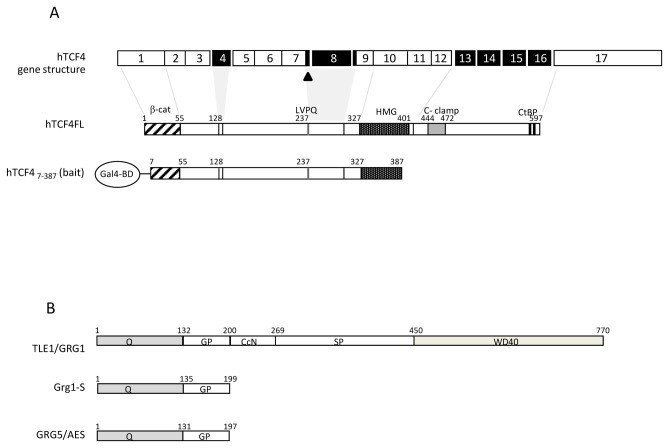
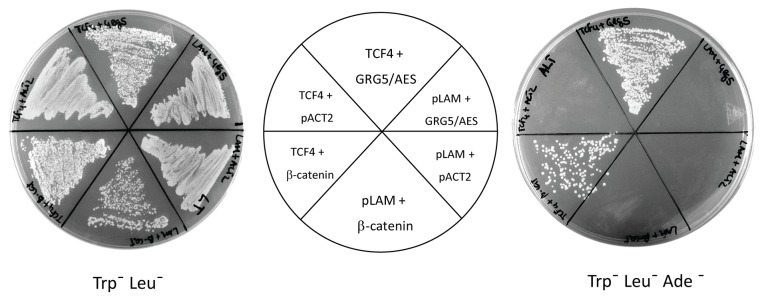
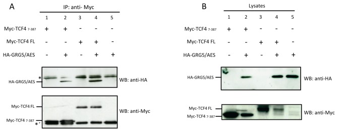
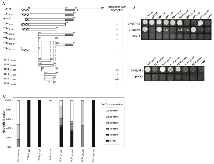
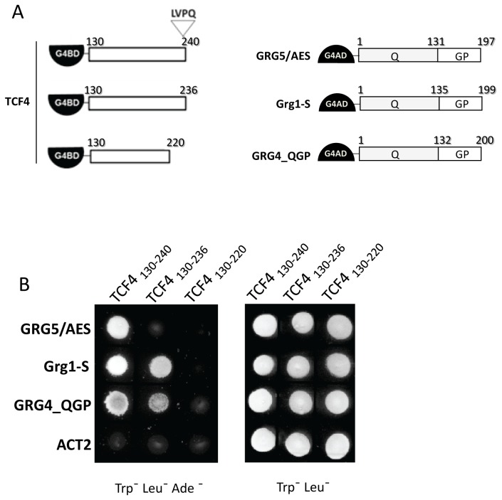
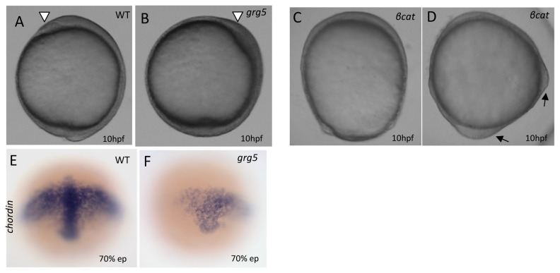
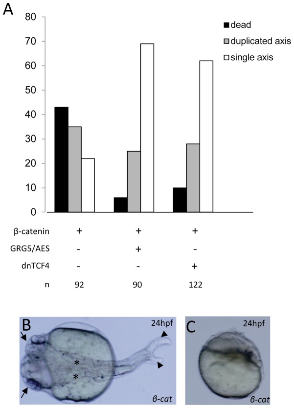
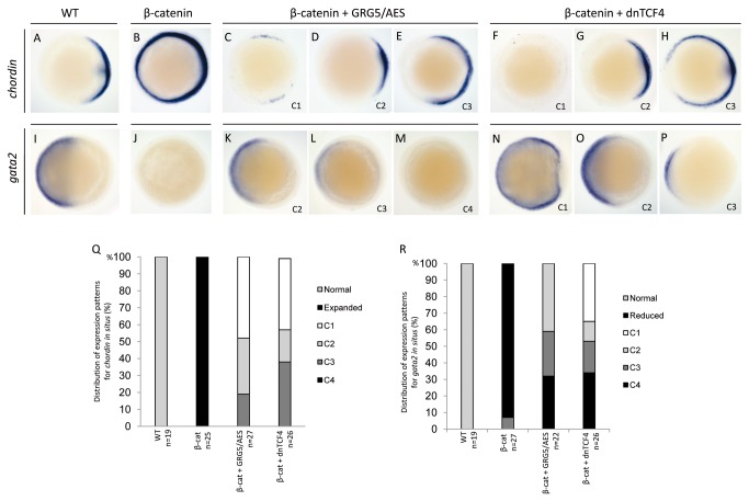
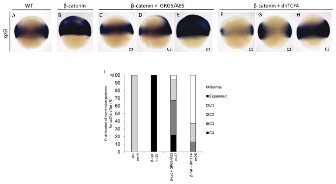
Transcriptional control by TCF/LEF proteins is crucial in key developmental processes such as embryo polarity, tissue architecture and cell fate determination. TCFs associate with β-catenin to activate transcription in the presence of Wnt signaling, but in its absence act as repressors together with Groucho-family proteins (GRGs). TCF4 is critical in vertebrate intestinal epithelium, where TCF4-β-catenin complexes are necessary for the maintenance of a proliferative compartment, and their abnormal formation initiates tumorigenesis. However, the extent of TCF4-GRG complexes' roles in development and the mechanisms by which they repress transcription are not completely understood. Here we characterize the interaction between TCF4 and GRG5/AES, a Groucho family member whose functional relationship with TCFs has been controversial. We map the core GRG interaction region in TCF4 to a 111-amino acid fragment and show that, in contrast to other GRGs, GRG5/AES-binding specifically depends on a 4-amino acid motif (LVPQ) present only in TCF3 and some TCF4 isoforms. We further demonstrate that GRG5/AES represses Wnt-mediated transcription both in human cells and zebrafish embryos. Importantly, we provide the first evidence of an inherent repressive function of GRG5/AES in dorsal-ventral patterning during early zebrafish embryogenesis. These results improve our understanding of TCF-GRG interactions, have significant implications for models of transcriptional repression by TCF-GRG complexes, and lay the groundwork for in depth direct assessment of the potential role of Groucho-family proteins in both normal and abnormal development.


Conflict of interest statement

Competing Interests: The authors have declared that no competing interests exist.

Figures

Figure 1
Figure 1. Schematic representation of the mammalian TCF4 and GRG domains organization.
A) The human TCF4 gene consists of 17 exons (top), some of which are subject to alternative splicing (black exons). The TCF4 variant used (Ex1-17) includes isoform specific sequences such as the LVPQ domain due to the use of an alternative splice donor site at the end of exon 7 (arrowhead). A TCF4 fragment lacking the C terminus (a.a.s 7-387), was fused to Gal4’s DNA-binding domain (Gal4-BD) and used as bait in a two hybrid screen. B) The GRG family comprises two distinct classes of proteins, based on their domain constitution: the “long Grouchos”, GRG/TLE 1-4, consisting of five different domains (Q, GP, CcN, SP and WD40); and the “short Grouchos”, such as the alternative splice-product Grg1-S or the GRG5/AES subfamily members, which contain only the Q and GP domains.
Figure 2
Figure 2. Physical interaction of TCF4 and GRG5/AES in yeast cells.
The AH109 yeast strain was co-transformed with the indicated constructs (central panel). Transformants were selected in medium lacking tryptophan and leucine (left panel) and activation of a Gal4-dependent ADE2 gene was examined by growth on selective plates additionally lacking adenine (right panel). Yeast cells containing both TCF4 and GRG5/AES grew in the selective media. pLAM and pACT2 plasmids were used as bait and prey negative controls respectively, while the β-catenin prey plasmid constitutes a positive control for the interaction assays.
Figure 3
Figure 3. Physical interaction of TCF4 and GRG5/AES in human cells.
A) Lysates of HEK293 cells transiently transfected with the indicated expression plasmids were subject to immunoprecipitation (IP) with anti-Myc antibody and the immunoprecipitated proteins were analyzed by Western blot (WB) with anti-HA and anti-Myc antibodies. Co-precipitation of HA-tagged GRG5/AES was detected only in the lysates of cells co-transfected with Myc-TCF47-387 (lane 2) or Myc-TCF4FL (full-length) (lane 4). B) 10% of each total cell lysate was loaded for WB with either anti-HA or anti-Myc antibodies. The asterisks indicate nonspecific binding to immunoglobulin light (*) and heavy (*´) chains.
Figure 4
Figure 4. Mapping of TCF4’s GRG5/AES-interacting region using yeast two hybrid assays.
A) Schematic diagram of the Gal4BD-TCF4 constructs tested for interaction with GRG5/AES. B) GRG5/AES-interacting TCF4 fragments activate an ADE2 reporter gene, conferring to yeast transformants the ability to grow in selective medium lacking tryptophan, leucine and adenine. C) Transformants were also tested for activation of a HIS3 reporter, as determined by growth in medium lacking histidine and with increasing doses of 3-aminotriazol (3AT). As defined by both reporter assays and summarized on the right panel of the diagrams, the region encompassing residues 130-240 is sufficient for the interaction, with residues 220-240 being strictly necessary. Two previously described fragments were included as internal controls: dnTCF4, that lacks the N terminal β-catenin binding domain [13]; and TCF1176-359 [49]; n.d.: not done.
Figure 5
Figure 5. Convergent and divergent sequence requirements for TCF4 binding to different GRGs.
A) Schematic view of TCF4 (left) and GRGs (right) fusion constructs used in yeast-two hybrid assays. B) Two-hybrid analysis of the importance of amino acids 130-240, 221-240 and the LVPQ motif for the interactions between TCF4 and GRGs. Co-transformant yeast cells were selected in medium lacking tryptophan and leucine and assayed for the indicated interactions using the adenine reporter gene (in Trp-Leu-Ade-medium). The same 111-amino acid region (residues 130-240) is sufficient for strong interaction with all three GRGs tested, and amino acids 221-240 are required for it, whereas removal of the LVPQ motif abrogates binding to GRG5/AES, but not the other GRGs, although it has some effect on the TCF4-GRG4 interaction. The ACT2 plasmid was used as a negative control.
Figure 6
Figure 6. GRG5/AES represses TCF-β-catenin mediated transcription and co-operates with both dnTCF4 and Grg1-L for transcriptional repression.
HEK293 cells were transfected with either the reporter construct TCF-Luc (containing three TCF consensus binding sites upstream of the firefly luciferase cDNA), or the control reporter TCF*-Luc (with mutations in the TCF-binding sites) and the indicated combinations of expression plasmids. A) Addition of GRG5/AES plasmid decreases the luciferase activity induced by transfected βcat and endogenous TCFs in a dose dependent manner. At the highest dose (1.2 μg/μl), GRG5/AES-mediated repression is comparable to the effect of dominant negative TCF4 (dnTCF4), a previous established repressor of the canonical Wnt pathway [13]. B) Repression of TCF-β-catenin mediated transcription by various Groucho-family proteins. In contrast to “long” Groucho Grg1 (Grg1-L), GRG5/AES, GRG4 and Grg1-S (a short GRG1 isoform [63]) effectively repressed TCF-dependent luciferase activity. 1.2 µg were used for transfection of each Groucho expression plasmid. C) Co-transfection of GRG5/AES with either dnTCF4 or Grg1L (0.4µg of each plasmid) results in higher repression of TCF-β-catenin dependent luciferase activity then transfections with each effector plasmid individually. β-galactosidase assays were performed as an internal control for transfection efficiency. All the assays were done in triplicate in three independent experiments.
Figure 7
Figure 7. GRG5/AES overexpression leads to ventralization of zebrafish embryos.
(A–D) Lateral view, dorsal to the right, of 10hpf-stage embryos. (A) Control uninjected; (B) Injected with 250pg of grg5/aes mRNA; arrowhead: anterior end of the neural plate with formation of the “polster” in the prospective head region; (C–D) Injected with 100pg of an activated β-catenin mRNA. Black arrows: ectopic dorsal axis; E-F) Whole-mount in situ hybridization for chordin expression at 70% epiboly uninjected (E) and grg5/aes-injected (F) embryos.
Figure 8
Figure 8. GRG5/AES reduces both mortality and axis duplication in β-catenin-overexpressing embryos.
A) One blastomere of 8-cell stage zebrafish embryos was injected with the indicated combinations of β-catenin (50 pg), grg5/aes (500 pg) and dntcf4 (500 pg) mRNAs. Partial and complete axis duplications were grouped to avoid ambiguity. The total number of scored embryos (n) is indicated at the lower part of the panel. B) Example of 24hpf embryo showing complete axis duplication (induced by β-catenin injection), with two discernible heads (arrows), notochords (asterisks) and tails (arrowheads). Dorsal view, anterior to the left. C) Example of 24hpf embryo injected with β-catenin showing extreme dorsalization.
Figure 9
Figure 9. GRG5/AES antagonizes β-catenin in dorsal-ventral patterning during early zebrafish embryogenesis.
Whole-mount in situ hybridizations for chordin (A–H) and gata2 (I–P) of double-injection experiments (columns). Animal pole views, dorsal to the right, of 70% epiboly stage-embryos. Injection of β-catenin strongly dorsalizes embryos, as shown by an expanded and circumferential expression of the dorsal-specific gene chordin (B) and a total absence of the ventral-specific gene gata2 (J). Co-injection of grg5/aes (C-E; K-M) rescues β-catenin-induced dorsalization and restores normal expression domains for both marker genes. dntcf4 co-injections (F–H; N–P) were used as a control for β-catenin antagonism. Co-injected embryos were grouped in four classes of marker gene expression: C1 (C, F) – abnormal expression consistent with embryo ventralization; C2 (D, G; K,O) − restoration of normal expression patterns (A,I); C3 (E, H; L, P) – partial reversion of β-catenin-induced abnormalities; C4 (M) − abnormal expression consistent with embryo dorsalization. Q–R: distribution of the embryos in the different classes of chordin (Q) and gata2 (R) expression. GRG5/AES antagonizes β-catenin effects on chordin and gata2 in 100% and 68% of the embryos respectively. n: number of embryos tested.
Figure 10
Figure 10. GRG5/AES represses Wnt signaling during early zebrafish embryogenesis.
Whole-mount in situ hybridizations for sp5l, a direct target of canonical Wnt signaling. Lateral view, dorsal to the right, of 70% epiboly stage-embryos. Injection of β-catenin induces broad ectopic domains of sp5l expression (B), which are reversed by co-injection of grg5/aes (C–E) or dntcf4 (F–H). Expression patterns in co-injected embryos were grouped in four classes, as above. AES reversed β-catenin-induced misexpression in 75% of the embryos (I). n: number of embryos tested.

Similar articles

Cited by

References

    1. Castrop J, van Norren K, Clevers H (1992) A gene family of HMG-box transcription factors with homology to TCF-1. Nucleic Acids Res 20: 611. doi:10.1093/nar/20.3.611. PubMed: 1741298. - DOI - PMC - PubMed
    1. Giese K, Amsterdam A, Grosschedl R (1991) DNA-binding properties of the HMG domain of the lymphoid-specific transcriptional regulator LEF-1. Genes Dev 5: 2567-2578. doi:10.1101/gad.5.12b.2567. PubMed: 1752444. - DOI - PubMed
    1. Giese K, Cox J, Grosschedl R (1992) The HMG domain of lymphoid enhancer factor 1 bends DNA and facilitates assembly of functional nucleoprotein structures. Cell 69: 185-195. doi:10.1016/0092-8674(92)90129-Z. PubMed: 1555239. - DOI - PubMed
    1. van de Wetering M, Clevers H (1992) Sequence-specific interaction of the HMG box proteins TCF-1 and SRY occurs within the minor groove of a Watson-Crick double helix. EMBO J 11: 3039-3044. PubMed: 1639073. - PMC - PubMed
    1. Travis A, Amsterdam A, Belanger C, Grosschedl R (1991) LEF-1, a gene encoding a lymphoid-specific protein with an HMG domain, regulates T-cell receptor alpha enhancer function [corrected]. Genes Dev 5: 880-894. doi:10.1101/gad.5.5.880. PubMed: 1827423. - DOI - PubMed

Publication types

MeSH terms

Grants and funding

This work was supported by Fundação para a Ciência e a Tecnologia (through grants POCTI/41854/MGI/2001 and Ciência2008-ICAAM to L.T.C., SFRH/BD/24402/2005 to Â.M.S.C. and SFRH/BD/44264/2008 to I.P.-C. and Strategic Project PEst-C/AGR/UI0115/2011) as well as FEDER Funds through the COMPETE programme. IPATIMUP is an Associate Laboratory of the Portuguese Ministry of Science, Technology and Higher Education and is partially supported by FCT. The funders had no role in study design, data collection and analysis, decision to publish, or preparation of the manuscript.

LinkOut - more resources