. 2013 Jul;25(7):2661-78.
doi: 10.1105/tpc.113.113118. Epub 2013 Jul 9.

Arabidopsis CURVATURE THYLAKOID1 proteins modify thylakoid architecture by inducing membrane curvature

Affiliations

Arabidopsis CURVATURE THYLAKOID1 proteins modify thylakoid architecture by inducing membrane curvature

Ute Armbruster et al. Plant Cell. 2013 Jul.

Abstract

Chloroplasts of land plants characteristically contain grana, cylindrical stacks of thylakoid membranes. A granum consists of a core of appressed membranes, two stroma-exposed end membranes, and margins, which connect pairs of grana membranes at their lumenal sides. Multiple forces contribute to grana stacking, but it is not known how the extreme curvature at margins is generated and maintained. We report the identification of the CURVATURE THYLAKOID1 (CURT1) protein family, conserved in plants and cyanobacteria. The four Arabidopsis thaliana CURT1 proteins (CURT1A, B, C, and D) oligomerize and are highly enriched at grana margins. Grana architecture is correlated with the CURT1 protein level, ranging from flat lobe-like thylakoids with considerably fewer grana margins in plants without CURT1 proteins to an increased number of membrane layers (and margins) in grana at the expense of grana diameter in overexpressors of CURT1A. The endogenous CURT1 protein in the cyanobacterium Synechocystis sp PCC6803 can be partially replaced by its Arabidopsis counterpart, indicating that the function of CURT1 proteins is evolutionary conserved. In vitro, Arabidopsis CURT1A proteins oligomerize and induce tubulation of liposomes, implying that CURT1 proteins suffice to induce membrane curvature. We therefore propose that CURT1 proteins modify thylakoid architecture by inducing membrane curvature at grana margins.


Figures
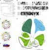
Figure 1.
Figure 1.
Characteristics of CURT1 Proteins. (A) Expression characteristics of CURT1 protein family genes. CURT1A, B, C, and D, together with the chloroplast protein coding genes TIC110, TOC33, PSBX, PSAD1, and VIPP1 and the nonchloroplast protein coding gene CRY2, were hierarchically clustered according to expression profile similarity using the gene coexpression database ATTEDII (http://atted.jp/) (Obayashi et al., 2007). Low distance values indicate high expression similarities. (B) Sequence alignment of the CURT1 proteins in Arabidopsis. Predicted chloroplast transit peptide sequences are shown in lowercase letters, positions of α-helices (H1-H4) are indicated, and the two TMs (∼) are highlighted in bold. Peptide sequences used for antibody generation are depicted in italics. Identical and closely related amino acids that are conserved in >50% of the sequences are highlighted by black and gray shading, respectively. (C) CURT1 proteins contain a conserved putative N-terminal amphipathic helix. Helical wheel representations of helix H1 (as defined in [B]) are shown for CURT1A (CURT1A-H1) and CURT1B (CURT1B-H1). In the helical wheel representation, the amino acids are colored according to the physicochemical properties of the side chains (yellow, hydrophobic; blue, polar, positively charged; pink, polar, negatively charged; green, polar, uncharged). Both helical wheel representations show a significant spatial separation of polar and charged from hydrophobic amino acid residues, indicative of an amphipathic helix. (D) Three-dimensional model of CURT1A-H1. The computed surfaces are gradually colored according to their hydrophobicity from dark red (highly hydrophobic) to deep blue (highly polar). (E) Phylogenetic analysis of the CURT1 protein family. Branch lengths reflect the estimated number of substitutions per 100 sites. The cyanobacterial and green algal clades are highlighted by blue and dark-green background/branch color, respectively. Bryophytes and gymnosperms are indicated by light-green background/branch color and magnoliophytes (flowering plants) by green color.
Figure 2.
Figure 2.
CURT1 Proteins Are Intrinsic Thylakoid Proteins. (A) Subcellular localization of CURT1 proteins. Full-length CURT1A-, CURT1C-, and CURT1D-dsRED fusions were transiently introduced into Arabidopsis protoplasts by polyethylene glycol–mediated DNA uptake and analyzed using fluorescence microscopy (Auto, chloroplasts revealed by chlorophyll autofluorescence; dsRED, fluorescence of the fusion protein; Merged, overlay of both images). Bar = 10 μm. (B) Suborganellar localization of CURT1 proteins. Chloroplasts from wild-type and oeCURT1D-HA plants were subfractionated into thylakoids (Thy), stroma (Str), and two envelope (Env) fractions. Aliquots (40 µg) of protein from each fraction were subjected to SDS-PAGE, followed by immunoblot analysis using antibodies raised against CURT1A, B, C, or HA. As controls for purity of the different fractions, antibodies recognizing Lhcb1, Tic110, and RbcL, which are located in thylakoids, envelope, and stroma, respectively, were used. (C) Extraction of thylakoid-associated proteins with chaotropic salt solutions or alkaline pH. Thylakoids from wild-type and oeCURT1D-HA plants were resuspended at 0.5 mg chlorophyll/mL in 10 mM HEPES/KOH, pH 7.5, containing either 2 M NaBr, 0.1 M Na2CO3, 2 M NaSCN, or no additive. After incubation, supernatants containing the extracted proteins (s) and membrane fractions (p) were separated by SDS-PAGE and immunolabeled with antibodies against CURT1A, B, C, and HA. As control for peripheral membrane proteins, antibodies raised against NdhH were used. To control for integral membrane proteins, antibodies specific for Lhcb1, cytochrome b6, and Rieske (PetC) were employed.
Figure 3.
Figure 3.
CURT1 Proteins Form Oligomers. (A) Abundance of CURT1 proteins in curt1 mutant plants. Total protein extracts from Col-0 and curt1 mutant plants corresponding to 3 µg of chlorophyll were fractionated by SDS-PAGE, and blots were probed with antibodies raised against CURT1A, B, and C. The Ponceau Red (P.R.)–stained protein blot served as loading control. (B) Absolute abundance of CURT1 proteins in wild-type (WT) plants. The four Arabidopsis CURT1 proteins were expressed in Escherichia coli as C-terminal fusions to the MBP, purified by affinity chromatography, and quantified. Adequate quantities of these four MBP fusions, as well as of the MBP fusions of PetC and PsaD as controls (see Supplemental Figure 3E online), were titrated against thylakoid membrane protein preparations and subjected to immunoblot analyses. Representative results from five experiments are shown. The calculated concentration of the respective CURT1 protein in thylakoid preparations is given below each panel. (C) Two-dimensional BN/SDS-PAGE separation of thylakoid protein complexes. Individual lanes from BN-PA gels like those shown on top were separated in a second dimension by SDS-PAGE. Blots were immunolabeled with antibodies raised against Lhcb1, PsaB, cytochrome b6, and CURT1A, B, and C. The positions of major thylakoid multiprotein complexes are indicated by Roman numerals (top) and the composition of the complexes by circled Arabic numbers (bottom) as in Supplemental Figure 3F online. f.p., free protein. (D) Chemical cross-linking. Thylakoid proteins from wild-type (Landsberg erecta [Ler]) and mutant (curt1b-1, curt1a-1, and curt1c-1) plants were cross-linked with bis(sulfosuccinimidyl) suberate, separated by SDS-PAGE, and subjected to immunoblot analysis with antibodies raised against CURT1A and B. On the right, the protein composition of cross-linked products is shown. As loading control, blots were stained with Ponceau Red (see Supplemental Figure 3H online). (E) CoIP. Thylakoid membranes from wild-type, CURT1A-HA curt1a-2, and oeCURT1D-HA plants were solubilized, and HA-tagged proteins were allowed to bind to α-HA affinity matrix. The matrix was recovered and the supernatant was collected. After washing, coimmunoprecipitated proteins were eluted. Thylakoids, supernatant (s.n.), and coimmunoprecipitated proteins (Co-IP HA) were then analyzed by SDS-PAGE followed by immunoblot analysis with antibodies raised against HA, CURT1A, B, and C, and Lhcb1.
Figure 4.
Figure 4.
Lack of CURT1 Proteins Does Not Disturb Photosynthetic Complex Accumulation but Affects Photosynthesis Pleiotropically. (A) The composition of thylakoid complexes from the wild type (Landsberg erecta [Ler]) and curt1 mutants was analyzed as in Figure 3A, except that aliquots corresponding to 5 µg chlorophyll were used. Antibodies specific for the PSII subunit CP43, cytochrome b6 from the cytochrome b6/f complex, the D subunit of PSI (PsaD), the γ-subunit of the chloroplast ATP synthase, NdhL from the NDH complex, and CURT1A were used. Ponceau Red (P.R.) staining served as the loading control. (B) Immunoblot analysis of leaf proteins (corresponding to 40 µg of total protein) from the wild type (Col-0 and Landsberg erecta), curt1a-1 and curt1b-1 mutants, and mutants devoid of PSII (hcf136), PSI (psad1 psad2), cytochrome b6/f (petc-2), or cpATPase (atpd-1). Antibodies specific for CURT1A, B, and C and the respective thylakoid multiprotein complexes, or actin as control, were used. (C) Effective quantum yield (ΦII), NPQ, and 1-qL (as listed in Supplemental Table 2 online) for wild-type and curt1 mutant plants are indicated on a gray scale. (D) Time course of chlorophyll a fluorescence during illumination with actinic light at 100 μmol photons m−2 s−1. Average values ± sd (bars) for three to five different plants are indicated by closed (wild type) or open (curt1abcd) circles. Relative units (r.u.) are shown. (E) Quantification of cyclic electron transport in situ. Increases in chlorophyll fluorescence were measured in ruptured chloroplasts after the addition of NADPH and Fd as described (Munekage et al., 2002). As controls, the mutants pgr5 (defective in Fd-dependent cyclic electron transport) and crr2 (defective in NDH-dependent cyclic electron transport) were employed.
Figure 5.
Figure 5.
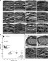
Thylakoid Architecture Depends on CURT1 Protein Levels. (A) TEM micrographs of ultrathin sections of leaves from the wild type (Landsberg erecta [Ler] and Col-0), curt1 mutants, and lines expressing tagged CURT1A proteins (CURT1A-HA curt1a-2 and CURT1A-cmyc curt1a-2). Note that the CURT1A-HA curt1a-2 and CURT1A-cmyc curt1a-2 lines express ∼50 and 400% of wild-type levels of CURT1A, respectively (see Supplemental Figure 1 online). (B) Scatterplot indicating the height (y axis) and diameter (x axis) of thylakoid stacks in the genotypes shown in (A) and (D). Average values (n ≥ 30) including the respective se are presented; a complete list of the P values, indicating the significance of the differences in grana stacking between the genotypes, is provided in Supplemental Data Set 1 online. WT, the wild type. (C) and (D) Sections of chloroplasts as in (A) from curt1ab and curt1abcd (C) and from stn7 stn8 and tap38 mutant plants (D). Circles indicate tubuli and vesicles characteristic for curt1 mutant chloroplasts.
Figure 6.
Figure 6.
CURT1A Levels Determine Thylakoid Architecture. (A) Topographic view of envelope-free chloroplasts from curt1abcd, wild-type (Col-0), and CURT1A-cmyc curt1a-2 leaves (that express tagged CURT1A proteins at ∼400% of wild-type CURT1A levels) by scanning electron microscopy. (B) Wild-type and CURT1A-cmyc curt1a-2 (oeCURT1A) thylakoids were cross-linked and analyzed as in Figure 3D, and CURT1A signals were quantified. Intensities for bands representing monomers (m), dimers (d), tetramers (t), and oligomers (o) are given in percentages next to the corresponding genotype. Asterisks indicate unspecific bands.
Figure 7.
Figure 7.
CURT1 Proteins Are Enriched at Grana Margins. (A) Scanning electron microscopy images of envelope-free chloroplasts from wild-type (Col-0), CURT1A-HA curt1a-2, and oeCURT1A-cmyc curt1a-2 plants, following immunogold labeling with antibodies raised against CURT1B, cytochrome f, HA, and cmyc. Bars = 500 nm. (B) Quantification of the distribution of immunogold-labeled CURT1 proteins. At least eight independent scanning electron microscopy pictures of each experiment, as exemplarily shown in (A), were analyzed for the distribution of signals from gold particles. “Margins” were defined as the area covering around 5 nm in both directions of the visual membrane bending zones. The residual surface was combined to represent the grana core and stroma lamellae regions. Note that the minor amounts of CURT1 proteins in Col-0 and oeCURT1A-cmyc plants assigned to the grana core or stroma lamellae regions were also mainly detected close to the curved membrane borders but just outside of the region arbitrarily defined as “margins.” Error bars represent the sd between the independent scanning electron microscopy images. WT, the wild type. (C) The distribution ratios of the CURT1 proteins and, as control, cytochrome f were determined based on the specific accumulation of gold particles in the margins or the remaining membrane area (grana core + stroma lamellae) of the same scanning electron microscopy images analyzed in (B). Error bars represent sd as in (B).
Figure 8.
Figure 8.
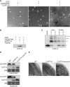
CURT1A Oligomers Tubulate Liposomes and Their Functions Are Evolutionary Conserved. (A) Liposomes with thylakoid-like lipid composition were incubated with E. coli cell-free extracts (CFEs) either containing a template (CURT1A) for de novo CURT1A synthesis or not. After purification, liposomes were analyzed by TEM. Tubular structures are indicated by white arrowheads. (B) Liposomal integration of CURT1A. Liposomes from reaction mixtures with indicated composition were purified and subjected to SDS-PAGE and protein gel blot analysis. Because the CURT1A antibody recognizes a stroma-exposed region of CURT1A (see Supplemental Figure 1C online), trypsination of proteoliposomes confirmed the surface-exposed and thylakoid-like topology of CURT1A. (C) CURT1A proteoliposomes were cross-linked with membrane-permeable [dithiobis(succinimidylpropionate) (DSP)] and -impermeable [3,3′-dithiobis(sulfosuccinimidylpropionate) (DTSSP)] cross-linkers. Cross-linking products (A-A to A-A-A-A) were detected by SDS-PAGE and protein gel blot analysis. (D) Heterologous expression of Arabidopsis CURT1A in Synechocystis alters thylakoid membrane structure. Different Synechocystis mutant strains that express CURT1A in the presence (CURT1A) or absence (CURT1A syncurt1) of endogenous Synechocystis CURT1 (synCURT1) were generated. Total proteins and thylakoid proteins of wild-type (WT) and mutant Synechocystis strains were analyzed by SDS-PAGE and immunoblot analysis, employing antibodies specific for CURT1A and synCURT1. Coomassie blue staining (CBB) served as the loading control. (E) The same genotypes as in (D) were analyzed regarding their thylakoid structure by TEM. Exemplary detached phycobilisomes are indicated by white arrowheads.
Figure 9.
Figure 9.
Hypothetic Model for the Effects of CURT1 Proteins and Their Phosphorylated Forms on Grana Stacking. CURT1 proteins are symbolized by black ellipses. Hypothetical phosphorylation-induced changes in CURT1 structure or oligomerization state are indicated by changes in ellipse size. Note that changes in grana stacking in the stn7 stn8 and tap38 mutants might alternatively be caused by changes in repulsion between membrane layers due to altered PSII phosphorylation (Fristedt et al., 2009), although this effect is overridden in curt1 mutants with increased thylakoid phosphorylation.

Similar articles

Cited by

References

    1. Albertsson P.A., Andreasson E. (2004). The constant proportion of grana and stroma lamellae in plant chloroplasts. Physiol. Plant. 121: 334–342 - PubMed
    1. Allen J.F., Forsberg J. (2001). Molecular recognition in thylakoid structure and function. Trends Plant Sci. 6: 317–326 - PubMed
    1. Anderson J.M. (1986). Photoregulation of the composition, function, and structure of thylakoid membranes. Annu. Rev. Plant Physiol. Plant Mol. Biol. 37: 93–136
    1. Anderson J.M., Chow W.S., De Las Rivas J. (2008). Dynamic flexibility in the structure and function of photosystem II in higher plant thylakoid membranes: The grana enigma. Photosynth. Res. 98: 575–587 - PubMed
    1. Armbruster U., Zühlke J., Rengstl B., Kreller R., Makarenko E., Rühle T., Schünemann D., Jahns P., Weisshaar B., Nickelsen J., Leister D. (2010). The Arabidopsis thylakoid protein PAM68 is required for efficient D1 biogenesis and photosystem II assembly. Plant Cell 22: 3439–3460 - PMC - PubMed

Publication types

MeSH terms