. 2013 Sep;12(9):1860-73.
doi: 10.1158/1535-7163.MCT-13-0157. Epub 2013 Jul 9.

Chk1/2 inhibition overcomes the cisplatin resistance of head and neck cancer cells secondary to the loss of functional p53

Affiliations

Chk1/2 inhibition overcomes the cisplatin resistance of head and neck cancer cells secondary to the loss of functional p53

Mayur A Gadhikar et al. Mol Cancer Ther. 2013 Sep.

Abstract

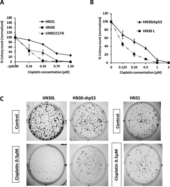
Despite the use of multimodality therapy using cisplatin to treat patients with advanced stage squamous cell carcinoma of the head and neck (HNSCC), there is an unacceptably high rate of treatment failure. TP53 is the most commonly mutated gene in HNSCC, and the impact of p53 mutation on response to cisplatin treatment is poorly understood. Here, we show unambiguously that wild-type TP53 (wtp53) is associated with sensitivity of HNSCC cells to cisplatin treatment, whereas mutation or loss of TP53 is associated with cisplatin resistance. We also show that senescence is the major cellular response to cisplatin in wtp53 HNSCC cells and that cisplatin resistance in p53-null or -mutant TP53 cells is due to their lack of senescence. Given the dependence on checkpoint kinase (Chk)1/2 kinases to mediate the DNA damage response in p53-deficient cells, there is potential to exploit this to therapeutic advantage through targeted inhibition of the Chk1/2 kinases. Treatment of p53-deficient HNSCC cells with the Chk inhibitor AZD7762 sensitizes them to cisplatin through induction of mitotic cell death. This is the first report showing the ability of a Chk kinase inhibitor to sensitize TP53-deficient HNSCC to cisplatin in a synthetic lethal manner, which has significance given the frequency of TP53 mutations in this disease and because cisplatin has become part of standard therapy for aggressive HNSCC tumors. These preclinical data provide evidence that a personalized approach to the treatment of HNSCC based on Chk inhibition in p53-mutant tumors may be feasible.


Figures

Figure 1
Figure 1. HNSCC cells respond to cisplatin in a p53 dependent manner
A) HN30, UMSCC17A (wtp53) and HN31 (mutp53) cells seeded for clonogenic assay were treated with cisplatin for 24hrs and surviving colonies were counted after imaging. B) HN30L (wtp53) and HN30-shp53 (p53 knockdown) cells seeded for clonogenic assay were treated with cisplatin for 24hrs at various concentrations. After drug wash out, colonies were allowed to form and counted. Surviving colonies at each cisplatin concentration were normalized to the control and plotted in the graphs. All cisplatin treatments were performed in triplicates and each experiment was repeated at least three times. Note in some cases error bars are not visible because they are smaller than marker symbols C) The images shown are representative of the differential response to cisplatin observed with HN30, HN30-shp53, and HN31 cells.
Figure 2
Figure 2. Apoptosis is not likely the main mode of cell death in HNSCC cells
A) HN30 and HN30-shp53 cells were treated with cisplatin for 24hrs. At 24, 48 and 72hrs, cells were fixed and stained with propidium iodide and sub G1 values were quantitated by flow cytometry. B) HN30, HN30-shp53 and HN31 cells were treated with cisplatin (1.5μM) and cell lysates were collected at 24 and 48hrs post treatment. *Lysates from staurosporine treated (1μM) HN30, HN31 and HN30-shp53 cells were collected at 8hrs and used as positive controls for apoptosis. C) HN30 and HN30-shp53 cells were treated with cisplatin (1.5μM) for 24hrs. Later, cells were fixed, stained and counter stained with DAPI and FITC phalloidin, respectively. Staurosporine (1μM) treated HN30-shp53 cells were used a positive control for apoptosis. D) HN30 cells were treated with cisplatin (20μM) for 24hrs and at given time points cells were processed for flow cytometry and subG1 values were quantitated as above. HN30 cells were treated with cisplatin (20μM) for 24hrs. After drug wash out, cell lysates were collected at 30 hrs and WB was performed to probe for PARP cleavage.
Figure 3
Figure 3. HNSCC cells undergo senescence in response to cisplatin treatment in a p53 dependent manner
A) HN30 and HN30-shp53 cells were treated with cisplatin for 24hrs and morphological changes were monitored 4 days later (20X magnification). B) HN30, UMSCC17A and HN30-shp53 cells were treated with cisplatin (1.5μM) for 24hrs, and 4 days later cells were assayed for β-Gal enzymatic activity. The β-Gal senescent staining observed in HN30 and UMSCC17A cells is illustrated in the photos (10X magnification). Comparatively, only a small proportion of HN30-shp53 cells stained positive for β-Gal (data not shown). C) The proportion of β-Gal positive cells in HN30, UMSCC17A and HN30shp53 cells were graphed. Similarly, HN31 cells treated with cisplatin were assessed for β-Gal staining. D) Cell lysates from cisplatin treated HN30 and HN30-shp53 cells were probed for the presence of p21 using western blot. E) Representative photos of cisplatin treated HN30L and HN30-shp21 cells were taken after β-Gal senescent staining (20X magnification) and percentage of β-Gal positive cells were graphed. Western blot was performed to confirm knockdown of p21. §, significantly different from cisplatin treated HN30 cells by two tailed student's t test, , significantly different from cisplatin treated HN30L cells by two tailed student's t test.
Figure 4
Figure 4. Inhibition of Chk1/2 sensitizes p53 mutant or p53 knockdown HNSCC cells to cisplatin
A) HN30 and HN31 cells were seeded for clonogenic assay and subjected to following treatments: - DMSO (CNT), AZD7762 alone (100nM for 48hrs), Cisplatin (0.4μM and 0.8μM) (24hrs), and cisplatin plus AZD7762 (combination for 24hrs, then PBS wash, AZD7762 treatment for another 24hrs). Treatments were performed in triplicate wells. Later, surviving colonies were stained and counted as described previously. Surviving colonies in each treatment were normalized to the control and the data plotted. The clonogenic survival of HN30L (empty lentiviral control) and HN30-shp53 cells in response to the individual treatments was determined and surviving colonies were counted and plotted. *, significantly different from single agent cisplatin or AZD7762 treatment in the same group using one way ANOVA and bonferroni's multiple comparison test. B) HN31 cells were electroporated with buffer alone (mock), control scrambled siRNA, Chk1 siRNA, Chk2 siRNA, or Chk1/Chk2 siRNA, and then seeded for clonogenic assay. 24hrs post electroporation, the cells were treated with cisplatin (0.4μM and 0.8μM) (24hrs). After drug wash out, colonies were allowed to form, stained and counted as described earlier. 24hrs after electroporation with different siRNAs, HN31 cells were treated with cisplatin (1.5 μM) and cell lysates were collected 24hrs later. The knockdown of target genes was confirmed by western blot (C). D) The structure formula of AZD7762 is shown. , significantly lower than scramble transfected cells treated with cisplatin (0.4μM), , significantly lower than Chk1 knockdown cells treated with cisplatin (0.4μM), §, significantly lower than scramble transfected cells treated with cisplatin (0.8μM), using one way ANOVA and bonferroni's multiple comparison test.
Figure 5
Figure 5. Cisplatin plus Chk inhibitor induces polyploidy and subsequent mitotic catastrophe in p53 mutant or p53 knockdown HNSCC cells
A) Inhibition of Chk1 activity was detected in HN31 cells by measuring the levels of Chk1 autophosphorylation on S296. B) Cell lysates from HN31, HN30, HN30-shp53 cells under individual treatments were collected at 24hrs and the levels of DNA damage response markers and also the levels of phospho-H3 (S10), a mitotic marker were examined by western blot. p21 levels were examined from cell lysates collected at 72 hrs. C) HN30, HN31 and HN30-shp53 cells were treated with DMSO (CNT), AZD7762 alone (Chk 100nM), cisplatin (Cisp 1.5μM), or cisplatin plus AZD7762 (Cisp plus Chk). The duration of individual treatments was similar to that in clonogenic assays. Cells were collected at 24, 48 and 72 hrs, fixed, stained with propidium iodide and FACSCAN analysis was performed to determine %8N or polyploidy values. Each cell cycle experiment was performed at least two times. In some cases, error bars may not be visible because they are smaller than marker symbols. , significantly greater than cisplatin treatment alone in each group, *, significantly greater than the combination treatment in HN30 cells at 72hrs time point, using one way ANOVA and bonferroni's multiple comparison test. D) HN31 and HN30-shp53 cells were subjected to individual treatments as described above. At four days post-treatment, cells were fixed, stained with DAPI and counterstained with FITC-Phalloidin. Light fluorescence microscopy was performed and representative images under each treatment were taken. Red arrow under combination treatment show multi-nucleated cells. Percentage of multinucleated cells under each treatment condition are plotted on graph. *, significantly greater than cisplatin treatment alone using two tailed student's t test. E) HN31 and HN30-shp53 cells were exposed to individual treatments as above and harvested at 24, 48, 72 and 96hrs, and sub G1 values were quantitated using flow cytomtery. , significantly greater than cisplatin treatment alone in each group using two tailed student's t test.
Figure 6
Figure 6. HNSCC cells harboring different p53 mutations are also sensitive to cisplatin plus Chk inhibitor treatment
SCC-61 (R110L) and Cal-27 (H193L) cells were seeded for clonogenic assay. Cells were then exposed to the following treatments: DMSO (Con), AZD7762 alone (100nM for 48hrs), cisplatin (0.5μM and 1μM) (24hrs), and cisplatin plus AZD7762 (combination for 24hrs, then PBS wash, AZD7762 treatment for another 24hrs). Each treatment was done in triplicates. Surviving colonies in each treatment were counted, normalized to the control and plotted on the graph as shown. *, significantly different from single agent cisplatin or AZD7762 treatment in the same group using one way ANOVA and bonferroni's multiple comparison test.

Similar articles

Cited by

References

    1. Siegel R, Naishadham D, Jemal A. Cancer statistics, 2012. CA Cancer J Clin. 2012;62:10–29. - PubMed
    1. Abuzeid WM, Davis S, Tang AL, Saunders L, Brenner JC, Lin J, et al. Sensitization of head and neck cancer to cisplatin through the use of a novel curcumin analog. Arch Otolaryngol Head Neck Surg. 2011;137:499–507. - PMC - PubMed
    1. Yang XH, Feng ZE, Yan M, Hanada S, Zuo H, Yang CZ, et al. XIAP is a predictor of cisplatin-based chemotherapy response and prognosis for patients with advanced head and neck cancer. PLoS One. 2012;7:e31601. - PMC - PubMed
    1. Jiffar T, Yilmaz T, Lee J, Hanna E, El-Naggar A, Yu D, et al. KiSS1 mediates platinum sensitivity and metastasis suppression in head and neck squamous cell carcinoma. Oncogene. 2011;30:3163–73. - PMC - PubMed
    1. Skinner HD, Sandulache VC, Ow TJ, Meyn RE, Yordy JS, Beadle BM, et al. TP53 disruptive mutations lead to head and neck cancer treatment failure through inhibition of radiation-induced senescence. Clin Cancer Res. 2012;18:290–300. - PMC - PubMed

Publication types

MeSH terms