

Masseter function and skeletal malocclusion
- PMID: 23838245
- PMCID: PMC3857144
- DOI: 10.1016/j.revsto.2013.01.015
Masseter function and skeletal malocclusion
Abstract
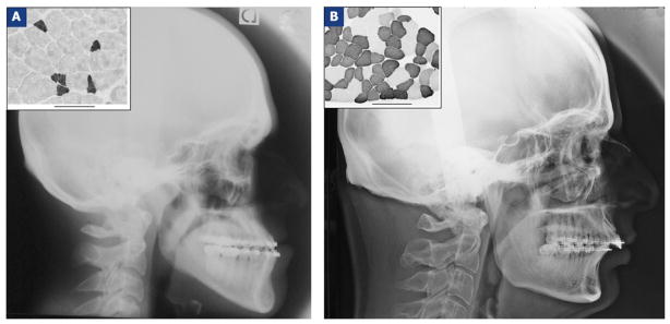
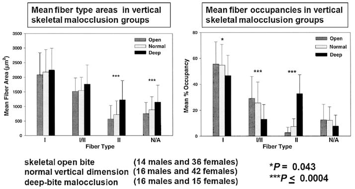
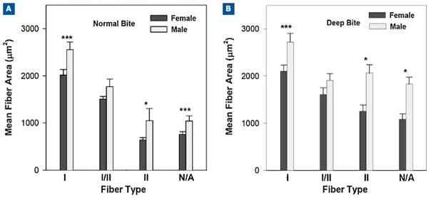
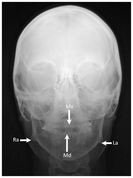
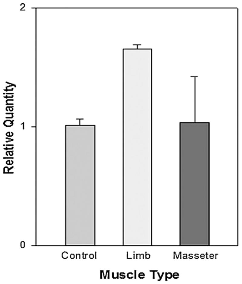
The aim of this work is to review the relationship between the function of the masseter muscle and the occurrence of malocclusions. An analysis was made of the masseter muscle samples from subjects who underwent mandibular osteotomies. The size and proportion of type-II fibers (fast) decreases as facial height increases. Patients with mandibular asymmetry have more type-II fibers on the side of their deviation. The insulin-like growth factor and myostatin are expressed differently depending on the sex and fiber diameter. These differences in the distribution of fiber types and gene expression of this growth factor may be involved in long-term postoperative stability and require additional investigations. Muscle strength and bone length are two genetically determined factors in facial growth. Myosin 1H (MYOH1) is associated with prognathia in Caucasians. As future objectives, we propose to characterize genetic variations using "Genome Wide Association Studies" data and their relationships with malocclusions.
Copyright © 2013 Elsevier Masson SAS. All rights reserved.
Conflict of interest statement
The authors declare that they have no conflicts of interest concerning this article.
Figures
Similar articles
-
Fiber-type differences in masseter muscle associated with different facial morphologies.Am J Orthod Dentofacial Orthop. 2005 Jan;127(1):37-46. doi: 10.1016/j.ajodo.2004.03.025. Am J Orthod Dentofacial Orthop. 2005. PMID: 15643413 Free PMC article.
-
Human masseter muscle fiber type properties, skeletal malocclusions, and muscle growth factor expression.J Oral Maxillofac Surg. 2012 Feb;70(2):440-8. doi: 10.1016/j.joms.2011.04.007. Epub 2011 Aug 6. J Oral Maxillofac Surg. 2012. PMID: 21821327 Free PMC article.
-
Masseter myosin heavy chain composition varies with mandibular asymmetry.J Craniofac Surg. 2011 May;22(3):1093-8. doi: 10.1097/SCS.0b013e3182107766. J Craniofac Surg. 2011. PMID: 21586952 Free PMC article.
-
Specialized cranial muscles: how different are they from limb and abdominal muscles?Cells Tissues Organs. 2003;174(1-2):73-86. doi: 10.1159/000070576. Cells Tissues Organs. 2003. PMID: 12784043 Free PMC article. Review.
-
A review of the management of anterior open bite malocclusion.Aust Orthod J. 1990 Mar;11(3):147-60. Aust Orthod J. 1990. PMID: 2152430 Review.
Cited by
-
ACTN3 R577X genotypes associate with Class II and deepbite malocclusions.Am J Orthod Dentofacial Orthop. 2014 Nov;146(5):603-11. doi: 10.1016/j.ajodo.2014.07.021. Epub 2014 Oct 28. Am J Orthod Dentofacial Orthop. 2014. PMID: 25439211 Free PMC article.
-
Introducing surface-to-surface matching technique to evaluate mandibular symmetry: A retrospective study.Heliyon. 2022 Jul 9;8(7):e09914. doi: 10.1016/j.heliyon.2022.e09914. eCollection 2022 Jul. Heliyon. 2022. PMID: 35855982 Free PMC article.
-
Epigenetic influence of KAT6B and HDAC4 in the development of skeletal malocclusion.Am J Orthod Dentofacial Orthop. 2013 Oct;144(4):568-76. doi: 10.1016/j.ajodo.2013.06.016. Am J Orthod Dentofacial Orthop. 2013. PMID: 24075665 Free PMC article.
-
Contribution of masticatory muscle pattern to craniofacial morphology in normal adults: A cross-sectional MRI study.Natl J Maxillofac Surg. 2023 May-Aug;14(2):213-220. doi: 10.4103/njms.njms_473_21. Epub 2023 Jul 13. Natl J Maxillofac Surg. 2023. PMID: 37661977 Free PMC article.
-
ENPP1 and ESR1 genotypes associated with subclassifications of craniofacial asymmetry and severity of temporomandibular disorders.Am J Orthod Dentofacial Orthop. 2017 Nov;152(5):631-645. doi: 10.1016/j.ajodo.2017.03.024. Am J Orthod Dentofacial Orthop. 2017. PMID: 29103441 Free PMC article.
References
-
- Kiliaridis S, Engström C, Thilander B. Histochemical analysis of masticatory muscle in the growing rat after prolonged alteration in the consistency of the diet. Arch Oral Biol. 1988;33:187–93. - PubMed
-
- Horowitz SL, Shapiro HH. Modification of skull and jaw architecture following removal of the masseter muscle in the rat. Am J Phys Anthropol. 1955;13:301–8. - PubMed
-
- Nanda SK, Merow WW, Sassouni V. Repositioning of the masseter muscle and its effect on skeletal form and structure. Angle Orthod. 1967;37:304–8. - PubMed
-
- McPherron AC, Lawler AM, Lee SJ. Regulation of skeletal muscle mass in mice by a new TGF-beta superfamily member. Nature. 1997;387:83–90. - PubMed
Publication types
MeSH terms
Grants and funding
LinkOut - more resources
Full Text Sources
Other Literature Sources
Research Materials
