. 2013 Jul 9:14:15.
doi: 10.1186/1471-2199-14-15.

Loss of CARM1 is linked to reduced HuR function in replicative senescence

Affiliations

Loss of CARM1 is linked to reduced HuR function in replicative senescence

Lijun Pang et al. BMC Mol Biol. .

Abstract

Background: The co-activator-associated arginine methyltransferase 1 (CARM1) catalyzes the methylation of HuR. However, the functional impact of this modification is not fully understood. Here, we investigated the influence of HuR methylation by CARM1 upon the turnover of HuR target mRNAs encoding senescence-regulatory proteins.

Results: Changing the methylation status of HuR in HeLa cells by either silencing CARM1 or mutating the major methylation site (R217K) greatly diminished the effect of HuR in regulating the turnover of mRNAs encoding cyclin A, cyclin B1, c-fos, SIRT1, and p16. Although knockdown of CARM1 or HuR individually influenced the expression of cyclin A, cyclin B1, c-fos, SIRT1, and p16, joint knockdown of both CARM1 and HuR did not show further effect. Methylation by CARM1 enhanced the association of HuR with the 3'UTR of p16 mRNA, but not with the 3'UTR of cyclin A, cyclin B1, c-fos, or SIRT1 mRNAs. In senescent human diploid fibroblasts (HDFs), reduced CARM1 was accompanied by reduced HuR methylation. In addition, knockdown of CARM1 or mutation of the major methylation site of HuR in HDF markedly impaired the ability of HuR to regulate the expression of cyclin A, cyclin B1, c-fos, SIRT1, and p16 as well to maintain a proliferative phenotype.

Conclusion: CARM1 represses replicative senescence by methylating HuR and thereby enhancing HuR's ability to regulate the turnover of cyclin A, cyclin B1, c-fos, SIRT1, and p16 mRNAs.


Figures

Figure 1
Figure 1
Methylation by CARM1 enhances the regulating of cyclin A, cyclin B1, c-fos, SIRT1, and p16 by HuR. (A) Forty-eighth after transfection of HeLa cells with CARM1siRNA (+) or a control siRNA (−), lysates were prepared for IP assays by using HuR antibody. The presence of total and methylated HuR in the IP materials was determined by Western blot analysis by using M/DMA and HuR antibodies, respectively. (B) Cell lysates described in Figure 1A were subjected to Western blot analysis to assess the protein levels of CARM1, HuR, cyclin A, cyclin B1, c-fos, SIRT1, p16, and GAPDH. Western blotting signals were quantified by densitometry. (C) HeLa cells were transfected with a vector expressing flag-HuR or flag-HuRΔ. Twenty fourh later, cells were further transfected with CARM1 siRNA or a control siRNA and cultured for an additional 48 h. Whole-cell lysates were prepared and subjected to IP assays by using anti-flag antibody (M2). Western blot analysis was performed to assess the total and methylation levels of flag-tagged HuR in the IP materials using M/MDA and flag antibodies, respectively. (D) HeLa cells were transfected with a vector expressing flag-HuR or flag-HuRΔ. Forty eighth later, lysates were prepared to assess the protein levels of CARM1, HuR, cyclin A, cyclin B1, c-fos, SIRT1, p16, and GAPDH by Western blot analysis, the signals of Western blotting were quantified by densitometry. (E) HeLa cells were either transfected with HuR or CARM1 siRNA or co-transfected with both siRNAs. Forty eighth later, Western blot analysis was performed to evaluate the levels of CARM1, HuR, cyclin A, cyclin B1, c-fos, SIRT1, p16, and GAPDH; Western blotting signals were quantified by densitometry. Data are representatives from 3 independent experiments.
Figure 2
Figure 2
Methylation by CARM1 influences the effect of HuR in regulating mRNA turnover. (A and B) RNA was prepared from cells described in Figure 1A (A) and 1D (B) to assess the mRNA levels of cyclin A, cyclin B1, c-fos, SIRT1, p16, and β-tublin by real-time qPCR against GAPDH. (C and D) Cells described in Figure 1A and 1D were exposed to actinomycin D (2 μg/ml), whereupon the cellular RNA was isolated at times indicated. Real-time qPCR against GAPDH was performed to assess the half-lives of cyclin A, cyclin B1, c-fos, SIRT1, p16, and β-tublin mRNA, as described in Methods. The real-time qPCR data are represented as means ± SEM from 3 independent experiments. The statistic significance was analyzed by Student’s t test.
Figure 3
Figure 3
Influences of CARM1-mediated methylation on the subcellular distribution and RNA-binding affinity of HuR. (A and B) Western blot analysis was performed to assess the presence of endogenous HuR (A) as well as flag-tagged HuR (flag-HuR and flag-HuRΔ) (B) in whole-cell (Total, 10 μg), cytoplasmic (Cyto., 40 μg), and nuclear fractions (Nuc., 5 μg) prepared from cells described in Figure 1A and 1D. Assessment of the levels of cytoplasmic-specific tubulin and nuclear-specific HDAC1 served to verify the quality and equal loading of the cytoplasmic and nuclear preparations, respectively. (C) Cytoplasmic extracts (100 μg) described in Figure 3A were subjected to RNA pull-down assays using biotinylated 3′UTR fragments of cyclin A, cyclin B1, c-fos, SIRT1, and p16 to detect bound endogenous HuR by Western blotting. A 10-μg aliquot of whole-cell lysates (Lys.), binding of HuR and GAPDH to the beads (Neg.), and binding of GAPDH to the cyclin A, cyclin B1, c-fos, SIRT1, and p16 3′UTR were also tested. (D) Cytoplasmic extracts (100 μg) described in Figure 3A were subjected to RNP IP assays using anti- HuR antibody. The presence of cyclin A, cyclin B1, c-fos, SIRT1, and p16 mRNAs in the IP materials were assessed by real-time qPCR. (E, F) Cytoplasmic extracts (100 μg) described in Figure 3B were either subjected to RNA pull-down assays (E) or RNP IP assays (F) to assess the association of flag-HuR and flag-HuRΔ with the mRNAs of cyclin A, cyclin B1, c-fos, SIRT1, and p16, as described in Figure 3C and 3D. (G, H) Cytoplasmic extracts described in Figure 3A and 3B were either used for RNA pull-down assays (G) or used for RNP IP assays (H) to assess the association of AUF1 with p16 mRNA, as described in Figure 3C and 3D.
Figure 4
Figure 4
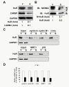
CARM1 and methylated HuR reduce replicative senescence. (A) Western blot analysis of CARM1, HuR, and GAPDH protein levels in early-passage (Young, ~27 pdl, Y) and late-passage (Senescent, ~60 pdl, S) 2BS cells. (B) HuR was immunoprecipitated from the whole cell lysates (100 μg) described in Figure 3A, whereupon the total or methylated HuR was assessed by Western blot analysis using HuR or M/DMA antibodies, respectively. Western blotting signals were quantified by densitometry. Data are representative from 3 independent experiments. (C) Cytoplasmic extracts prepared from cells described in Figure 4A were subjected to RNA pull-down assays using biotinylated 3′UTR fragments of cyclin A, cyclin B1, c-fos, SIRT1, and p16 to detect bound HuR by Western blotting. A 10-μg aliquot of whole-cell lysates (Input) and binding of GAPDH to the cyclin A, cyclin B1, c-fos, SIRT1, and p16 3′UTR were also tested. (D) Cytoplasmic extracts described in Figure 4C were subjected to RNP IP assays using anti-flag antibody, the presence of cyclin A, cyclin B1, c-fos, SIRT1, and p16 mRNAs in the IP materials were assessed by real-time qPCR.
Figure 5
Figure 5
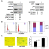
Knockdown of CARM1 reduces the levels of HuR target mRNAs and accelerates cell senescence. (A) Human diploid fibroblasts (2BS) were stably transfected with a vector expressing CARM1 shRNA or control shRNA. The levels of CARM1, cyclin A, cyclin B1, c-fos, SIRT1, p16, and GAPDH were assessed by Western blotting and the signals quantified by densitometry. (B, C) Cells described in Figure 5A were subjected to FACS analysis (B) and SA-β-gal staining (C) to assess the cell cycle distribution and senescent status. The SA-β-gal staining was represented as means ± SDs from three independent experiments. The statistic significance was analyzed by Student’s t test.
Figure 6
Figure 6
Mutation of the methylation site attenuates the effect of HuR in regulating mRNA turnover and cell senescence. (A) Human diploid fibroblasts were stably transfected with a vector expressing flag-HuR (W) or flag-HuRΔ (M), or an empty vector (−). Western blot analysis was performed to assess the protein levels of HuR, cyclin A, cyclin B1, c-fos, SIRT1, p16, and GAPDH, and quantified by densitometry. (B,C) Cells described in Figure 6A were subjected to FACS analysis (B) and SA-β-gal staining (C) to analyze the cell cycle distribution and cell senescent status, as described in Figure 5B and 5C. Values of the SA-β-gal staining represent means ± SDs of the results from three independent experiments. Statistical significance was analyzed by Student’s t test.

Similar articles

Cited by

References

    1. Ma W, Cheng S, Campbell C, Wright A, Furneaux HM. Cloning and characterization of HuR, a ubiquitously expressed elav-like protein. J Biol Chem. 1996;271:8144–8151. doi: 10.1074/jbc.271.14.8144. - DOI - PubMed
    1. Brennan CM, Gallouzi IE, Steitz JA. Protein ligands to HuR modulate its interaction with target mRNAs in vivo. J Cell Biol. 2000;151:1–14. - PMC - PubMed
    1. Srikantan S, Gorospe M. HuR function in disease. Front Biosci. 2012;17:189–205. doi: 10.2741/3921. - DOI - PMC - PubMed
    1. Wang W. Regulatory RNA-binding proteins in senescence. Ageing Res Rev. 2012;11:485–490. doi: 10.1016/j.arr.2012.02.006. - DOI - PubMed
    1. Wang W, Yang X, Cristofalo VJ, Holbrook NJ, Gorospe M. Loss of HuR is linked to reduced expression of proliferative genes during replicative senescence. Mol Cell Biol. 2001;21:5889–5898. doi: 10.1128/MCB.21.17.5889-5898.2001. - DOI - PMC - PubMed

Publication types

MeSH terms